Abstract
Background
In pediatric patients, the common cold coronavirus (ccCoV) usually causes mild respiratory illness. There are reports of coronavirus causing central nervous system (CNS) infection in experimental animal models. Some immunocompromised patients have also been reported to have fatal CNS infections with ccCoV. The aim of this study was to investigate the clinical characteristics of CNS complications related to ccCoV infection.
Methods
From January 2014 to December 2019, a retrospective analysis was performed of medical records from hospitalized patients under 19 years of age whose ccCoV was detected through polymerase chain reaction in respiratory specimens. The CNS complications were defined as clinically diagnosed seizure, meningitis, encephalopathy, and encephalitis.
Results
A total of 436 samples from 420 patients were detected as ccCoV. Among the 420 patients, 269 patients were immunocompetent and 151 patients were immunocompromised. The most common type of ccCoV was OC43 (52% in immunocompetent, 37% in immunocompromised). CNS complications were observed in 9.4% (41/436). The most common type of CNS complication was the fever-provoked seizure under pre-existing neurologic disease (42% in immunocompetent and 60% in immunocompromised patients). Among patients with CNS complications, two immunocompetent patients required intensive care unit admission due to encephalitis. Three patients without underlying neurological disease started anti-seizure medications for the first time at this admission. There was no death related to ccCoV infection.
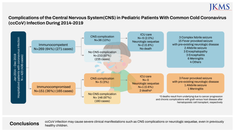
Graphical Abstract
The common cold coronavirus (ccCoV, previous human coronavirus) is a common cause of colds and is also associated with severe lower respiratory tract infections in pediatric patients with an immunocompromised state.1 ccCoV-OC43 and 229E were discovered in the 1960s.23 In the early 2000s, when polymerase chain reaction (PCR) testing was introduced, new viruses were rapidly identified. Five coronaviruses, including NL63, HKU1, severe acute respiratory syndrome coronavirus (SARS-CoV), Middle East respiratory syndrome coronavirus (MERS-CoV), and SARS-CoV-2 that infect humans were additionally discovered in the last 20 years.45678
ccCoV, including ccCoV-OC43, 229E, NL63, and HKU1, cause upper and lower respiratory infections in children.910 Some studies have suggested a possible association between ccCoV and neurologic manifestations such as febrile seizure or encephalitis in pediatric patients.911121314 Fatal encephalopathy and encephalitis cases with OC43 infections were reported in two infants with severe combined immunodeficiency and acute lymphoblastic leukemia (ALL), respectively.914 The SARS-CoV-2 is responsible for the coronavirus disease 2019 (COVID-19) pandemic that started in 2020. A study from the Korea reported that most SARS-CoV-2 infections in children had resulted in mild illnesses.15 However, some patients with underlying diseases needed hospitalization or intensive care unit (ICU) care.1617 Recent studies have reported the association between SARS-CoV-2 and complications in the central nervous system (CNS).181920
The aim of this study was to investigate the clinical characteristics of CNS complications in patients with ccCoV infection. Research on CNS complications of ccCoV may improve our understanding of newly discovered coronaviruses, including SARS-CoV, MERS-CoV, and SARS-CoV-2.
We retrospectively investigated the clinical features of pediatric patients under 19 years of age who were admitted to Samsung Medical Center (SMC) from January 2014 to December 2019 by reviewing electronic medical records. It was a tertiary care center with 127 beds dedicated to pediatrics, and an average of 6,783 pediatric patients were admitted each year. For hospitalized patients presenting with fever or respiratory symptoms, routine care included respiratory virus PCR testing. Patients with positive ccCoV in the respiratory virus PCR test from the nasopharyngeal swab were included. As a part of routine clinical care, ccCoV was identified in clinical nasopharyngeal sample using multiplex reverse transcription PCR (LG AdvanSure™ RV-plus real-time PCR [LG Life Science, Seoul, Korea] that can detect 12 respiratory viruses, including ccCoV 229E, NL63 and OC43 or FilmArray [BioFire Diagnostics, Salt Lake City, UT, USA] that can detect 20 respiratory viruses and bacteria, including 229E, NL63, OC43, and HKU1). If the same subtype of coronavirus was detected within two months in one patient, it was counted as one case.
The following data were collected from medical records: age, sex, underlying medical conditions, types of CNS symptoms (Supplementary Table 1), laboratory findings, images including brain ultrasonography and brain magnetic resonance imaging (MRI), electroencephalogram, ICU admission, respiratory viral co-infection, and clinical outcome with coronavirus infections. The severity of respiratory symptoms was categorized as asymptomatic, upper respiratory infection, and lower respiratory infection (diagnosed as bronchitis, bronchiolitis, or pneumonia, including new infiltration on radiologic finding or oxygen requirement).
When the pathogen was not clearly identified even though the patient showed a clinically serious course, the residual cerebrospinal fluid (CSF) sample was placed in a falcon tube and stored in a freezer at −80°C. Sample was thawed at room temperature prior to testing. RNA was extracted from 200 μL of CSF sample with QIAamp® MinElute® (QIAGEN, Hilden, Germany) following the manufacturer’s instructions. The library was constructed using the Nextera XT DNA sample preparation kit (Illumina, San Diego, CA, USA), followed by 5′ and 3′ adapter ligation. Adapter-ligated fragments were PCR amplified and gel purified. Trimmomatic was used to remove adapter sequences and low quality reads to reduce biases in the analysis.21 The library was sequenced using Illumina sequencing by synthesis technology. Through K-mer analysis, information of K-mer coverage, heterozygosity, and estimated genome size was provided. To assemble the fragmented sequencing, the De Bruijn Graph method was used by SPAdes (v3.13.0).22 The assembled genome was validated using a self-mapping strategy and Benchmarking Universal Single-Copy Orthologs analysis. Filtered reads were aligned against the assembled genome, and the insert size was estimated for validation. Basic Local Alignment Search Tool analysis was carried out to find regions of local similarity between sequences and identify matching species.23 Best hit and top five hit results were identified using the National Center for Biotechnology Information NT database (Supplementary Fig. 1).
Data for numerical variables are shown as median (range). Categorical data are shown as frequencies and percentages. The comparisons of clinical manifestations were evaluated by the Mann–Whitney test for numerical variables and the χ2 test or Fisher’s exact test for categorical variables. Variables with a P value of less than 0.05 were statistically significant. Statistical analysis was performed using SPSS software version 27 (IBM Corp., Armonk, NY, USA).
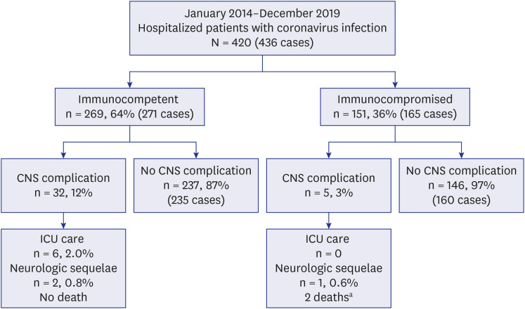
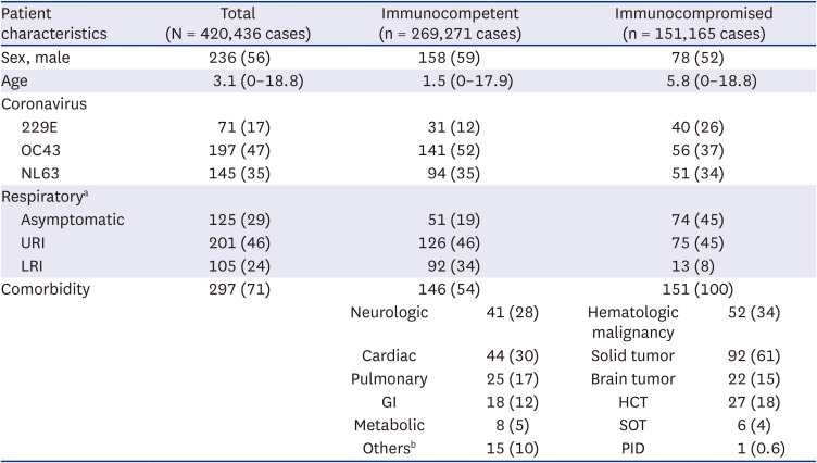
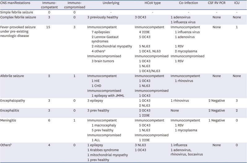
From January 2014 to December 2019, a total of 420 hospitalized patients had ccCoV detected in respiratory samples (Fig. 1). Among them, 41 patients (9.8%) had CNS complications. The specific patient demonstrations are shown in Table 1. CNS complications occurred in 10.4% (13/125) of patients without respiratory symptoms, 10.0% (20/201) of patients with upper respiratory symptoms, and 7.6% (8/105) of lower respiratory infections. The seasonality analysis showed that most patients with ccCoV infection distributed in winter. There was no statistically significant difference in seasonality between groups with or without immunocompromised conditions and between groups with or without CNS complications (Supplementary Fig. 2). Although there was no statistical significance (P = 0.226), the most common subtype of ccCoV was OC43 in the groups with and without CNS complications. The results were also similar in the immunocompetent and immunocompromised groups (Supplementary Fig. 3).
Data are presented as median (range) or number (%).
URI = upper respiratory infection, LRI = lower respiratory infection, GI = gastrointestinal, HCT = hematopoietic cell transplant, SOT = solid organ transplant, PID = primary immunodeficiency.
aData are presented as cases, 5 cases were not available by retrospective chart review.
bOthers include metabolic disease (n = 8, 5%), genitourinary (n = 5, 3%), and chronic kidney disease (n = 2, 1%).
Among the 269 immunocompetent patients, 36 patients (12.8%, 36/269) had CNS complications, while five immunocompromised patients (3%, 5/151) had CNS complications (P < 0.001) (Supplementary Table 2). Both groups included approximately 60% of male patients (P = 1.0). The median patient age in the immunocompetent and immunocompromised patient groups was 2.8 years and 8.1 years, respectively, and the age was significantly higher in immunocompromised patients (P = 0.035). Underlying neurologic disease in the immunocompetent group was observed in 58% (21/36). In addition, there were five immunocompromised patients with CNS complications, including three patients with a brain tumor, one with ALL, and one with lung graft versus host disease (GVHD) after allogenic hematopoietic cell transplant (HCT) due to juvenile myelomonocytic leukemia (JMML). Concomitant respiratory viral infection was found in 14 (39%) immunocompetent patients (3 with adenovirus, 3 with influenza, 2 with rhinovirus, 2 with respiratory syncytial virus (RSV), 3 with mycoplasma), and 1 immunocompromised patient with RSV (Supplementary Table 3). Six (2.0%) immunocompetent patients required intensive care due to neurological symptoms. However, there were no immunocompromised patients who needed ICU care. Two immunocompromised host died due to cancer progression and chronic complications with GVHD after HCT, respectively.
The most common CNS complication was the fever-provoked seizure under pre-existing neurologic disease in both groups (Table 2). In the immunocompetent patients, and meningitis (17%, 6/36) was the second most frequent events, followed by complex febrile seizures (8%, 3/36), encephalopathy (8%, 3/36), encephalitis (8%, 3/36), and afebrile seizure (6%, 2/36).
CNS = central nervous system, HCoV = human coronavirus, CSF = cerebrospinal fluid, RV = respiratory virus, PCR = polymerase chain reaction, ICU = intensive care unit, RSV = respiratory syncytial virus, HIE = hypoxic ischemic encephalopathy, CHD = congenital heart disease, ALL = acute lymphoblastic leukemia, JMML = juvenile myelomonocytic leukemia.
aHemimegalencephaly (n = 1), Rett's syndrome (n = 1), developmental delay (n = 1), cerebral infarction (n = 1).
bCerebellar dysfunction (n = 1), myoclonus (n = 1), rigidity (n = 2).
In the immunocompetent patients, three patients with encephalopathy had underlying epilepsy. They were admitted to PICU for altered mentality because of status epilepticus. Recovery of consciousness was observed in all three patients, and the time interval ranged from one day to six days. Three patients underwent CSF study. There were no pleocytosis, no bacteria grew, and no virus was detected (herpes simplex virus, enterovirus). Among the patients, two tested respiratory virus PCR using CSF sample and showed negative results.
In the immunocompromised patients, three patients with brain tumor developed seizures. Two had aggravated underlying seizures with fever and tumor bleeding. The other patient developed generalized tonic-clonic seizure with upper respiratory symptoms and was diagnosed with brain neoplasm through workup for seizure. The patient with ALL developed neck stiffness, headache, and vomiting during maintenance chemotherapy. The examination of CSF revealed pleocytosis, and he received antibiotics for ten days under the diagnosis of meningitis. No bacteria were identified in the CSF culture and blood culture, and 229E was confirmed in the respiratory virus PCR test, which was performed because of a fever three days before the onset of the headache. However, the result of respiratory virus PCR using the CSF sample was negative. One of the immunocompromised patients with JMML and lung GVHD developed seizures without a provocation factor. At that time, he developed recurrent pneumothorax, and OC43 was detected in the respiratory virus PCR test.
In most cases of CNS complications without underlying neurologic disease, the patients had no more neurologic symptoms at discharge, and no anti-seizure medications (ASMs) were required. Three patients required ASM upon discharge, and they were considered to have neurologic sequelae. Among these three, while one patient had lung GVHD after allogeneic HCT for JMML, the other two patients were previously healthy (Supplementary Table 4). OC43 type was detected in all three patients, and there was no co-infection with other respiratory viruses. The initial symptoms were seizures in all three patients. Further clinical manifestations are described (Supplementary Data 1).
Respiratory virus real-time reverse transcription PCR was performed using CSF samples in four patients in whom the sample was available among the six patients with encephalopathy or encephalitis, but all CSF samples were negative for ccCoV. To identify the pathogen from a patient with critical encephalitis, we conducted the NGS using the residual CSF sample. However, there was no proven respiratory virus from the CSF sample.
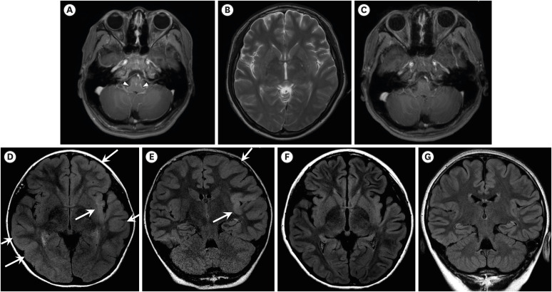
Among the patients with CNS complications, 14 underwent brain MRIs (14/41, 34%). Brain MRI was performed in six patients with fever-provoked seizure under pre-existing neurologic disease (6/18, 33%), two patients with meningitis (2/7, 29%), three patients with encephalopathy (3/3, 100%), and three patients with encephalitis (3/3, 100%). Among these 14 patients, there were no significant lesions except for three encephalitis patients. Two patients with encephalitis exhibited high signal intensity in brain lesions: one focally in the left cerebellum on T2/fluid-attenuated inversion recovery (FLAIR) images, and the other in the bilateral insular cortex, basal ganglia, thalamus, splenium, and cerebellum on T2-weighted image (Fig. 2B). In addition, the second patient showed leptomeningeal enhancement along the brainstem surface (Fig. 2A and C). The third patient with encephalitis showed left insula temporal gyrus, frontal, and right temporo-occipital area swelling on FLAIR images (Fig. 2D-G).
Among the 18 patients with fever provoked seizures under pre-existing neurologic disease, 11 patients (52%) underwent EEG. The findings were normal (n = 3), focal epileptiform discharge (n = 6), diffuse slow wave (n = 1), and focal epileptiform discharge with diffuse slow wave (n = 1). Two of three patients with afebrile seizure tested EEG and the results showed diffuse slow wave (n = 1), and focal epileptiform discharge with diffuse slow wave (n = 1). Three patients with encephalopathy showed epileptiform discharges that appeared temporal area (n = 2) and focal epileptiform discharges with background slow (n = 1). Two encephalitis patients with neurologic sequelae (who required ASM upon discharge) underwent EEG that showed focal epileptiform discharge and diffuse slow wave.
This is a retrospective study of CNS complications in pediatric patients with ccCoV infections. The proportion of CNS complications in hospitalized pediatric patients with ccCoV was 9.8% (41/420). The proportion of immunocompetent patients with CNS complications was 13% (36/269), and immunocompromised patients with CNS complications was 3% (5/151). The most common CNS complication was a fever provoked seizure in patients having an underlying neurologic disease. Three patients started ASMs for the first time at this admission with ccCoV infection. Six patients received ICU care (2.4%, 6/420) of whom two patients were previously healthy (33.3%, 2/6). There was no death related to ccCoV.
The proportion of CNS complications in hospitalized pediatric patients with ccCoV was 9.8% in this study. Although the prevalence of CNS complications associated with ccCoV is unknown, some cases have been reported.111424 In a prospective study, 9.9% of patients hospitalized for febrile seizures were reported to be positive for ccCoV in a PCR using respiratory samples. However, co-infection was identified in 52.6%.12 In China, anti-CoV IgM antibodies were detected in 22/183 (12%) with acute encephalitis-like syndrome.25 In a literature review conducted by Ellul et al.,18 the calculated prevalence rates of CNS complications associated with SARS-CoV and MERS-CoV were 0.04% and 0.2%, respectively.
In this study, OC43 was the most common subtype in patients with CNS complications. In addition, in all patients with neurologic sequelae, OC43 was the only pathogen identified from a nasopharyngeal swab. In a prospective study,12 among hospitalized patients with febrile seizures, nasopharyngeal swabs detected ccCoV in 19 patients, and 12 (63%) cases were OC43. We also performed PCR and deep sequencing in CSF from a patient with sequelae, but ccCoV was not detected. In several cases,111424 OC43 was detected in brain tissue or CSF. Among the ccCoV, which subtype is related to CNS complications is unknown. Recently, different febrile seizure incidences with COVID-19 were reported between the era of the Omicron variant of concern (VOC) and Delta VOC.26 Further research is needed to determine whether each subtype or VOC affects CNS complications differently.
In a prospective study, among 19 ccCoV-positive patients with febrile seizures, co-infection with other respiratory viruses occurred in 10 cases (52.6%), and adenovirus and RSV were the most common pathogens in five cases each.12 In our study, co-infection occurred in 15 patients (36.6%) of patients with CNS complications, and adenovirus was the most frequent pathogen in four cases. In these patients, the complex febrile seizure or fever-provoked seizure under pre-existing neurologic disease could be caused by another respiratory viral infection. However, it is difficult to determine which virus was mainly responsible.
The association of CNS complications with coronavirus has been suggested in several studies. Various mechanisms have been proposed for ccCoV as an etiologic factor to CNS complications.2728 As neurologic symptoms of COVID-19 have emerged, neuroinvasion of coronavirus is being continuously discussed.181920 A study by Abdelaziz and Waffa20 classified the mechanisms of neuronal cell injury from coronavirus into direct injury and indirect injury. Direct injury refers to virus-induced neuropathology and includes hematogenous invasion and olfactory route. In addition, the distribution of the receptors for viral entry in CNS tissue has been reported.192930 Indirect injury means virus-induced neuro-immunopathology, including misdirected autoimmune responses such as antigen-antibody reaction and inflammatory response.20
The mechanism of neurologic complications with ccCoV infection depends on the types of CNS complications. Encephalopathy and encephalitis may be related to viral direct invasion,11142028 autoimmune reaction after viral infection,31 or host factor.111314 However, viral infection often causes fever, which can lead to febrile seizure and fever-provoked seizure.32 In this study, the most common CNS complications were fever-provoked seizures in pre-existing neurologic disease patients followed by complex febrile seizures. Because the study population included patients admitted to a tertiary care institution, there were no patients with simple febrile convulsions. In the case of seizures with fever, a threshold to febrile seizure dependent on the height of body temperature was previously suggested32 as well as the mechanism of direct injury from coronavirus. Febrile convulsions with the influenza virus mainly occur in east Asia.3233 Several chromosomal loci related to febrile seizure were identified, such as FEB1, -2, -3, and -4, on chromosomes 8q13, 19p, 2q23-q24, and 5q14-q15.32 Various sodium channels and γ-aminobutyric acid receptors are also associated with febrile seizures. Therefore, genetic etiology may also involve febrile seizures with coronavirus infection. In addition, cytokine secretion, including interleukin (IL) 1β, IL-6, tumor necrosis factor α, and IL-10, or immune response to viral infection might be considered etiologic factors for febrile seizures.32
The frequencies of CNS complications of influenza and COVID-19 were comparable to that of this study (9.8%). In Australia, neurologic complications of pandemic influenza A (H1N1) was prospectively investigated in hospitalized pediatric patients. Seizures and encephalitis/encephalopathy was reported with frequency of 7.5% and 1.4%, respectively.34 In US, influenza-associated neurologic complication was identified in 10.8% of hospitalized children with laboratory-confirmed influenza and the most common neurologic complications were seizure and encephalopathy.35 Other retrospective study for neurologic complications of influenza in Korea reported 8.1% of hospitalized pediatric patients developed seizure, meningitis, encephalopathy, or encephalitis.33 In addition, neurologic complications of COVID-19 had occurred 7.0% of hospitalized children with SARS-CoV-2 infection in the US and the most frequent neurologic symptoms were febrile seizure, afebrile seizure, and encephalopathy.36 However, CNS complications with adenovirus were identified in 3.3% of hospitalized children with adenoviral infection, the most common symptom was febrile seizure.37 In addition, a systematic review and meta-analysis have been reported to occurred with a pooled prevalence of 30 cases of febrile seizure per 1000 RSV cases (I2=88.5%).38
To our knowledge, there was little study comparing CNS complications between immunocompetent and immunocompromised patients with ccCoV infection. A retrospective study was reported to investigate of characteristics of ccCoV between immunocompetent and immunocompromised children.1 Among the main diagnosis acute illness of ccCoV infected children, febrile seizures were reported in 3% of non-immunocompromised children, but no immunocompromised child presented with febrile seizure.1 A national surveillance study for neurological manifestations of influenza reported that there was no immunocompromised patient with CNS complications during the study period.39 However, there were fatal case reports such as encephalitis in immunocompromised patients caused by ccCoV-OC43.111314 These results suggest that CNS complications in an immunocompromised patient could be fatal but rare. In our study, the frequency of CNS complications was higher in the immunocompetent group than in the immunocompromised. Even some previously healthy patients showed a critical course with ICU admission. The number of patients in this single center study was insufficient to demonstrate such a difference between the two groups. In addition, the frequency of neurological complications was significantly higher in the group with underlying neurological diseases (P < 0.001). Some studies have reported neurologic disorders as a risk factor for CNS complications with influenza.3940 Therefore, it may be considered whether not only the immune status but also the underlying neurological disease affects the occurrence of CNS complications.
Increased intensity in the insula on brain MRI was observed in two patients with encephalitis (Fig. 2). However, this may also be due to a secondary change of prolonged seizure.41 Although a recent report suggested that SARS-CoV-2 could enter the brain through the human olfactory bulb,19 there is no evidence in previous studies that ccCoV invades a specific part of the brain. However, the influenza virus was demonstrated to exist in the olfactory bulb and hippocampus through several experiments.4243 In addition, brain images from patients with influenza infection showed gyral or cerebral swelling.44 Further evaluation is needed to determine whether the increased intensity of the insula in the present study is related to direct invasion of ccCoV or reactive change after seizures.
Metagenome NGS (mNGS) is an emerging approach for diagnosing pathogens, antibiotic resistance, the microbiome, and oncology.45 We examined patients with severe progression with ccCoV infection using residual CSF samples by mNGS. However, no viral genome was detected. According to recent studies, no etiology was found in more than half of the encephalitis cases despite comprehensive laboratory evaluation.46 mNGS is currently applied in the research field and is expected to benefit diagnosis and treatment if applied to the clinical field.
This study has some limitations, involving the small sample size and retrospective nature of the study design. First, this study was based on a single referral hospital with hospitalized patients, and there might be selection bias. Most patients with a common cold by ccCoV do not require laboratory or image study due to mild symptoms. Consequently, the frequency of CNS complications with ccCoV infection might be higher than the actual prevalence. Second, considering that long-term sequelae of COVID-19 patients have been recently reported,47 follow-up and additional data are needed, but this study lacks the long-term course of CNS complications related to ccCoV infection. Third, cytokines that may affect CNS complications are not measured in routine clinical care. Finally, none of the subjects had proven CNS infection by ccCoV in our study. However, the possibility cannot be ruled out that ccCoV might have been associated CNS complications through direct or indirect mechanisms, as reviewed in the literature. To prove the association or causality between ccCoV and CNS complications, prospective studies are required, including mNGS for etiology, in vivo or in vitro experiments for viral receptors, and evaluation of cytokines related to CNS complications. Nevertheless, this study provides basic epidemiology and clinical characteristics of CNS complications in pediatric patients with ccCoV infection because only a few studies have examined this issue in ccCoV infection so far. In addition, considering the increasing reports on CNS complications during this COVID-19 pandemic era, our study provides valuable additional information for comparison.
Our findings show that ccCoV infection may be associated with severe clinical manifestations such as CNS complications or neurologic sequelae, even in previously healthy children. Further study is necessary to clarify the pathogenesis and prognosis of CNS complications with ccCoV infection.
ACKNOWLEDGMENTS
We thank Ji Youn Kim from Samsung Medical Center for assistance with the particular technique for nucleic acid extraction.
Notes
Author Contributions:
Conceptualization: Park H, Kim YJ.
Data curation: Park H, Kim KR, Yoon Y, Park E.
Formal analysis: Park H, Investigation.
Methodology: Kim YJ, Huh HJ.
Software: Park H.
Validation: Park H, Kim KR, Yoon Y, Park E.
Visualization: Park H.
Writing - original draft: Park H.
Writing - review & editing: Park H, Huh HJ, Cho J, Lee J1, Lee J2, Kim JH, Kim YJ.
Lee J1, Jiwon Lee; Lee J2, Jeehun Lee.
References
1. Ogimi C, Englund JA, Bradford MC, Qin X, Boeckh M, Waghmare A. Characteristics and outcomes of coronavirus infection in children: the role of viral factors and an immunocompromised state. J Pediatric Infect Dis Soc. 2019; 8(1):21–28. PMID: 29447395.
2. Tyrrell DA, Bynoe ML. Cultivation of a novel type of common-cold virus in organ cultures. BMJ. 1965; 1(5448):1467–1470. PMID: 14288084.
3. McIntosh K, Dees JH, Becker WB, Kapikian AZ, Chanock RM. Recovery in tracheal organ cultures of novel viruses from patients with respiratory disease. Proc Natl Acad Sci U S A. 1967; 57(4):933–940. PMID: 5231356.
4. Ksiazek TG, Erdman D, Goldsmith CS, Zaki SR, Peret T, Emery S, et al. A novel coronavirus associated with severe acute respiratory syndrome. N Engl J Med. 2003; 348(20):1953–1966. PMID: 12690092.
5. van der Hoek L, Pyrc K, Berkhout B. Human coronavirus NL63, a new respiratory virus. FEMS Microbiol Rev. 2006; 30(5):760–773. PMID: 16911043.
6. Woo PC, Lau SK, Chu CM, Chan KH, Tsoi HW, Huang Y, et al. Characterization and complete genome sequence of a novel coronavirus, coronavirus HKU1, from patients with pneumonia. J Virol. 2005; 79(2):884–895. PMID: 15613317.
7. Zaki AM, van Boheemen S, Bestebroer TM, Osterhaus AD, Fouchier RA. Isolation of a novel coronavirus from a man with pneumonia in Saudi Arabia. N Engl J Med. 2012; 367(19):1814–1820. PMID: 23075143.
8. Zhu N, Zhang D, Wang W, Li X, Yang B, Song J, et al. A novel coronavirus from patients with pneumonia in China, 2019. N Engl J Med. 2020; 382(8):727–733. PMID: 31978945.
9. Ogimi C, Kim YJ, Martin ET, Huh HJ, Chiu CH, Englund JA. What’s new with the old coronaviruses? J Pediatric Infect Dis Soc. 2020; 9(2):210–217. PMID: 32314790.
10. Choi YY, Kim YK, Choi EH. Clinical and epidemiological characteristics of common human coronaviruses in children: a single center study, 2015–2019. Pediatr Infect Vaccine. 2021; 28(2):101–109.
11. Morfopoulou S, Brown JR, Davies EG, Anderson G, Virasami A, Qasim W, et al. Human coronavirus OC43 associated with fatal encephalitis. N Engl J Med. 2016; 375(5):497–498. PMID: 27518687.
12. Jevšnik M, Steyer A, Pokorn M, Mrvič T, Grosek Š, Strle F, et al. The role of human coronaviruses in children hospitalized for acute bronchiolitis, acute gastroenteritis, and febrile seizures: a 2-year prospective study. PLoS One. 2016; 11(5):e0155555. PMID: 27171141.
13. Kasereka MC, Hawkes MT. Neuroinvasive potential of human coronavirus OC43: case report of fatal encephalitis in an immunocompromised host. J Neurovirol. 2021; 27(2):340–344. PMID: 33405204.
14. Nilsson A, Edner N, Albert J, Ternhag A. Fatal encephalitis associated with coronavirus OC43 in an immunocompromised child. Infect Dis (Lond). 2020; 52(6):419–422. PMID: 32067542.
15. Seon JY, Jeon WH, Bae SC, Eun BL, Choung JT, Oh IH. Characteristics in pediatric patients with coronavirus disease 2019 in Korea. J Korean Med Sci. 2021; 36(20):e148. PMID: 34032033.
16. Choi JH, Choi SH, Yun KW. Risk factors for severe COVID-19 in children: a systematic review and meta-analysis. J Korean Med Sci. 2022; 37(5):e35. PMID: 35132841.
17. Lee H, Choi S, Park JY, Jo DS, Choi UY, Lee H, et al. Analysis of critical COVID-19 cases among children in Korea. J Korean Med Sci. 2022; 37(1):e13. PMID: 34981683.
18. Ellul MA, Benjamin L, Singh B, Lant S, Michael BD, Easton A, et al. Neurological associations of COVID-19. Lancet Neurol. 2020; 19(9):767–783. PMID: 32622375.
19. Fodoulian L, Tuberosa J, Rossier D, Boillat M, Kan C, Pauli V, et al. SARS-CoV-2 receptors and entry genes are expressed in the human olfactory neuroepithelium and brain. iScience. 2020; 23(12):101839. PMID: 33251489.
20. Abdelaziz OS, Waffa Z. Neuropathogenic human coronaviruses: a review. Rev Med Virol. 2020; 30(5):e2118. PMID: 32687681.
21. Bolger AM, Lohse M, Usadel B. Trimmomatic: a flexible trimmer for Illumina sequence data. Bioinformatics. 2014; 30(15):2114–2120. PMID: 24695404.
22. Bankevich A, Nurk S, Antipov D, Gurevich AA, Dvorkin M, Kulikov AS, et al. SPAdes: a new genome assembly algorithm and its applications to single-cell sequencing. J Comput Biol. 2012; 19(5):455–477. PMID: 22506599.
23. Altschul SF, Gish W, Miller W, Myers EW, Lipman DJ. Basic local alignment search tool. J Mol Biol. 1990; 215(3):403–410. PMID: 2231712.
24. Yeh EA, Collins A, Cohen ME, Duffner PK, Faden H. Detection of coronavirus in the central nervous system of a child with acute disseminated encephalomyelitis. Pediatrics. 2004; 113(1 Pt 1):e73–e76. PMID: 14702500.
25. Li Y, Li H, Fan R, Wen B, Zhang J, Cao X, et al. Coronavirus infections in the central nervous system and respiratory tract show distinct features in hospitalized children. Intervirology. 2016; 59(3):163–169. PMID: 28103598.
26. Iijima H, Kubota M, Ogimi C. Change in seizure incidence in febrile children with COVID-19 in the era of omicron variant of concern. J Pediatric Infect Dis Soc. 2022; 11(11):514–517. PMID: 35984115.
27. Bohmwald K, Gálvez NMS, Ríos M, Kalergis AM. Neurologic alterations due to respiratory virus infections. Front Cell Neurosci. 2018; 12:386. PMID: 30416428.
28. Desforges M, Le Coupanec A, Dubeau P, Bourgouin A, Lajoie L, Dubé M, et al. Human coronaviruses and other respiratory viruses: underestimated opportunistic pathogens of the central nervous system? Viruses. 2019; 12(1):14. PMID: 31861926.
29. Hoffmann M, Kleine-Weber H, Schroeder S, Krüger N, Herrler T, Erichsen S, et al. SARS-CoV-2 cell entry depends on ACE2 and TMPRSS2 and is blocked by a clinically proven protease inhibitor. Cell. 2020; 181(2):271–280.e8. PMID: 32142651.
30. Morgello S. Coronaviruses and the central nervous system. J Neurovirol. 2020; 26(4):459–473. PMID: 32737861.
31. Lin JE, Asfour A, Sewell TB, Hooe B, Pryce P, Earley C, et al. Neurological issues in children with COVID-19. Neurosci Lett. 2021; 743:135567. PMID: 33352286.
32. Millichap JG, Millichap JJ. Role of viral infections in the etiology of febrile seizures. Pediatr Neurol. 2006; 35(3):165–172. PMID: 16939854.
33. Choi GJ, Park JY, Choi JS, Choi SR, Kim D, Lee JH, et al. Influenza-associated neurologic complications in hospitalized pediatric patients: a multicenter retrospective study in Republic of Korea. Pediatr Infect Dis J. 2021; 40(12):e466–e471. PMID: 34609108.
34. Khandaker G, Zurynski Y, Buttery J, Marshall H, Richmond PC, Dale RC, et al. Neurologic complications of influenza A(H1N1)pdm09: surveillance in 6 pediatric hospitals. Neurology. 2012; 79(14):1474–1481. PMID: 22993280.
35. Frankl S, Coffin SE, Harrison JB, Swami SK, McGuire JL. Influenza-associated neurologic complications in hospitalized children. J Pediatr. 2021; 239:24–31.e1. PMID: 34293371.
36. Antoon JW, Hall M, Howard LM, Herndon A, Freundlich KL, Grijalva CG, et al. COVID-19 and acute neurologic complications in children. Pediatrics. 2022; 150(5):e2022058167. PMID: 35949041.
37. Huang YC, Huang SL, Chen SP, Huang YL, Huang CG, Tsao KC, et al. Adenovirus infection associated with central nervous system dysfunction in children. J Clin Virol. 2013; 57(4):300–304. PMID: 23619053.
38. Riccò M, Cerviere MP, Corrado S, Ranzieri S, Marchesi F. Respiratory syncytial virus: an uncommon cause of febrile seizures-results from a systematic review and meta-analysis. Pediatr Rep. 2022; 14(4):464–478. PMID: 36412662.
39. Goenka A, Michael BD, Ledger E, Hart IJ, Absoud M, Chow G, et al. Neurological manifestations of influenza infection in children and adults: results of a National British Surveillance Study. Clin Infect Dis. 2014; 58(6):775–784. PMID: 24352349.
40. Antoon JW, Hall M, Herndon A, Johnson DP, Brown CM, Browning WL, et al. Prevalence, risk factors, and outcomes of influenza-associated neurologic complications in children. J Pediatr. 2021; 239:32–38.e5. PMID: 34216629.
41. Kim JA, Chung JI, Yoon PH, Kim DI, Chung TS, Kim EJ, et al. Transient MR signal changes in patients with generalized tonicoclonic seizure or status epilepticus: periictal diffusion-weighted imaging. AJNR Am J Neuroradiol. 2001; 22(6):1149–1160. PMID: 11415912.
42. Hosseini S, Wilk E, Michaelsen-Preusse K, Gerhauser I, Baumgärtner W, Geffers R, et al. Long-term neuroinflammation induced by influenza A virus infection and the impact on hippocampal neuron morphology and function. J Neurosci. 2018; 38(12):3060–3080. PMID: 29487124.
43. van Riel D, Leijten LM, Verdijk RM, GeurtsvanKessel C, van der Vries E, van Rossum AM, et al. Evidence for influenza virus CNS invasion along the olfactory route in an immunocompromised infant. J Infect Dis. 2014; 210(3):419–423. PMID: 24550441.
44. Chen LW, Teng CK, Tsai YS, Wang JN, Tu YF, Shen CF, et al. Influenza-associated neurological complications during 2014–2017 in Taiwan. Brain Dev. 2018; 40(9):799–806. PMID: 29891404.
46. Glaser CA, Honarmand S, Anderson LJ, Schnurr DP, Forghani B, Cossen CK, et al. Beyond viruses: clinical profiles and etiologies associated with encephalitis. Clin Infect Dis. 2006; 43(12):1565–1577. PMID: 17109290.