Journal List > Ann Surg Treat Res > v.105(5) > 1516084753

Bong, Kang, and Park: Multicenter study of prognostic factors in paraaortic lymph node dissection for metastatic colorectal cancer

Abstract

Purpose

The role of paraaortic lymph node dissection (PALND) in colorectal cancer (CRC) has been less evaluated than surgical treatments for other distant metastases. We evaluated surgical outcomes after PALND and identified prognostic factors.

Methods

The medical records of patients who underwent PALND for paraaortic lymph node metastasis (PALNM) were reviewed retrospectively. All patients were categorized into the M1a group (isolated PALNM, n = 27), and the M1bc group (distant metastases other than PALNM, n = 26). Three severity factors (PALNM-SF: number of harvested paraaortic lymph nodes [hLN], ≥14; number of metastatic paraaortic lymph nodes [mLN], ≥5; and lymph nodes ratio [mLN/hLN], ≥0.5) were defined to determine their effects on survival.

Results

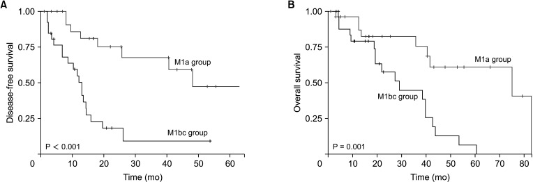
The 5-year overall survival (OS) of the M1a and M1bc groups were 61.1% and 6.4%, respectively (P = 0.0013). The 5-year disease-free survival (DFS) of the M1a group was 47.4%, and the 3-year DFS of the M1bc group was 9.1% (P < 0.001). Patients with 2 or more PALNM-SFs showed worse OS than those with 1 PALNM-SF (P = 0.017). In multivariate analysis, M1bc (non-isolated PALNM) was the only significant factor for survival. In the M1a group, patients with 2 or more PALNM-SFs showed significantly worse survival than those with a single PALNM-SF. In multivariate analysis, 2 or more PALNM-SF was a significant factor for survival.

Conclusion

PALND for CRC provided favorable outcomes in the survival of an isolated PALNM, although this was uncertain for non-isolated PALNMs. The PALNM-SFs helped assess the prognosis after PALND.

INTRODUCTION

Twenty percent of patients with colorectal cancer (CRC) have stage IV, metastatic colon cancer at the initial diagnosis, with a reported 5-year survival rate of 12.5% [1]. Recently, with the development of various treatment options such as surgery, chemotherapy, and radiation therapy, the mortality rate of CRC, including that of patients with stage IV, has been steadily decreasing [2]. Among them, the essential treatment option is whether resection of the metastasized area is possible. This is the only method to achieve a cure [3]. In particular, patients with metastases in the lung and liver can expect an improvement of more than 50% in overall survival (OS) through complete resection of the metastatic area.
While surgical treatment is recommended for liver and lung metastases, there is no straightforward recommended treatment for paraaortic lymph node metastasis (PALNM) of CRC. Although several studies have shown favorable outcomes regarding paraaortic lymph node dissection (PALND) [45], these studies’ cohorts have a small number of patients enrolled, and there aren’t many randomized control trials. This is because the incidence of PALNM is significantly low and is reported to be less than 2% in all CRCs. A systematic review revealed that PALND can improve survival outcomes with minimal complications and several factors, including well-differentiated histology, complete (R0) resection, a low number of PALNM, and metachronous metastasis with primary cancer, were reported as significant factors associated with good prognoses [6].
When considering the surgical risks of PALND, it is crucial to identify the prognostic factors that enable the selection of patients who would benefit from PALND. Based on the results of PALND conducted at 2 tertiary hospitals, we aimed to verify the prognostic factors proven in previous studies and suggest new factors.

METHODS

The Institutional Review Boards of the Korea University Guro Hospital and Korea University Anam Hospital approved the ethical clearances of this study (2021GR0332 and 2022AN0573). This study was performed in accordance with the Declaration of Helsinki and written informed consent was waived due to its retrospective nature.

Patients

The medical records of patients who underwent PALND for CRC between January 2013 and July 2022 were collected retrospectively from the clinical data warehouse of the Korea University Guro Hospital and Korea University Anam Hospital. PALND was defined as radical peritoneal and retroperitoneal lymphadenectomy of at least 1 of 5 areas: the right inferior vena cava (IVC), aortocaval, left paraaorta, right common iliac, and left common iliac area (below the renal vessels and above the common iliac bifurcations) (Fig. 1). Patients who underwent PALND for a diagnostic biopsy were excluded. Patients with <1 month of follow-up after PALND were also excluded. Age, sex, body mass index, American Society of Anesthesiologists physical status (ASA PS) grade, primary cancer characteristics including T and N categories, CEA levels, histologic types, microsatellite instability status, and neoadjuvant chemotherapy (NAC) were evaluated.

Paraaortic lymph node dissection

PALND was performed in patients with the possibility of complete resection of the metastatic site, including PALNM. PALNM was preoperatively evaluated based on radiological modalities, including abdominopelvic CT or PET-CT. Radiologists and nuclear radiologists in the multidisciplinary team of each institution reviewed all preoperative PALNM diagnoses. PALND was performed concurrently with primary tumor resection and metastasectomy for other metastases in the M1bc group that were determined as completely resectable by preoperative imaging based on the multidisciplinary team’s decision. Furthermore, we investigated whether they had synchronous or metachronous metastasis with primary cancer (SYNCH-PALNM or METACH-PALNM, respectively). In addition, the number of harvested lymph nodes (hLN) and metastasized lymph nodes (mLN) were analyzed, and the lymph node ratio (LNR) was derived by dividing mLN by hLN. Based on the results of statistical differences in survival rates, the severity factors for 3 PALNMs (PALNM-SF) were derived: hLN of ≥14, mLN of ≥5, and LNR of ≥0.5 (Supplementary Fig 1). The number of PALNM-SF (1, 2, or more) that patients had was assessed to analyze the disseminated status of PALNM. The clinical features of lymph nodes (LNs) for PALNM-SF were evaluated based on the outcomes of LNs harvested after radical lymphadenectomy in paraaortic lymph node (PALN) areas. The information on LNs harvested in the regional area of the primary tumor was not included.
The patients were divided into 2 groups according to the presence of distant metastases. The M1a group consisted of patients with isolated PALNM, whereas the M1bc group consisted of patients with PALNM and distant CRC metastases. OS represents the period from the time of surgery for PALND to death, and disease-free survival (DFS) represents the period from the time of surgery for PALND to disease progression.

Statistical analysis

All statistical analyses were performed using the R software version 4.1.0 (R Foundation for Statistical Computing). Proportional survival analysis, including OS and DFS, was performed using the Kaplan-Meier method and log-rank test. Risk factors and hazard ratios for OS and DFS were evaluated using the Cox proportional regression analysis in the univariate and multivariate analysis. LNR was not independent of mLN and hLN, and PALNM-SFs were not included in the univariate analysis. The statistical significance was defined as a 2-sided P-value of <0.05.

RESULTS

Patient characteristics and postoperative results

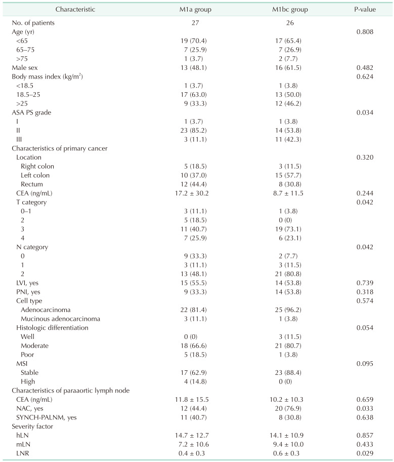
The clinical data of 98 patients who underwent PALND were collected. Forty-five patients were excluded due to the following factors: biopsy only (n = 5), pelvic lateral side LN dissection only (n = 25), less than 1 month of follow-up after PALND (n = 4), and insufficient information (n = 11). Therefore, 53 patients were included in the analysis. The M1a group consisted of 27 individuals, while the M1bc group consisted of 26 patients. The clinical characteristics of the 2 groups are presented in Table 1. The T and N categories of the primary tumors in the M1bc group were more aggressive than those in the M1a group. NAC was performed more frequently in the M1bc group. The hLN was 14.7 ± 12.7 and 14.1 ± 10.9 in the M1a and M1bc groups, respectively. The mLN in the M1bc group (9.4 ± 10.0) was higher than that in the M1a group (7.2 ± 10.6); however, the difference was not statistically significant. LNR of the M1bc group was higher than that of the M1a group (0.6 ± 0.3 vs. 0.4 ± 0.3, P = 0.029). PALND was mainly performed in 3 areas: the aortocaval (n = 42, 79.2%), right IVC (n = 41, 77.3%), and left aorta (n = 42, 79.2%). There was no significant difference in the PALND area between the 2 groups (Supplementary Table 1). Postoperative complications occurred in 5 (18.5%) and 2 (7.6%) patients in the M1a and M1bc groups, respectively. One case of mortality (3.7%) occurred in the M1a group due to pneumonia. Table 2 shows the proportion of patients with PALNM-SF in both groups; no statistically significant difference was observed.

Survivals

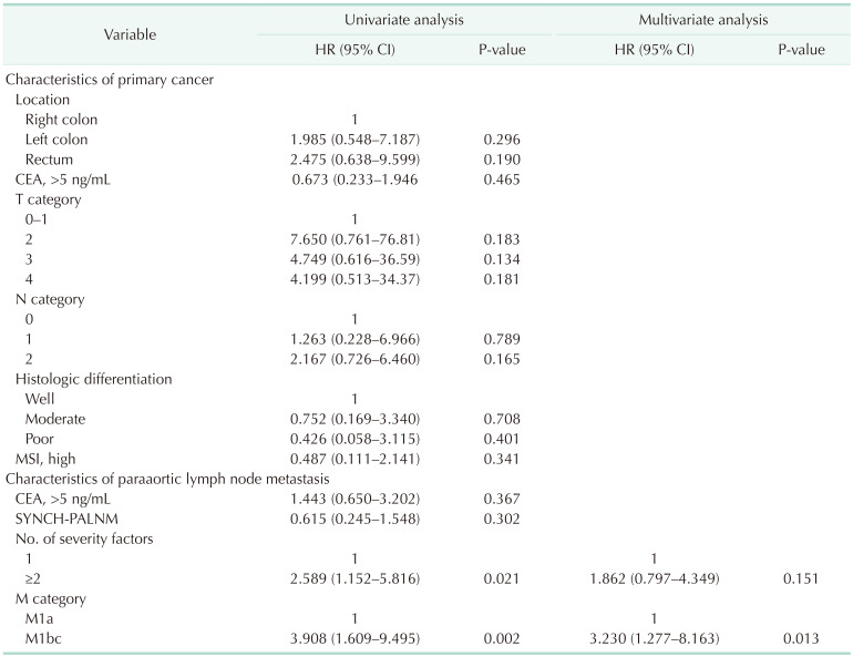
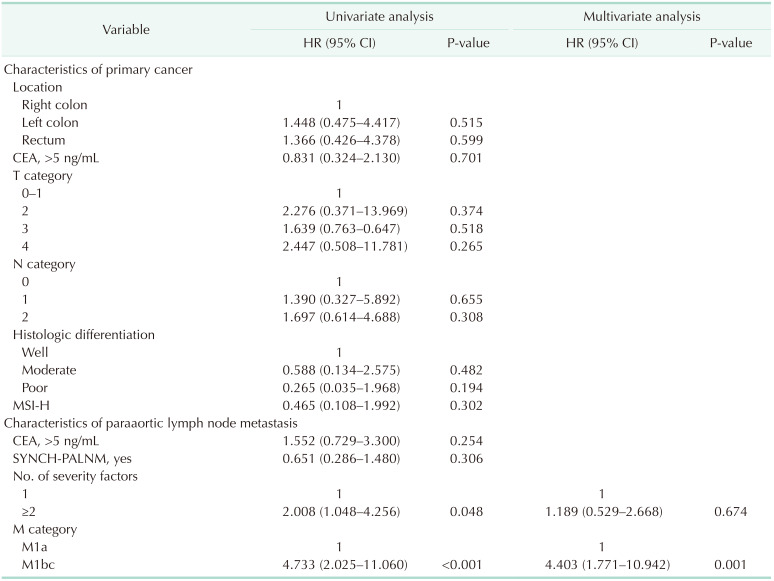
Tables 3 and 4 show that the number of PALNM-SF and M categories was a significant factor for OS and DFS in the univariate analysis. In multivariate analysis, only the M category was significantly related to OS and DFS. The 5-year OS rates of the M1a and M1bc groups were 61.1% and 6.4%, respectively (P = 0.0013) (Fig. 2). The 5-year DFS rate of patients with M1a was 47.4%. The 5-year DFS of the M1bc group was not available, and the 3-year DFS rate was 9.1% (P < 0.001). As shown in Fig. 3, patients with 2 or more PALNM-SFs had worse OS than those with 1 PALNM-SF (P = 0.017). The DFS of patients with 2 or more PALNM-SFs was also worse than that of patients with 1 PALNM-SF; however, the statistical difference was marginal (P = 0.065).
In the univariate and multivariate analyses for OS and DFS of the M1a group, the number of PALNM-SFs was significant (Supplementary Tables 1 and 2). Patients in the M1a group with 2 or more PALNM-SFs showed significantly worse OS and DFS than those with 1 PALNM-SF (Fig. 4).
For subgroup analysis, we divided the M1bc group into the ‘M1bc-resection’ group and the ‘M1bc-no resection’ group. The M1bc-resection group consisted of 15 patients who had undergone metastasectomy for distant metastases before or at the time of PALND. The M1bc-no resection group consisted of 11 patients who received palliative chemotherapy without metastasectomy for other distant metastases due to disease progression after PALND. The OS and DFS of the M1bc-resection and M1bc-no resection groups showed no significant difference (Supplementary Fig. 2).

DISCUSSION

In the current study, survival after PALND for patients with CRC with an isolated PALNM yielded acceptable outcomes. In addition, 3 PALNM-SFs were suggested to increase prognosis precision after PALND. Previous research indicated that PALND should be selectively considered as a treatment option for PALNM [78]. Researchers recommended that, if R0 resection can be achieved, PALND should be considered when an enlarged PALN is identified during surgery. Several researchers have reported that the median OS of patients with non-resected PALNM is lower (12–33 months) than that of patients with resected PALNM (34–64 months) [49]. PALND may offer a survival advantage for specific patient subgroups; hence, it is imperative to define appropriate criteria and anticipate survival outcomes subsequent to PALND.
In our study, we set 3 PALNM-SFs related to the prognosis of patients with PALNM according to the subgroup analysis in Supplementary Fig 1. The first factor was the mLN. The 5-year OS rates for patients with mLN of <5 and ≥5 were 51.2% and 15.8%, respectively (P = 0.049), while the 5-year DFS rates for patients with mLN of <5 and ≥5 were 39.4% and 18.1%, respectively (P = 0.16). Bae et al. [10] reported that mLN of >7 was significantly related to worse survival on an isolated PALNM. Song et al. [7] also reported that only mLN of ≥4 was associated with survival after PALND. Although the cut-off values of mLN vary in previous studies, greater mLN represents a worse prognosis [11]. The second factor was the hLN. In our study, >14 harvested PALNs were associated with worse survival. We hypothesized that this was because of the association between hLN and the distribution of PALNM. Unlike the dissection of regional LNs in primary cancer, the principle of PALND has not been thoroughly established. The areas of PALND were selectively determined according to preoperative or operational findings. The scope of PALND may vary depending on the patient’s condition. In our study, the number of dissected areas in patients with hLNs of ≥14 were greater than those in patients with hLNs of <14 (Supplementary Fig. 3). Thus, greater hLNs may represent a more broadly disseminated distribution of PALN over the PALN areas. The third factor was the LNR. The role of LNR has been elucidated in previous research on the regional LNs of CRC [12]. Through their systematic review analysis, Ceelen et al. [13] reported that the prognostic effect of LNR on patients with stage III CRC was superior to that of the N category in the conventional TNM staging system. However, the effects of parameters related to the distribution of LNs have not yet been evaluated for PALNM. In our study, we applied LNR to PALNM and found that LNR was a significant prognostic factor (Supplementary Fig. 1C). Moreover, PALNM with 2 or more PALNM-SFs showed significantly worse survival than PALNM with a single PALNM-SF. Therefore, in this study, we evaluated the severity of PALNM more accurately by considering the distribution, ratio, and absolute PALNM count.
There are more conflicting reports on the non-isolated PALNM (M1bc group) than on the isolated PALNM (M1a group) in patients with CRC. In a series of 25 patients, Gagnière et al. [5] demonstrated that the 5-year OS of patients with isolated and non-isolated PALNM was 56% and 51%, respectively. Nakai et al. [14] reported in their study on the prognosis of 30 patients with PALNM that adjuvant chemotherapy, high CEA level, and the presence of lateral LNs were related to survival. However, simultaneous distant metastasis was not associated with poor survival. On the other hand, there are reports that isolated PALNM has a better prognosis. Ushigome et al. [15] did not show statistical significance of the M1bc categories for worse survival than the M1a category. However, the authors specified the early recurrence and poor OS of M1bc after PALND and emphasized that distant metastases other than PALNM may have prognostic potential for PALNM. Yamada et al. [16] reported that the recurrence-free survival of patients in the M1bc group was significantly worse than that of patients in the M1a group (27.6% vs. 0.0%). In our study, patients in the M1bc group showed substantially worse survival than those with isolated PALNM (M1a group). In addition, the subgroup analysis did not demonstrate the usefulness of other metastasectomies. This result contradicted that of previous studies in which survival rate improvements required repeated resection for complete resection of PALNM and distant metastasis [1112171819].
SYNCH-PALNM and METACH-PALNM are also significant factors in determining the treatment strategy for PALNM. In our study, the survival outcomes of patients with SYNCH-PALNM were better than those of patients with METACH-PALNM, although a statistical difference was not observed (Tables 3, 4, 5, Supplementary Fig. 4). Although the survival difference between synchronous and metachronous liver metastasis in CRC is debatable, there is a trend that metachronous liver metastasis correlates with better survival than synchronous liver metastasis [1120]. Arimoto et al. [21] showed a similar trend regarding those with SYNCH-PALNM which had a significantly worse OS than those with METACH-PALNM. Choi et al. [9] also reported that the median OS of SYNCH-PALNM was worse than that of METACH-PALNM (29 months vs. 61 months). This may be because SYNCH-PALNM has a more significant tumor burden than METACH-PALNM. In our study, SYNCH-PALNM appeared to elicit a better prognosis than METACH-PALNM. However, there was no statistical difference, and the sizes of the 2 groups were too small to generate conclusive evidence.
PALND is not commonly performed because of the low incidence and risk of surgery, as well as the evolution of chemotherapeutic agents [17]. In our institutions, the NAC regimens were based on combination therapy of target agents, including cetuximab or bevacizumab plus irinotecan or oxaliplatin. In our study, the proportion of patients who received NAC was significantly higher in the M1bc group than in the M1a group (76.9% vs. 44.4%). The purpose of NAC was to reduce the tumor burden to render the metastasis resectable. In addition, we could predict survival after surgery according to the response to NAC, and it helps clinicians select patients who could benefit from invasive surgery [2223]. Previous studies reported improved curability and survival after good response, or even complete response, in patients who underwent NAC for PALNM of CRC [2425]. In our study, 5 patients were diagnosed with a complete response to NAC (3 in the M1a group and 2 in the M1bc group), and 1 received neoadjuvant chemoradiotherapy (NCRT) for PALNM. Similar to the recent developments in total neoadjuvant therapy for advanced rectal cancer, the effect of NAC or NCRT for PALNM on the survival of patients with PALNM needs to be further elucidated [26].
In our study, 6 patients (13.2%) showed 7 morbidities (1 ileus, 1 intraabdominal abscess, 1 pneumonia, 1 wound infection, and 3 bleeding), which was comparable with other reports (8%–38.9%) and 1 patient died in the M1a group (5, 7, 16). There is no consensus on the anatomical areas to be dissected by PALND, which requires invasive procedures that can potentially injure retroperitoneal organs, including ureters and large vessels. Since CT scans and PET-CT have 80.3%–89.4% accuracy for PALNM, a multidisciplinary team is required to more correctly and safely plan for PALND and reduce postoperative morbidity and mortality [17]. In addition, the recent application of an indocyanine green fluorescence imaging-guided approach for PALND of rectal cancer showed a potentially safer method of performing PALND and yielded a sufficient harvest [27].
This study has several limitations. The first is the heterogeneous characteristics of the patients in each group. The pattern of metastasis varies according to the organ and extent. Although we divided the patients into M1a and M1bc groups, heterogeneity may exist in each group, which may hinder the reproducibility of the statistical analysis. The second limitation is cancer-sidedness. In this study, most primary cancers were on the left side, including the left sigmoid colon and rectum (81.4% in the M1a group and 88.8% in the M1bc group). Right-sided colon cancer is known to have different immune responses than left-sided CRC, and tumor aggressiveness of right-sided colon cancer is worse than that of left-sided CRC, especially in stage IV [28]. In this study, there was no statistical relationship. However, we cannot exclude an association between tumor-sidedness and PALNM prognosis owing to each group’s composition and reduced size. Third, patients who received PALND other than the aortic area (including the aortocaval and both the right IVC and left aorta) were also included. Pelvic lateral LN metastases are a significant prognostic factor, particularly in advanced rectal cancer [29]. In this study, 10 patients (18.8%) received PALND and pelvic lateral LN dissection simultaneously. In addition, 7 patients (14%) underwent radical retroperitoneal lymphadenectomy over the common iliac area without aortic area dissection. This might make the patient characteristics in this study less reliable. Finally, only after surgery could PALNM-SF be obtained. As a result, PALNM-SF has a relatively limited role in determining indications for PANLD.
According to the present results, PALND showed favorable outcomes in isolated PALNM; however, its survival benefits in PALNM with other distant metastases are uncertain. In addition, this study suggests that the PALNM-SF is a new prognostic factor for PALND.

Notes

Fund/Grant Support: This work was supported by Korea University (No.K2023001) and the National Research Foundation of Korea (NRF) grant funded by the Korea government (MSIT) (No. NRF-2022R1F1A1072455).

Conflict of Interest: No potential conflict of interest relevant to this article was reported.

Author Contribution:

  • Conceptualization, Methodology: JWB, PP.

  • Formal Analysis: JWB, SK.

  • Investigation: JWB.

  • Funding Acquisition: SK, PP.

  • Supervision: PP.

  • Writing – Original Draft: JWB.

  • Writing – Review & Editing: JWB, SK.

References

1. Hu CY, Bailey CE, You YN, Skibber JM, Rodriguez-Bigas MA, Feig BW, et al. Time trend analysis of primary tumor resection for stage IV colorectal cancer: less surgery, improved survival. JAMA Surg. 2015; 150:245–251. PMID: 25588105.
crossref
2. Jawed I, Wilkerson J, Prasad V, Duffy AG, Fojo T. Colorectal cancer survival gains and novel treatment regimens: a systematic review and analysis. JAMA Oncol. 2015; 1:787–795. PMID: 26181239.
crossref
3. Lee RM, Cardona K, Russell MC. Historical perspective: two decades of progress in treating metastatic colorectal cancer. J Surg Oncol. 2019; 119:549–563. PMID: 30806493.
crossref
4. Min BS, Kim NK, Sohn SK, Cho CH, Lee KY, Baik SH. Isolated paraaortic lymph-node recurrence after the curative resection of colorectal carcinoma. J Surg Oncol. 2008; 97:136–140. PMID: 17963247.
crossref
5. Gagnière J, Dupré A, Chabaud S, Peyrat P, Meeus P, Rivoire M. Retroperitoneal nodal metastases from colorectal cancer: curable metastases with radical retroperitoneal lymphadenectomy in selected patients. Eur J Surg Oncol. 2015; 41:731–737. PMID: 25899983.
crossref
6. Wong JS, Tan GH, Teo MC. Management of para-aortic lymph node metastasis in colorectal patients: a systemic review. Surg Oncol. 2016; 25:411–418. PMID: 27916174.
crossref
7. Song SH, Park SY, Park JS, Kim HJ, Yang CS, Choi GS. Laparoscopic para-aortic lymph node dissection for patients with primary colorectal cancer and clinically suspected para-aortic lymph nodes. Ann Surg Treat Res. 2016; 90:29–35. PMID: 26793690.
crossref
8. Lee J, Park HM, Lee SY, Kim CH, Kim HR. Prognostic significance of enlarged paraaortic lymph nodes detected during left-sided colorectal cancer surgery: a single-center retrospective cohort study. World J Surg Oncol. 2021; 19:9. PMID: 33430884.
crossref
9. Choi PW, Kim HC, Kim AY, Jung SH, Yu CS, Kim JC. Extensive lymphadenectomy in colorectal cancer with isolated para-aortic lymph node metastasis below the level of renal vessels. J Surg Oncol. 2010; 101:66–71. PMID: 19842140.
crossref
10. Bae SU, Hur H, Min BS, Baik SH, Lee KY, Kim NK. Which patients with isolated para-aortic lymph node metastasis will truly benefit from extended lymph node dissection for colon cancer? Cancer Res Treat. 2018; 50:712–719. PMID: 28707461.
crossref
11. Zizzo M, Dorma MP, Zanelli M, Sanguedolce F, Bassi MC, Palicelli A, et al. Long-term outcomes of surgical resection of pathologically confirmed isolated para-aortic lymph node metastases in colorectal cancer: a systematic review. Cancers (Basel). 2022; 14:661. PMID: 35158930.
crossref
12. Kim HJ, Choi GS. Clinical implications of lymph node metastasis in colorectal cancer: current status and future perspectives. Ann Coloproctol. 2019; 35:109–117. PMID: 31288500.
crossref
13. Ceelen W, Van Nieuwenhove Y, Pattyn P. Prognostic value of the lymph node ratio in stage III colorectal cancer: a systematic review. Ann Surg Oncol. 2010; 17:2847–2855. PMID: 20559741.
crossref
14. Nakai N, Yamaguchi T, Kinugasa Y, Shiomi A, Kagawa H, Yamakawa Y, et al. Long-term outcomes after resection of para-aortic lymph node metastasis from left-sided colon and rectal cancer. Int J Colorectal Dis. 2017; 32:999–1007. PMID: 28382511.
crossref
15. Ushigome H, Yasui M, Ohue M, Haraguchi N, Nishimura J, Sugimura K, et al. The treatment strategy of R0 resection in colorectal cancer with synchronous para-aortic lymph node metastasis. World J Surg Oncol. 2020; 18:229. PMID: 32859208.
crossref
16. Yamada K, Tsukamoto S, Ochiai H, Shida D, Kanemitsu Y. Improving selection for resection of synchronous para-aortic lymph node metastases in colorectal cancer. Dig Surg. 2019; 36:369–375. PMID: 30045044.
crossref
17. Albandar MH, Cho MS, Bae SU, Kim NK. Surgical management of extra-regional lymph node metastasis in colorectal cancer. Expert Rev Anticancer Ther. 2016; 16:503–513. PMID: 26950286.
crossref
18. Park J, Lee SD, Han SS, Kim SH, Park SJ, Oh JH, et al. Repeat hepatectomy for recurred colorectal liver metastasis: is it justified? Ann Surg Treat Res. 2019; 97:7–14. PMID: 31297347.
crossref
19. Guo M, Jin N, Pawlik T, Cloyd JM. Neoadjuvant chemotherapy for colorectal liver metastases: a contemporary review of the literature. World J Gastrointest Oncol. 2021; 13:1043–1061. PMID: 34616511.
crossref
20. Engstrand J, Strömberg C, Nilsson H, Freedman J, Jonas E. Synchronous and metachronous liver metastases in patients with colorectal cancer-towards a clinically relevant definition. World J Surg Oncol. 2019; 17:228. PMID: 31878952.
crossref
21. Arimoto A, Uehara K, Kato T, Nakamura H, Kamiya T, Nagino M. Clinical significance of para-aortic lymph node dissection for advanced or metastatic colorectal cancer in the current era of modern chemotherapy. Dig Surg. 2015; 32:439–444. PMID: 26402333.
crossref
22. Folprecht G, Gruenberger T, Bechstein WO, Raab HR, Lordick F, Hartmann JT, et al. Tumour response and secondary resectability of colorectal liver metastases following neoadjuvant chemotherapy with cetuximab: the CELIM randomised phase 2 trial. Lancet Oncol. 2010; 11:38–47. PMID: 19942479.
crossref
23. Primrose J, Falk S, Finch-Jones M, Valle J, O'Reilly D, Siriwardena A, et al. Systemic chemotherapy with or without cetuximab in patients with resectable colorectal liver metastasis: the New EPOC randomised controlled trial. Lancet Oncol. 2014; 15:601–611. PMID: 24717919.
crossref
24. Miyazawa T, Ebe K, Koide N, Fujita N. Complete response of isolated para-aortic lymph node recurrence from rectosigmoid cancer treated by chemoradiation therapy with capecitabine/oxaliplatin plus bevacizumab: a case report. Case Rep Oncol. 2012; 5:216–221. PMID: 22679426.
crossref
25. Yasuda S, Kamata H, Machida T, Okada K, Tanaka A, Suzuki T, et al. A case of isolated paraaortic lymph node recurrence from colon cancer successfully treated with chemoradiotherapy. Tokai J Exp Clin Med. 2012; 37:47–50. PMID: 22763827.
26. Kasi A, Abbasi S, Handa S, Al-Rajabi R, Saeed A, Baranda J, et al. Total neoadjuvant therapy vs standard therapy in locally advanced rectal cancer: a systematic review and meta-analysis. JAMA Netw Open. 2020; 3:e2030097. PMID: 33326026.
27. Sun Y, Xu Z, Yu L, Lin H, Huang Y, Chi P. Indocyanine green f luorescence imaging guided laparoscopic para-aortic lymphadenectomy for rectal cancer with para-aortic lymph node metastasis-a video vignette. Colorectal Dis. 2023; 25:340–341. PMID: 36050596.
crossref
28. Takasu C, Nishi M, Yoshikawa K, Tokunaga T, Kashihara H, Yoshimoto T, et al. Impact of sidedness of colorectal cancer on tumor immunity. PLoS One. 2020; 15:e0240408. PMID: 33045001.
crossref
29. Lim BL, Park IJ, Kim YI, Kim CW, Lee JL, Yoon YS, et al. Difference in prognostic impact of lateral pelvic lymph node metastasis between pre- and post-neoadjuvant chemoradiotherapy in rectal cancer patients. Ann Surg Treat Res. 2023; 104:205–213. PMID: 37051159.
crossref

SUPPLEMENTARY MATERIALS

Supplementary Tables 1 and 2 and Supplementary Figs. 1–4 can be found via https://doi.org/10.4174/astr.2023.105.5.271.

Supplementary Table 1

Postoperative outcomes of paraaortic lymph node dissection
astr-105-271-s001.pdf

Supplementary Table 2

Univariate analysis of overall survival and disease-free survival after paraaortic lymph node dissection in the M1a group
astr-105-271-s002.pdf

Supplementary Fig. 1

Overall and disease-free survival of patients according to severity factors. mLN, number of metastatic paraaortic lymph nodes; hLN, number of harvested paraaortic lymph nodes; LNR, lymph node ratio (mLN/hLN).
astr-105-271-s003.pdf

Supplementary Fig. 2

Comparison of the mean number of areas where paraaortic lymph node dissection (PALND) was performed between patients with the number of harvested paraaortic lymph nodes (hLN) of <14 and those with hLN of ≥14.
astr-105-271-s004.pdf

Supplementary Fig. 3

Subgroup analysis for overall and disease-free survival of patients with paraaortic lymph node dissection. M1a, patients with isolated paraaortic lymph node metastasis; M1bc-resection, patients with paraaortic lymph node metastasis with metastasectomy for other distant metastasis; M1bc-no resection, patients with paraaortic lymph node metastasis without metastasectomy for other distant metastasis.
astr-105-271-s005.pdf

Supplementary Fig. 4

Overall and disease-free survival of patients with synchronous paraaortic metastasis and primary cancer. (A) All patients and (B) M1a group. SYNCH, synchronous metastasis with primary cancer; METACH, metachronous metastasis with primary cancer; M1a group, patients with isolated paraaortic lymph node metastasis.
astr-105-271-s006.pdf
Fig. 1

Surgical areas of paraaortic lymph node dissection. IVC, inferior vena cava.

astr-105-271-g001
Fig. 2

Disease-free survival and overall survival of patients according to the M category. M1a group, patients with isolated paraaortic lymph node metastasis; M1bc group, patients with paraaortic lymph node metastasis with other distant metastases.
astr-105-271-g002
Fig. 3

Disease-free survival and overall survival after paraaortic lymph node dissection according to the number of severity factors of paraaortic lymph node metastasis. Severity factors: number of metastatic paraaortic lymph nodes (mLN) of ≥5, number of harvested paraaortic lymph nodes (hLN) of ≥14, and lymph node ratio (LNR; mLN/hLN) of ≥0.5.

astr-105-271-g003
Fig. 4

Disease-free survival and overall survival of the M1a group according to the number of severity factors of paraaortic lymph node metastasis. M1a group, patients with isolated paraaortic lymph node metastasis. Severity factors: number of metastatic paraaortic lymph nodes (mLN) of ≥5, number of harvested paraaortic lymph nodes (hLN) of ≥14, and lymph node ratio (LNR; mLN/hLN) of ≥0.5.

astr-105-271-g004
Table 1

Characteristics of colorectal cancer patients with paraaortic lymph node dissection
astr-105-271-i001

Values are presented as number only, number (%), or mean ± standard deviation.

M1a group, patients with isolated paraaortic lymph node metastasis; M1bc group, patients with metastasis of paraaortic lymph node and other distant organs; ASA PS, American Society of Anesthesiologists physical status; LVI, lymphovascular invasion; PNI, perineural invasion; MSI, microsatellite instability; NAC, neoadjuvant chemotherapy; SYNCH-PALNM, synchronous paraaortic metastasis with primary cancer; hLN, number of harvested paraaortic lymph nodes; mLN, number of metastatic paraaortic lymph nodes; LNR, lymph node ratio (mLN/hLN).

Severity factors were analyzed with only paraaortic lymph nodes, not including regional nodes of primary cancer.

Table 2

Severity factors of paraaortic lymph node metastasis

astr-105-271-i002

Values are presented as number only, number (%), or mean ± standard deviation.

M1a group, patients with isolated paraaortic lymph node metastasis; M1bc group, patients with metastasis of paraaortic lymph node and other distant organs; hLN, number of harvested paraaortic lymph nodes; mLN, number of metastatic paraaortic lymph nodes; LNR, lymph node ratio (mLN/hLN).

Severity factors were analyzed with only paraaortic lymph nodes, not including regional nodes of primary cancer.

Table 3

Univariate and multivariate analysis for overall survival after paraaortic lymph node dissection
astr-105-271-i003

HR, hazard ratio; CI, confidence interval; MSI, microsatellite instability; SYNCH-PALNM, synchronous paraaortic lymph node metastasis with primary cancer; M1a, isolated paraaortic lymph node metastasis; M1bc, metastasis of paraaortic lymph node and other distant organs.

Severity factors were analyzed with only paraaortic lymph nodes, not including regional nodes of primary cancer.

Table 4

Univariate and multivariate analysis for disease-free survival after paraaortic lymph node dissection
astr-105-271-i004

HR, hazard ratio; CI, confidence interval; MSI, microsatellite instability; SYNCH-PALNM, synchronous paraaortic lymph node metastasis with primary cancer; M1a, isolated paraaortic lymph node metastasis; M1bc, metastasis of paraaortic lymph node and other distant organs.

Severity factors were analyzed with only paraaortic lymph nodes, not including regional nodes of primary cancer.

Table 5

Multivariate analysis for overall survival and disease-free survival after paraaortic lymph node dissection of the M1a group

astr-105-271-i005

M1a group, patients with isolated paraaortic lymph node metastasis; HR, hazard ratio; CI, confidence interval; SYNCH-PALNM, synchronous paraaortic lymph node metastasis with primary cancer.

Severity factors were analyzed with only paraaortic lymph nodes, not including regional nodes of primary cancer.

TOOLS
Similar articles