Journal List > Clin Endosc > v.56(2) > 1516081890

Giri, Afzalpurkar, Angadi, Varghese, and Sundaram: Influence of biliary stents on the diagnostic outcome of endoscopic ultrasound–guided tissue acquisition from solid pancreatic lesions: a systematic review and meta-analysis

Abstract

Background/Aims

This meta-analysis analyzed the effect of an indwelling biliary stent on endoscopic ultrasound (EUS)–guided tissue acquisition from pancreatic lesions.

Methods

A literature search was performed to identify studies published between 2000 and July 2022 comparing the diagnostic outcomes of EUS-tissue acquisition (TA) in patients with or without biliary stents. For non-strict criteria, samples reported as malignant or suspicious for malignancy were included, whereas for strict criteria, only samples reported as malignant were included in the analysis.

Results

Nine studies were included in this analysis. The odds of an accurate diagnosis were significantly lower in patients with indwelling stents using both non-strict (odds ratio [OR], 0.68; 95% confidence interval [CI], 0.52–0.90) and strict criteria (OR, 0.58; 95% CI, 0.46–0.74). The pooled sensitivity with and without stents were similar (87% vs. 91%) using non-strict criteria. However, patients with stents had a lower pooled sensitivity (79% vs. 88%) when using strict criteria. The sample inadequacy rate was comparable between groups (OR, 1.12; 95% CI, 0.76–1.65). The diagnostic accuracy and sample inadequacy were comparable between plastic and metal biliary stents.

Conclusions

The presence of a biliary stent may negatively affect the diagnostic outcome of EUS-TA for pancreatic lesions.

INTRODUCTION

Pancreatic masses can occur in autoimmune pancreatitis, chronic pancreatitis (CP), pancreatic ductal adenocarcinoma, neuroendocrine tumor, solid pseudopapillary tumor, or commonly metastasis.1 Computed tomography (CT) is the most widely available modality for assessing distant metastases and resectability. However, it is unable to detect small lesions of <20 mm.2 In a systematic review of nine studies by Dewitt et al.,3 endoscopic ultrasound (EUS) was more sensitive than CT for diagnosing pancreatic adenocarcinoma. In a previous series, 6.5% to 10.4% of all patients who underwent pancreaticoduodenectomy had a benign histopathology.4,5 In the presence of a solid mass suggestive of malignancy, a previous consensus suggested that biopsy proof was not required before resection. However, confirmation of malignancy was considered mandatory for patients with borderline resectable disease to be treated with neoadjuvant therapy before exploration for resection.6 EUS with fine-needle aspiration (FNA) has a pooled sensitivity and specificity of 90.8% and 96.5%, respectively, for the histological diagnosis of malignancy in patients with solid pancreatic masses.7 Furthermore, needle track seeding and peritoneal carcinomatosis have been reported more frequently with percutaneous sampling than with EUS-guided biopsy.8
Patients with a pancreatic mass may present with obstructive jaundice, which is conventionally managed by endoscopic placement of plastic or metal biliary stents. However, the optimal timing of EUS in such cases remains unclear. In centers with facilities for EUS and endoscopic retrograde cholangiopancreatography (ERCP), both biliary stenting and EUS-guided tissue acquisition are conveniently performed during the same session. They are especially beneficial in elderly patients who are unlikely to tolerate repeated procedures.9,10 EUS before ERCP has the added advantage of assessing resectability of pancreatic tumors and aids in stent selection.
Traditional studies hypothesized that biliary stent placement before EUS-TA has certain disadvantages due to stent-induced material artifacts, inflammatory reaction induced by the stent, pneumobilia, acoustic reverberation, and shadowing, resulting in the understaging of periampullary tumors.11,12 The presence of a stent may lead to mis-staging of pancreatic adenocarcinomas, resulting in unnecessary laparotomies.13 However, the impact of a biliary stent on histologic evaluation of pancreatic head masses with EUS-TA is unclear. Therefore, this systematic review and meta-analysis aimed to compare the diagnostic outcomes of EUS-TA in the presence or absence of an indwelling biliary stent.

METHODS

Information sources and search strategy

A comprehensive search of all suitable studies was conducted using the Medline, Embase, and ScienceDirect databases from January 2000 to June 2022. The keywords used were (Pancreas OR Pancreatic) AND (EUS OR “Endoscopic Ultrasound”) AND Stent. To ensure that no potentially relevant items were overlooked, the reference lists of the included studies were also manually searched. The study methodology adhered to the Preferred Reporting Items for Systematic Review and Meta-Analyses (PRISMA) guidelines.14

Study selection

The PICO criteria used for the comparative studies included (1) Patients: pancreatic mass undergoing EUS-TA; (2) Intervention: patients with biliary stent; (3) Comparison: patients without biliary stent; and (4) Outcomes: diagnostic outcomes including sample inadequacy, sensitivity, accuracy, and number of passes. In accordance with the selection criteria described above, the titles and abstracts of all the studies were independently reviewed by two authors. Disagreements were resolved by a third reviewer. As long as the study outcomes were mentioned in the text, no restrictions on language were applied. The exclusion criteria were non-comparative studies, case series, and studies involving persons aged <18 years.

Data extraction

Two independent reviewers performed data extraction, and a third reviewer resolved any disagreements. Data were collected under the following headings: study author and year, country of study, study design, number of patients, age and sex distribution, details of pancreatic lesions, availability of rapid onsite evaluation (ROSE), type of intervention used, and diagnostic outcomes.

Definition of outcomes

The primary outcome of the analysis was diagnostic accuracy, defined as the proportion of correct diagnoses made with or without a stent. For the analysis using strict criteria cutoffs, only samples reported as malignant were categorized as positive for malignancy. For the analysis using non-strict criteria, samples reported as malignant or suspicious for malignancy were classified as positive for malignancy. The secondary outcomes of the study were sample inadequacy, sensitivity for the diagnosis of malignancy, and the number of passes. Inadequacy was defined as the proportion of cases in which a tissue specimen could not be obtained for cytological examination. All outcomes were also compared between patients with plastic and metal stents.

Risk of bias in individual studies

After data extraction, the same two reviewers performed a risk of bias (quality) assessment using the validated tools. The Cochrane risk-of-bias tool was used for randomized controlled trials,15 and the Cochrane Collaboration’s risk of bias in non-randomized studies of interventions (ROBINS‑I) tool was used for non-randomized studies.16

Statistical analysis

Odds ratios (ORs) with 95% confidence intervals (CIs) were calculated for dichotomous outcomes. Continuous variables were analyzed using mean differences (MDs). Regardless of heterogeneity, the Mantel-Haenszel test for random effects was used. Cochran's Q test and I2 statistics were used to determine the heterogeneity between the studies. A p-value of Q test <0.1 or I2 value >50% was considered significant. Funnel plots were visually inspected to assess publication bias. Sensitivity analysis was performed using a leave-one-out meta-analysis, in which one study was excluded from each analysis to analyze each study's influence on the overall effect-size estimate and identify influential studies. The RevMan software (ver. 5.4.1; Cochrane Collaboration), Metadisc (ver. 1.4; Unit of Clinical Biostatistics, Ramón y Cajal Hospital, Madrid, Spain), and STATA software (ver. 17; StataCorp., College Station, TX, USA) were used for the statistical analysis.

RESULTS

Study characteristics and study quality analysis

The search strategy yielded 1,584 studies, of which nine studies17-25 were included after screening and exclusion. Figure 1 presents the PRISMA diagram of the study selection and inclusion process. Table 1 presents the baseline characteristics of the included studies. All the studies were retrospective, with sample sizes varying from 72 to 842. Most of the studies were from the USA,17-20,22 and four studies were from Europe.21,23-25 The mean age of the patients varied from 65.9 to 69.7 years. A male predominance was observed in all the studies, except for the study by Fisher et al.17. The mean tumor size varied from 27 mm to 33.1 mm. The 22-G or 25-G needle was the most commonly used. Only four studies used strict criteria to define malignancy.17,23-25 Tables 2 and 3 summarize the diagnostic outcomes for all studies based on the presence or absence of stents and stent type, respectively.17-25 Supplementary Figure 1 presents the risk of bias assessment using the ROBINS-I tool. Of the nine studies, only three had a moderate risk of bias,18,21,25 while the remaining six had a high risk of bias.17,19,20,22-24

Diagnostic accuracy

A comparison of accurate diagnoses was reported in eight studies with 2,531 patients. The odds of an accurate diagnosis with EUS-TA were significantly lower in patients with indwelling stents than in those without a stent, both using the non-strict criteria (OR, 0.68; 95% CI, 0.52–0.90; I2=0.0%) (Fig. 2A) and the strict criteria (OR, 0.58; 95% CI, 0.46–0.74; I2=0.0%) (Fig. 2B). Among patients with metal and plastic stents, the odds of diagnostic accuracy were comparable using the non-strict criteria (OR, 1.18; 95% CI, 0.58–2.42; I2=53.0%) (Fig. 2C).

Sample inadequacy

Seven studies with 2,458 patients reported sample inadequacy. The sample inadequacy rate was comparable between patients with and without stents (OR, 1.12; 95% CI, 0.76–1.65; I2=0.0%) without any heterogeneity among the studies (Supplementary Fig. 2). Among patients with stents in situ, the inadequacy rate of the sample was comparable between plastic and metal stents with an OR of 0.83 (95% CI, 0.40–1.71; I2=0.0%) (Supplementary Fig. 3).

Sensitivity

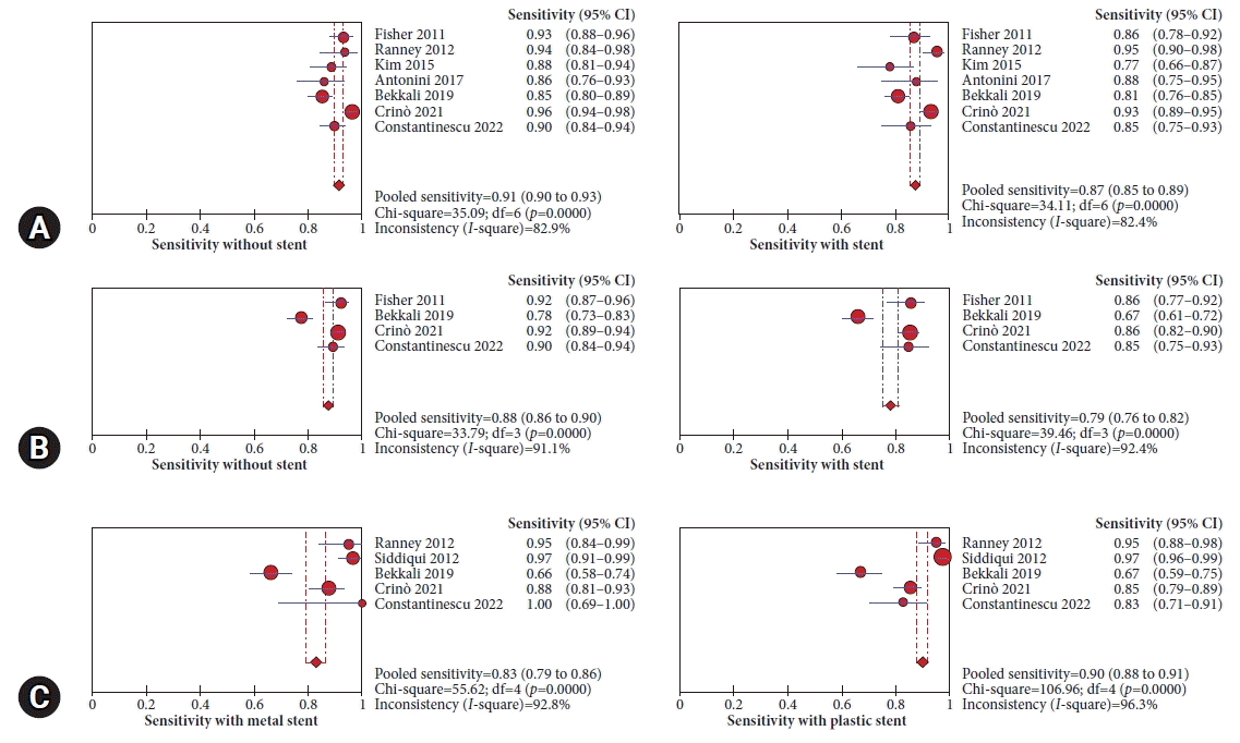
The pooled sensitivity with and without stents using the non-strict criteria was comparable (87%; 95% CI, 85%–89%; I2=82.4% vs. 91%; 95% CI, 90%–93%; I2=82.9%, respectively) (Fig. 3A). However, using strict criteria, the pooled sensitivity was higher in those without stents (88%; 95% CI, 86%–90%; I2=91.1% vs. 79%; 95% CI, 0.76–0.82; I2=92.4%) (Fig. 3B). Using non-strict criteria in patients with stents, the sensitivity was lower with metal stents than with plastic stents (83%; 95% CI, 79%–86%; I2=92.8% vs. 90%, 95% CI, 88%–91%; I2 = 96.3%) (Fig. 3C).

Number of passes

Five studies have reported differences in the number of passes between the two groups. Patients with stents required more needle passes than those without stents, with an MD of 0.31 (95% CI, 0.05–0.57; I2=83.0%), with significant heterogeneity among the studies (Supplementary Fig. 4).

Publication bias and sensitivity analysis

No publication bias in the assessment of funnel plots was observed for all assessed outcomes (Supplementary Fig. 5). In the leave-one-out meta-analysis, no significant change in the OR of sample inadequacy or diagnostic accuracy was also noted. However, with the exclusion of studies by Kim et al.,20 Kulkarni et al.,22 and Crinò et al.,24 one at a time, the number of passes was comparable between patients with and without stents. Table 4 summarizes the findings based on the grade of evidence.

DISCUSSION

Currently, no clear consensus on whether ERCP should precede EUS-TA or vice versa exists. This is mainly because of the lack of reliable data regarding the influence of biliary stents on the diagnostic yield of EUS-TA. Bile duct stents can obscure pancreatic and ampullary mass visualization during EUS because of inflammation, pneumobilia, or acoustic shadowing.11,13,26 The international consensus on endoscopic management of distal biliary strictures has proposed the approach of performing ERCP and EUS-TA in a single session as the diagnostic yield of EUS-TA is not affected by the indwelling stent, and repeated sedation can be avoided.27 However, the Canadian Society for EUS stated that EUS‑TA should precede ERCP.28 No data on the endoscopist's preference for the first intervention, EUS, or ERCP were available. The present analysis aimed to assess the impact of indwelling biliary stents on the diagnostic outcomes of EUS-TA. The pooled sensitivities with and without stents using the non-strict criteria were comparable (87% vs. 91%). However, the pooled sensitivity was higher in those without stents (88% vs. 79%) when using the strict criteria. The diagnostic accuracy of EUS-TA was significantly lower in patients with biliary stents than in those without stents, both using the non-strict (OR, 0.68; 95% CI, 0.52–0.90) and strict criteria (OR, 0.56; 95% CI, 0.44–0.73). The sensitivity of EUS-TA in patients with and without stents and its effect on the stent type were analyzed. Using non-strict criteria, the pooled sensitivity was lower with metal stents than with plastic stents (83% vs. 92.8%); however, the diagnostic accuracy was comparable (OR, 1.18; 95% CI, 0.58–2.42). A higher degree of acoustic shadowing with metal stents than with plastic stents may explain the negative impact of metal stents on diagnostic outcomes.
Samples that did not exhibit definitive malignant features were considered suspicious. Some authors have sub-classified samples using strict and non-strict criteria based on whether the samples suspicious for malignancy were considered negative or positive, respectively. Bekkali et al.23 and Crinò et al.24 performed two different analyses using two criteria and reported conflicting results, as the type of stent impacted the diagnostic accuracy of EUS-TA in the former study but not in the later study. In our analysis, the odds of an accurate diagnosis with EUS-TA were significantly lower in patients with indwelling stents than in those without stents, using both criteria.
The European Society of Gastrointestinal Endoscopy guidelines suggest performing three to four needle passes with an FNA needle or two to three passes with an fine-needle biopsy needle when onsite pathological evaluation is unavailable.29 However, some studies on the approach to patients with solid pancreatic lesions have indicated that three to four passes with the FNA needle and two to three passes with the reverse-bevel needle are adequate.30-32 However, robust data or guidelines on the impact of an indwelling biliary stent on the number of passes during EUS-TA of a pancreatic mass are lacking. Although significant heterogeneity in this meta-analysis was observed, the patients with stents required more needle passes than those without stents with an MD 0.31 (95% CI, 0.05–0.57). The increased number of passes may reflect the uncertainty among endoscopists in obtaining an adequate sample with a stent in situ.
Two studies have reported the effect of the time interval between ERCP and EUS-TA on diagnostic outcomes in patients with biliary stents. Fisher et al.17 have reported reduced diagnostic accuracy of EUS-FNA when performed within one day of biliary stenting compared to when performed after one day. Similarly, Crinò et al.24 also identified a significant correlation between a shorter interval time between ERCP and EUS-TA and decreased diagnostic accuracy. The inflammation caused by CBD instrumentation may likely affect the visualization of the mass on EUS adversely. Kulkarni et al.22 compared the procedural time between the two groups and have reported a higher procedural time in patients with indwelling stents. However, other studies did not compare the procedural times of both groups; thus, we could not analyze the difference in procedural duration between the two groups.
Fusaroli et al.13 analyzed tumor (T) and nodal (N) staging data using EUS in patients with periampullary cancer and compared them with surgical T and N staging. Correct T staging by EUS was achieved in 85% of patients without biliary stents compared to 47% in those with stents. Subsequently, Oppong et al.33 have reported successful vascular staging in 97% of patients with a plastic stent compared with only 54% in those with a metal stent. Our systematic review could not analyze the impact of stents on tumor staging using EUS.
This meta-analysis had several limitations that warrant further investigation. First, significant heterogeneities in the definitions of malignancy across the included studies were noted. Only four studies used strict criteria to classify patients with a positive malignancy. Thus, using strict criteria is essential because oncologists generally do not consider suspicious samples sufficient for initiating chemotherapy. According to Bekkali et al.,23 additional factors associated with improved accuracy were tumor size, number of passes, and using fork-tip needles. Significant variations in tumor size, needle size (22- or 25-gauge), application of ROSE, and number of passes in the included studies were also observed. However, we were unable to evaluate the effects of these variables. Lastly, we could not assess the impact of associated CP. Constantinescu et al.25 have reported that patients with associated CP had a higher risk of obtaining false-negative results (OR, 1.92; 95% CI, 1.13–3.26). EUS elastography has been used to differentiate between inflammatory head masses and malignant lesions in patients with CP.34 Whether EUS elastography can be used to guide tissue acquisition in suspected areas remains an area for future research.
In conclusion, the presence of an indwelling biliary stent may be associated with lower diagnostic accuracy and sensitivity of EUS-TA for pancreatic masses, with an increased number of passes resulting in prolonged procedural time. EUS should precede ERCP with biliary stenting whenever possible. Further prospective multicenter randomized studies using strict criteria are needed to validate these findings in real-time.

Notes

Ethical Statements

Not applicable.

Conflicts of Interest

The authors have no potential conflicts of interest.

Funding

None.

Author Contributions

Conceptualization: SG, SAf; Methodology: SG, SAf, SAn, SS; Formal analysis: SG, SAf, JV; Project administration: SG, SS; Visualization: SG, SAf, SAn, JV; Software: SG; Validation: SG, SS; Writing–original draft: SG, SAf, SAn, JV; Writing–review & editing: SG, SAf, SAn, SS.

Supplementary Material

Supplementary Fig. 1. Risk of bias assessment using the ROBINS-I tool for non-randomized studies
ce-2022-282-suppl1.pdf
Supplementary Fig. 2. Forest plot comparing the rate of sample inadequacy with EUS-guided pancreatic tissue acquisition with and without biliary stent
ce-2022-282-suppl2.pdf
Supplementary Fig. 3. Forest plot comparing the rate of sample inadequacy with EUS-guided pancreatic tissue acquisition comparing plastic and metal stent
ce-2022-282-suppl3.pdf
Supplementary Fig. 4. Forest plot comparing the number of passes with EUS-guided pancreatic tissue acquisition with and without biliary stent
ce-2022-282-suppl4.pdf
Supplementary Fig. 5. Funnel plot for assessment of publication bias
ce-2022-282-suppl5.pdf
Supplementary materials related to this article can be found online at https://doi.org/10.5946/ce.2022.282.

REFERENCES

1. Guarneri G, Gasparini G, Crippa S, et al. Diagnostic strategy with a solid pancreatic mass. Presse Med. 2019; 48(3 Pt 2):e125–e145.
2. Dietrich CF, Jenssen C. Modern ultrasound imaging of pancreatic tumors. Ultrasonography. 2020; 39:105–113.
3. Dewitt J, Devereaux BM, Lehman GA, et al. Comparison of endoscopic ultrasound and computed tomography for the preoperative evaluation of pancreatic cancer: a systematic review. Clin Gastroenterol Hepatol. 2006; 4:717–725.
4. Gomes RM, Bal M, Patkar S, et al. Unexpected benign histopathology after pancreatoduodenectomy for presumed malignancy: accepting the inevitable. Langenbecks Arch Surg. 2016; 401:169–179.
5. Manzia TM, Toti L, Lenci I, et al. Benign disease and unexpected histological findings after pancreaticoduodenectomy: the role of endoscopic ultrasound fine needle aspiration. Ann R Coll Surg Engl. 2010; 92:295–301.
6. Asbun HJ, Conlon K, Fernandez-Cruz L, et al. When to perform a pancreatoduodenectomy in the absence of positive histology?: a consensus statement by the International Study Group of Pancreatic Surgery. Surgery. 2014; 155:887–892.
7. Banafea O, Mghanga FP, Zhao J, et al. Endoscopic ultrasonography with fine-needle aspiration for histological diagnosis of solid pancreatic masses: a meta-analysis of diagnostic accuracy studies. BMC Gastroenterol. 2016; 16:108.
8. Micames C, Jowell PS, White R, et al. Lower frequency of peritoneal carcinomatosis in patients with pancreatic cancer diagnosed by EUS-guided FNA vs. percutaneous FNA. Gastrointest Endosc. 2003; 58:690–695.
9. Ross WA, Wasan SM, Evans DB, et al. Combined EUS with FNA and ERCP for the evaluation of patients with obstructive jaundice from presumed pancreatic malignancy. Gastrointest Endosc. 2008; 68:461–466.
10. Gardner TB, Gordon SR. Combined EUS with FNA and ERCP for the evaluation of pancreatic adenocarcinoma in the very elderly. Gastrointest Endosc. 2009; 69:AB250.
11. Cannon ME, Carpenter SL, Elta GH, et al. EUS compared with CT, magnetic resonance imaging, and angiography and the influence of biliary stenting on staging accuracy of ampullary neoplasms. Gastrointest Endosc. 1999; 50:27–33.
12. Chen CH, Tseng LJ, Yang CC, et al. Preoperative evaluation of periampullary tumors by endoscopic sonography, transabdominal sonography, and computed tomography. J Clin Ultrasound. 2001; 29:313–321.
13. Fusaroli P, Manta R, Fedeli P, et al. The influence of endoscopic biliary stents on the accuracy of endoscopic ultrasound for pancreatic head cancer staging. Endoscopy. 2007; 39:813–817.
14. Page MJ, McKenzie JE, Bossuyt PM, et al. The PRISMA 2020 statement: an updated guideline for reporting systematic reviews. BMJ. 2021; 372:n71.
15. Higgins JP, Altman DG, Gøtzsche PC, et al. The Cochrane Collaboration’s tool for assessing risk of bias in randomised trials. BMJ. 2011; 343:d5928.
16. Sterne JA, Hernán MA, Reeves BC, et al. ROBINS-I: a tool for assessing risk of bias in non-randomised studies of interventions. BMJ. 2016; 355:i4919.
17. Fisher JM, Gordon SR, Gardner TB. The impact of prior biliary stenting on the accuracy and complication rate of endoscopic ultrasound fine-needle aspiration for diagnosing pancreatic adenocarcinoma. Pancreas. 2011; 40:21–24.
18. Ranney N, Phadnis M, Trevino J, et al. Impact of biliary stents on EUS-guided FNA of pancreatic mass lesions. Gastrointest Endosc. 2012; 76:76–83.
19. Siddiqui AA, Fein M, Kowalski TE, et al. Comparison of the influence of plastic and fully covered metal biliary stents on the accuracy of EUS-FNA for the diagnosis of pancreatic cancer. Dig Dis Sci. 2012; 57:2438–2445.
20. Kim JJ, Walia S, Lee SH, et al. Lower yield of endoscopic ultrasound-guided fine-needle aspiration in patients with pancreatic head mass with a biliary stent. Dig Dis Sci. 2015; 60:543–549.
21. Antonini F, Fuccio L, Giorgini S, et al. Biliary plastic stent does not influence the accuracy of endoscopic ultrasound-guided sampling of pancreatic head masses performed with core biopsy needles. Dig Liver Dis. 2017; 49:898–902.
22. Kulkarni AA, Singh S, Rao B, et al. Presence of an indwelling biliary stent in patients with pancreatic head masses: does it impact endoscopic ultrasound evaluation? Gastrointest Endosc. 2017; 85:AB439.
23. Bekkali NL, Nayar MK, Leeds JS, et al. Impact of metal and plastic stents on endoscopic ultrasound-guided aspiration cytology and core histology of head of pancreas masses. Endoscopy. 2019; 51:1044–1050.
24. Crinò SF, Conti Bellocchi MC, Antonini F, et al. Impact of biliary stents on the diagnostic accuracy of EUS-guided fine-needle biopsy of solid pancreatic head lesions: a multicenter study. Endosc Ultrasound. 2021; 10:440–447.
25. Constantinescu A, Plotogea OM, Stan-Ilie M, et al. Impact of biliary stenting in endoscopic ultrasound-guided tissue acquisition among patients with pancreatic cancer. J Clin Ultrasound. 2022; 50:844–849.
26. Bao PQ, Johnson JC, Lindsey EH, et al. Endoscopic ultrasound and computed tomography predictors of pancreatic cancer resectability. J Gastrointest Surg. 2008; 12:10–16.
27. Nakai Y, Isayama H, Wang HP, et al. International consensus statements for endoscopic management of distal biliary stricture. J Gastroenterol Hepatol. 2020; 35:967–979.
28. Arya N, Wyse JM, Jayaraman S, et al. A proposal for the ideal algorithm for the diagnosis, staging, and treatment of pancreas masses suspicious for pancreatic adenocarcinoma: results of a working group of the Canadian Society for Endoscopic Ultrasound. Endosc Ultrasound. 2020; 9:154–161.
29. Polkowski M, Jenssen C, Kaye P, et al. Technical aspects of endoscopic ultrasound (EUS)-guided sampling in gastroenterology: European Society of Gastrointestinal Endoscopy (ESGE) Technical Guideline: March 2017. Endoscopy. 2017; 49:989–1006.
30. Kamata K, Kitano M, Yasukawa S, et al. Histologic diagnosis of pancreatic masses using 25-gauge endoscopic ultrasound needles with and without a core trap: a multicenter randomized trial. Endoscopy. 2016; 48:632–638.
31. Gimeno-García AZ, Elwassief A, Paquin SC, et al. Randomized controlled trial comparing stylet-free endoscopic ultrasound-guided fine-needle aspiration with 22-G and 25-G needles. Dig Endosc. 2014; 26:467–473.
32. Fabbri C, Polifemo AM, Luigiano C, et al. Endoscopic ultrasound-guided fine needle aspiration with 22- and 25-gauge needles in solid pancreatic masses: a prospective comparative study with randomisation of needle sequence. Dig Liver Dis. 2011; 43:647–652.
33. Oppong KW, Nayar MK, Bekkali NL, et al. Impact of prior biliary stenting on diagnostic performance of endoscopic ultrasound for mesenteric vascular staging in patients with head of pancreas and periampullary malignancy. BMJ Open Gastroenterol. 2022; 9:e000864.
34. Ignee A, Jenssen C, Arcidiacono PG, et al. Endoscopic ultrasound elastography of small solid pancreatic lesions: a multicenter study. Endoscopy. 2018; 50:1071–1079.
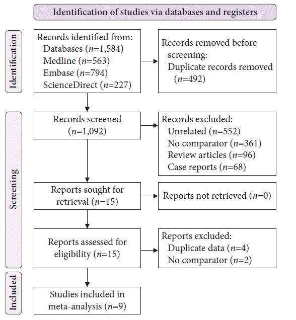
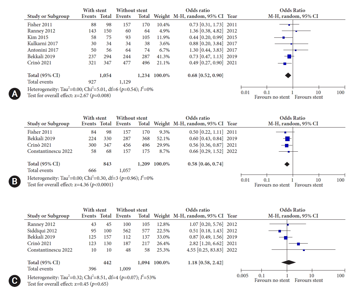
Fig. 1.
Preferred Reporting Items for Systematic Review and Meta-Analyses flowchart for the study selection and inclusion process.
ce-2022-282f1.tif
Fig. 2.
Forest plot comparing the odds of accurate diagnosis with endoscopic ultrasound–guided tissue acquisition in (A) patients with and without stents using the non-strict criteria, (B) patients with and without stents using the strict criteria, and (C) patients with metal and plastic stents using the non-strict criteria. M-H, Mantel-Haenszel; CI, confidence interval.
ce-2022-282f2.tif
Fig. 3.
Forest plot demonstrating pooled sensitivity of endoscopic ultrasound–guided tissue acquisition in (A) patients with and without stents using the non-strict criteria, (B) patients with and without stents using the strict criteria, and (C) patients with metal and plastic stents using the non-strict criteria. CI, confidence interval.
ce-2022-282f3.tif
Table 1.
Baseline characteristics of included studies
Study Country Patient (n) Age (yr) Male/female (n) Location Mass size (mm) Needle size (type) ROSE Passes (n) Criteria for malignancy with FNA/B Final diagnosis
Fisher et al. (2011)17 USA 268 68.4 120/148 Head 33.1 22-G/25-G FNA + Both Pathologically confirmed adenocarcinoma
Ranney et al. (2012)18 USA 214 67.9±12.3 137/77 Head 27±5.0 22-G/25-G FNA + 2.1±1.7 Non-strict Surgical cytopathology, death resulting from disease, or long-term follow-up
Siddiqui et al. (2012)19 USA 677 65.9 345/332 Head, uncinate 29.2±9.1 22-G FNA + 5±1.3 Non-strict Surgical cytopathology, death resulting from disease, or long-term follow-up
Kim et al. (2015)20 USA 180 65±12 108/72 Head >30 (42%) 22-G/25-G FNA 0.45 Non-strict Histologic diagnosis of malignancy on EUS-FNA, surgically resected specimen, and/or other tissue acquisition from endoscopic or percutaneous modalities
Antonini et al. (2017)21 Italy 130 69.7±10.1 79/51 Head 30.8±10.4 22-G/25-G FNA + 3.3±0.8 Non-strict Surgical cytopathology, death resulting from disease, or long-term follow-up (>6 months)
Kulkarni et al. (2017)22 USA 72 667±12.5 37/35 Head 24.1±9.1 22-G/25-G FNA/B 0.967 3.0±1.2 Non-strict
Bekkali et al. (2019)23 UK 631 65.6±10.8 345/286 Head 31.2±10.6 22-G/25-G FNA/B 0.162 Both Surgical cytopathology, death resulting from disease, or long-term follow-up (>12 months)
Crinò et al. (2021)24 Italy 842 68.9±11.1 493/349 Head, uncinate 30.7±8.9 20-/22-/25-G FNB 0.132 2.9±0.7 Both Surgical cytopathology, death resulting from disease, or long-term follow-up (>12 months)
Constantinescu et al. (2022)25 Romania 243 62.1±12.6 138/105 Head, body, tail 20–40 (61.7%), >40 (31.3%) 22-G FNA/B 2.1±0.6 Strict Pathologically confirmed adenocarcinoma

Both indicate analyses using strict as well as non-strict criteria.

FNA, fine-needle aspiration; EUS, endoscopic ultrasound; FNB, fine-needle biopsy.

Table 2.
Summary of outcomes by study
Study Stent No. of patients Inadequate sample TP FP TN FN
Fisher et al. (2011)17 With stent 98 4 84 (88) 0 0 14 (10)
Without stent 170 5 157 (158) 0 0 13 (12)
Ranney et al. (2012)18 With stent 150 7 128 0 15 7
Without stent 64 4 58 0 2 4
Kim et al. (2015)20 With stent 75 5 55 1 3 16
Without stent 105 5 92 0 1 12
Antonini et al. (2017)21 With stent 56 2 42 0 8 6
Without stent 74 7 61 0 3 10
Kulkarni et al. (2017)22 With stent 34 Diagnostic accuracy: 30/34
Without stent 38 Diagnostic accuracy: 34/38
Bekkali et al. (2019)23 With stent 294 20 196 (237) 0 0 98 (57)
Without stent 287 21 224 (244) 0 0 63 (43)
Crinò et al. (2021)24 With stent 347 6 288 (310) 0 (1) 12 (11) 47 (25)
Without stent 495 3 440 (461) 0 (0) 16 (16) 39 (18)
Constantinescu et al. (2022)25 With stent 68 10 58 0 0 10
Without stent 175 18 157 0 0 18

Numbers in brackets indicate values with non-strict criteria.

TP, true positive; FP, false positive; TN, true negative; FN, false negative.

Table 3.
Summary of outcomes by stent type
Study Stent type Patient (n) TP FP TN FN
Ranney et al. (2012)18 Metal 45 40 0 3 2
Plastic 105 88 0 12 5
Siddiqui et al. (2012)19 Metal 100 91 0 6 3
Plastic 577 512 1 50 14
Bekkali et al. (2019)23 Metal 157 104 (125) 0 0 53 (32)
Plastic 137 92 (112) 0 0 45 (25)
Crinò et al. (2021)24 Metal 130 114 (123) 0 (0) 0 (0) 16 (7)
Plastic 217 174 (187) 0 (1) 12 (11) 31 (18)
Constantinescu et al. (2022)25 Metal 10 10 0 0 0
Plastic 58 48 0 0 10

Numbers in brackets indicate values with non-strict criteria.

TP, true positive; FP, false positive; TN, true negative; FN, false negative.

Table 4.
Summary of findings and grade of evidence
Population: Patients with pancreatic mass undergoing EUS-guided tissue acquisition
Intervention: Patients with biliary stents
Comparison: Patients without biliary stents
Outcome Anticipated absolute effects (95% CI)
Relative effect (OR, 95% CI) No. of patients (study) Certainty assessment Overall certainty of evidence
Without stents With stents Risk of bias Inconsistency Indirectness Imprecision
Sample inadequacy 46 per 1,000 <5 per 1,000 (<11 to >30) 1.12 (0.76–1.65) 2,458 (7 studies) + Moderate
●●●○
Sensitivity (non-strict criteria) 0.91 (0.90–0.93) 0.87 (0.85–0.89) 2,458 (7 studies) + Moderate
●●●○
Sensitivity (strict criteria) 0.88 (0.86–0.90) 0.79 (0.76–0.82) 1,935 (4 studies) + Moderate
●●●○
Diagnostic accuracy (non-strict criteria) 915 per 1,000 <293 per 1,000 (<436 to <92) 0.68 (0.52–0.90) 2,288 (7 studies) + Moderate
●●●○
Diagnostic accuracy (strict criteria) 790 per 1,000 <332 per 1,000 (<427 to <205) 0.58 (0.46–0.74) 2,052 (4 studies) + Moderate
●●●○
No. of passes Mean no. of passes=2.83 >0.31 passes >0.05 to >0.57 1,438 (5 studies) + + Low
●●○○
Population: Patients with pancreatic mass undergoing EUS-guided tissue acquisition
Intervention: Patients with plastic stent
Comparison: Patients with metal stent
Outcome Plastic stent Metal stent
Sample inadequacy 56 per 1,000 <10 per 1,000 (<34 to >40) 0.83 (0.40–1.71) 859 (4 studies) + + Low
●●○○
Sensitivity (non-strict criteria) 0.90 (0.88–0.91) 0.83 (0.79–0.86) 1,536 (5 studies) + + Low
●●○○
Diagnostic accuracy (non-strict criteria) 922 per 1,000 >166 per 1,000 (<387 to >1309) 1.18 (0.58–2.42) 1,536 (5 studies) + + Low
●●○○

EUS, endoscopic ultrasound; CI, confidence interval; OR, odds ratio.

TOOLS
Similar articles