

 ePub
ePub Citation
 Print
Print


| Korean J Leg Med. 2016 May;40(2):44-47. English. Published online May 31, 2016. https://doi.org/10.7580/kjlm.2016.40.2.44 | |
| © Copyright 2016 by the Korean Society for Legal Medicine | |
|
Sohee Cho,1
Hee Jin Seo,2
Ji Hyun Lee,2
Hongil Ha,3
and Soong Deok Lee | |
|
1Institute of Forensic Science, Seoul National University College of Medicine, Seoul, Korea. | |
|
2Department of Forensic Medicine, Seoul National University College of Medicine, Seoul, Korea. | |
|
3Division of Forensic Medicine, National Forensic Service Busan Institute, Yangsan, Korea. | |
| Received March 07, 2016; Revised April 30, 2016; Accepted May 09, 2016. | |
|
This is an Open Access article distributed under the terms of the Creative Commons Attribution Non-Commercial License (http://creativecommons.org/licenses/by- | |
|
Abstract
| |
|
The declining tendency of signal joint T-cell receptor excision circles (sjTRECs) in peripheral blood is known to be age-dependent, and their quantification in blood or bloodstains has recently been introduced as a tool for age estimation. Lymphoid tissues such as the thymus and spleen represent potential candidates for age estimation because they undergo age-related structural and functional changes. In the present study, the correlation between age and sjTREC levels in human lymphoid tissues, namely the thymus, spleen, and blood, obtained from autopsy cases were investigated, with the goal of establishing a reliable age estimation model. Results showed negative regression curves with coefficient values of r=-0.410, r=-0.611, and r=-0.584 for thymus, spleen, and blood, respectively. In addition, this model was testing using thymus samples from the torsos of dismembered bodies from two real forensic cases, and results showed the predicted ages to be close to the actual ages of the victims. Further study will be required to improve accuracy and reduce estimation error, particularly within the lower age range. Nonetheless, these results suggest that quantification of sjTRECs in not only blood but also in other lymphoid tissues could be a useful tool for age estimation in forensic cases. |
|
Keywords: Age determination; Signal-joint T cell receptor excision circle; Thymus gland; Spleen |
|
|
Introduction
|
Identification of individuals based on short tandem repeat profiling is limited when no DNA profiles or references are available for comparison. Prediction of phenotypic traits such as eye, hair and skin color or ancestry could provide clues to narrow the range of investigation. Age is proving to be a promising factor in the forensic science field and can be estimated through various methods based on morphological and genetic changes [1, 2]. Among them, age estimation by signal joint T-cell receptor excision circle (sjTREC) quantification has demonstrated considerable prediction accuracy and can be performed using blood or blood stains found at crime scenes [3, 4]. Although several lymphoid organs related to lymphopoiesis and T-cell circulation, such as the thymus and spleen, could also be used for age estimation, sjTRECs levels in these tissues have not yet been measured for this purpose. Thus, this method could be extended to assist with solving unusual murder cases that lack blood remains, such as those involving decapitation or where only a torso remains, because it utilizes other (lymphocyte-containing) body parts as an alternate template for estimating age.
The thymus is a primary lymphoid organ responsible for T-lymphocyte development, including the generation of diverse T-cell receptors. Degeneration of the thymus, called thymic involution, is a process in which the perivascular spaces are gradually replaced with adipose and fibrous tissues, and it is known to progress with age [5]. sjTRECs found in the periphery, known as recent thymic emigrants, are indirect indicators of thymic output, which reflects thymic function, and has also been found to be correlated with age [6]. Therefore, it follows that thymic tissue could be used to estimate age based on sjTREC measurements. Other secondary lymphoid tissues, such as the spleen, could also be used in this way. In the current study, the application of lymphoid organ samples for age estimation using sjTREC levels was investigated for use in forensic science and was applied to samples from real torso murder cases.
|
Materials and Methods
|
1. Sample collection
To establish age estimation models, blood, thymus, and spleen samples were obtained from 33 forensic autopsy cases (range, 4 to 70 years). Thymus samples were sectioned in the region of the parenchyma, and spleen samples were collected from the middle of the organ. All samples were stored at -80℃ until DNA extraction.
2. DNA extraction, quantification of DNA, and sjTREC quantification
Genomic DNA was extracted from blood using a Maxwell 16 Blood DNA Purification Kit (Promega, Madison, WI, USA), and from tissue specimens using a QIAamp DNA Mini Kit (Qiagen, Valencia, CA, USA) following the manufacturer's protocols. Subsequent procedures were performed as previously described [7].
3. Statistical analysis
A regression analysis was performed using IBM SPSS Statistics ver. 20 (IBM Co., Armonk, NY, USA) and Microsoft Excel. Plots were generated using SigmaPlot ver. 12.0 software (Systat Software Inc., San Jose, CA, USA). Correlation was determined using Pearson's correlation coefficient, and age was estimated using Microsoft Excel.
|
Results
|
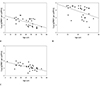
1. Correlation between sjTREC content and chronological age in blood, thymus, and spleen samples
All sjTRECs contents measured in the blood, thymus, and spleen showed a negative correlation with age, and the coefficients of correlation (r) were -0.584, -0.410, and -0.611, respectively (Table 1, Fig. 1). This was supported by a statistically significant P-value (P<0.05). Possible factor that could explain the lower re-squared values in this blood model than previously reported [7] is a smaller sample size.
|
|

2. Application of age estimation model to casework thymus tissue samples
Thymic tissues applied to age estimation in this experiment were obtained from two real forensic caseworks, which are described below.
(1) Case 1
Thymus extracted from the trunk of a body that was found without any major organs such as the heart, liver, or stomach. The thymus was formalin-fixed and washed in water for several days. Two 10-mg tissue samples were removed from different areas and used for DNA extraction.
(2) Case 2
Thymus extracted from a body where only the upper torso was found. The thymus stored at 4℃ for 2 days prior to the experiment. Two samples, each of 20-30 mg, from within the parenchyma were collected for DNA extraction.
Both of these samples were recovered from cases where the remaining body parts contained no other major organs or blood that could be used for identification. Ages were predicted from the equation established in the thymus age estimation model (y=78.734-7.167x, y age [year], XLog[sjTREC per µg DNA]), and were calculated for each sample. For the case 1 samples the age was determined to be 43.6 and 48.0 years old, and for the case 2 samples was 45.3 and 46.2 years old (Table 2). The actual age of the victims in cases 1 and 2, as identified after the criminal investigations, were confirmed to be 48 and 42 years old, respectively. Thus, the estimated ages in this experiment were reasonably accurate.
|

|
Discussion
|
It is known that the level of sjTRECs in blood shows a negative correlation with age. The current study investigated whether such a correlation also exists for other human lymphoid organs, namely thymus and spleen. Previous studies using a regression model showed that sjTREC levels were higher in the thymus than in blood or spleen samples [8], and that these levels were maintained in thymocytes until the subjects reached over 60 years of age [8]. This suggests that thymopoiesis may still be ongoing in adults [9], but further studies, with a larger sample size and wider age distribution, would be necessary to confirm this. In particular, this study lacked data from subjects aged in their twenties and below.
When this model was applied to two thymus samples from the torsos of dismembered bodies from real forensic cases, the victims' ages were predicted to be close to their actual age. Considering the fact that the reported standard error of estimation for human blood samples is known to be ±8, the predicted ages from the above two cases were deemed significant. However, further study will be needed to reduce the estimation error and improve the accuracy of the predicted ages. Although thymic involution is known to occur uniformly throughout the organ, it would still be worth investigating whether there are regional differences within the thymic tissues [5], given the fact that on-site sample collection may be limited, depending on the condition of the crime scene and the availability of evidence.
In conclusion, the results from this study demonstrate that measuring sjTREC levels in human thymus and spleen may be a valuable tool for age estimation in forensic applications, particularly in challenging cases, but also as a supplementary tool to existing methods.
|
Notes
|
Conflicts of Interest:No potential conflict of interest relevant to this article was reported.
|
Acknowledgments
|
This research was supported by the National Forensic Service (NFS) and the Bio & Medical Technology Development Program of the National Research Foundation (NRF) funded by the Ministry of Science, ICT & Future Planning (NRF-2014M3A9E1069989) in Korea.
|
References
|
| 1. | Lee SS, Byun YS, Park MJ, et al. The chronology of second and third molar development in Koreans and its application to forensic age estimation. Int J Legal Med 2010;124:659–665. 
|
| 2. | Bocklandt S, Lin W, Sehl ME, et al. Epigenetic predictor of age. PLoS One 2011;6:e14821.
|
| 3. | Zubakov D, Liu F, van Zelm MC, et al. Estimating human age from T-cell DNA rearrangements. Curr Biol 2010;20:R970–R971.
|
| 4. | Ou XL, Gao J, Wang H, et al. Predicting human age with bloodstains by sjTREC quantification. PLoS One 2012;7:e42412.
|
| 5. | Strobel P, Moritz R, Leite MI, et al. The ageing and myasthenic thymus: a morphometric study validating a standard procedure in the histological workup of thymic specimens. J Neuroimmunol 2008;201-202:64–73.
|
| 6. | Zhang L, Lewin SR, Markowitz M, et al. Measuring recent thymic emigrants in blood of normal and HIV-1-infected individuals before and after effective therapy. J Exp Med 1999;190:725–732.
|
| 7. | Cho S, Ge J, Seo SB, et al. Age estimation via quantification of signal-joint T cell receptor excision circles in Koreans. Leg Med (Tokyo) 2014;16:135–138.
|
| 8. | Jamieson BD, Douek DC, Killian S, et al. Generation of functional thymocytes in the human adult. Immunity 1999;10:569–575.
|
| 9. | Douek DC, McFarland RD, Keiser PH, et al. Changes in thymic function with age and during the treatment of HIV infection. Nature 1998;396:690–695.
|