Journal List > Endocrinol Metab > v.38(4) > 1516083550

Moon, Kim, Oh, Moon, Kwak, Park, Jang, Choi, and Cho: Triglyceride-Glucose Index Predicts Future Atherosclerotic Cardiovascular Diseases: A 16-Year Follow-up in a Prospective, Community-Dwelling Cohort Study

Abstract

Background

While the triglyceride-glucose (TyG) index is a measure of insulin resistance, its association with cardiovascular disease (CVD) has not been well elucidated. We evaluated the TyG index for prediction of CVDs in a prospective large community-based cohort.

Methods

Individuals 40 to 70 years old were prospectively followed for a median 15.6 years. The TyG index was calculated as the Ln [fasting triglycerides (mg/dL)×fasting glucose (mg/dL)/2]. CVDs included any acute myocardial infarction, coronary artery disease or cerebrovascular disease. We used a Cox proportional hazards model to estimate CVD risks according to quartiles of the TyG index and plotted the receiver operating characteristics curve for the incident CVD.

Results

Among 8,511 subjects (age 51.9±8.8 years; 47.5% males), 931 (10.9%) had incident CVDs during the follow-up. After adjustment for age, sex, body mass index, diabetes mellitus, hypertension, total cholesterol, smoking, alcohol, exercise, and C-reactive protein, subjects in the highest TyG quartile had 36% increased risk of incident CVD compared with the lowest TyG quartile (hazard ratio, 1.36; 95% confidence interval, 1.10 to 1.68). Carotid plaque, assessed by ultrasonography was more frequent in subjects in the higher quartile of TyG index (P for trend=0.049 in men and P for trend <0.001 in women). The TyG index had a higher predictive power for CVDs than the homeostasis model assessment of insulin resistance (HOMA-IR) (area under the curve, 0.578 for TyG and 0.543 for HOMA-IR). Adding TyG index on diabetes or hypertension alone gave sounder predictability for CVDs.

Conclusion

The TyG index is independently associated with future CVDs in 16 years of follow-up in large, prospective Korean cohort.

INTRODUCTION

Metabolic perturbations including insulin resistance and hyperglycemia lead to the development of cardiovascular disease (CVD) [1]. To identify subjects at risk of CVD, a number of working groups including the American College of Cardiology, the American Heart Association, and the Framingham Heart Study have developed different formulae to estimate future CVD risk [2,3]. However, the equations are complex that makes difficult to calculate the risk manually and require more than five variables to be input. Still, the area under the receiver operating characteristic (ROC) curve is below 0.75 which is not satisfactory enough considering its complexity, and the model does not fit well in Asians which requires recalibration of coefficients or developing a new equation for certain ethnicity [4,5]. Therefore, an unmet clinical need remains for simpler and effective tool to estimate the risk of CVD.
Insulin resistance is a part of the proatherogenic milieu and accompanies activation of proinflammatory cytokines, altered coagulation, and other pathways [6]. The homeostasis model assessment of insulin resistance (HOMA-IR) has been used to evaluate the extent of insulin resistance in epidemiologic studies and has been shown to be associated with increased CVD risk [7,8]. However, its accuracy and clinical use is limited in subjects with normal or low body mass index (BMI) [9], and standardized insulin measurement is not always available in clinical settings. Recent studies have shown that the triglyceride-glucose (TyG) index is highly correlated with insulin resistance and incident diabetes [10]. For instance, normoglycemic subjects in the highest quartile of the TyG index had seven times the risk of developing diabetes in an 8.8-year follow-up study [11]. A number of cross-sectional studies have demonstrated that the TyG index is associated with cardiovascular risk surrogates including arterial stiffness [12] and coronary artery calcification [13]. Given that non-diabetic hyperglycemia and high triglycerides (TGs) contribute to CVD [14-16], the TyG index has clinical relevance for the screening of people without evident metabolic disorders for the future risk of CVD. The predictive value of TyG index for CVD as a primary outcome has not been well evaluated in a prospective manner.
This study aimed to assess the utility of the TyG index as a predictor for future CVD in the Korean population. Additionally, the study examined the potential association between a high TyG index and advanced atherosclerosis, which may contribute to an elevated risk of future CVD events. The Ansung and Ansan cohorts are prospective community-based cohorts that represent rural and urban areas of South Korea, respectively [17, 18]. In this study, after 16 years of follow-up, we recorded CVDs and mortality events and assessed their event rates in association with the baseline TyG index. Furthermore, we performed carotid ultrasonography to evaluate whether a higher TyG index is associated with atherosclerosis measured as carotid plaque and intima-media thickness.

METHODS

Study participants

The Ansung–Ansan Cohort Study is an ongoing prospective, community-based cohort study that has been described in detail previously [17,19,20]. The present study is part of the Korean Genome and Epidemiology Study, a Korean Government-funded epidemiological survey that investigates trends in chronic diseases. The baseline survey began in 2001–2002 and there have been follow-up examinations every 2 years thereafter. Data from 2001–2002 to 2017–2018 were analyzed in this study. Eligible participants were 40 to 70 years old at baseline and lived in the Ansung or Ansan regions that represent rural and urban areas of Korea. Among 7,192 eligible residents in Ansung, 5,018 were surveyed using cluster sampling, with stratification by age, sex, and residential district. A total of 5,020 of 124,775 eligible individuals were recruited from Ansan using a random sampling of the local telephone directory.
In the present study, participants with a history of cancer (n=102), with a history of CVD (n=249), with estimated glomerular filtration rate <30 mL/min/1.73 m2 calculated using the Modification of Diet in Renal Disease study equation (n=16), taking dyslipidemia medication (n=56), and with missing data (n=1,172) were excluded. A total of 8,511 participants (4,259 from Ansung and 4,252 from Ansan) were eligible for inclusion. The study protocol was approved by the ethics committee of the Korean Center for Disease Control and the Institutional Review Board of Ajou University School of Medicine (IRB No. AJIRBBMR-SMP-17-477). All participants provided written informed consent. This study was conducted according to the guidelines specified in the Declaration of Helsinki.

Assessment of metabolic parameters

Anthropometric parameters and blood pressure were measured by standard methods. Blood was drawn after an overnight fast, and glucose and lipids (total cholesterol, high-density lipoprotein cholesterol [HDL-C], and TG) were measured enzymatically. The level of low-density lipoprotein cholesterol (LDL-C) was calculated using Friedewald’s formula [21]. Glycated hemoglobin was measured using high-performance liquid chromatography. High-sensitivity C-reactive protein was measured using an automated analyzer.
The definition of diabetes mellitus was based on plasma glucose concentrations during a 75 g oral glucose tolerance test defined by the 1997 American Diabetes Association criteria [22]. Hypertension was defined as systolic blood pressure ≥140 mm Hg, diastolic blood pressure ≥90 mm Hg, or taking an antihypertensive medication. Metabolic syndrome was diagnosed if ≥ 3 of the criteria were met, according to the National Cholesterol Education Program Adult Treatment panel III revised criteria [23]. Central obesity was defined as waist circumference >90 cm in men and >80 cm in women. Smokers were divided into current smokers versus past or never smokers. Alcohol intake was divided into moderate (<420 kcal per week) versus heavy intake (≥420 kcal per week). Exercise was classified into none versus moderate exercise (at least one session per week). One session of exercise was defined as exercising for at least 30 minutes. The HOMA-IR and homeostasis model assessment of β-cell function were also calculated [8]. The TyG index was calculated as the Ln [fasting TG (mg/dL)×fasting glucose (mg/dL)/2].

Definitions of outcomes

A CVD event was defined as any acute myocardial infarction, coronary artery disease (CAD), or cerebrovascular disease that occurred during the follow-up period. Participants were considered to have CAD if they received coronary bypass surgery, coronary angioplasty, or insertion of a coronary stent. Data on CVD events were obtained from the participants’ reports and were corroborated by in-depth interviews and interviews repeated at each biennial follow-up.
Mortality data were only available for the Ansung cohort. Researchers contacted all participants who did not attend the follow-up examination by telephone or made a personal visit, and all deaths were reported to local district office by participants’ families. Information about deaths, including the date, place, and cause, was obtained through interviews with families and reference to death certificates. The interview or death certificate was used to classify a death as CVD-related or cancer-related.

Carotid ultrasonography

A total of 2,376 participants (27.9%) underwent carotid ultrasonography in 2017 to 2018. Carotid ultrasonography was acquired according to a standard protocol with a high-resolution linear transducer (GE LOGIQ 400 PRO, GE Healthcare, Seoul, Korea). An experienced technician registered as a certified diagnostic medical sonographer performed all studies. Participants were examined in the supine position. Images were obtained bilaterally of the proximal to distal common carotid artery, the carotid bifurcations, and the origin of the internal carotid arteries.
Carotid intima-media thickness was defined as the distance from the lumen-intima interface to the media–adventitia interface. The carotid intima-media thickness was measured at three points of the segmentation of the common carotid artery at a site without any discrete plaque along a 10-mm-long segment of the far wall. We screened the longitudinal image for plaque and measured the thickest point of the segment from the short-axis image.

Statistical analysis

Data are presented as mean±standard error for continuous variables and as numbers (percentages) for dichotomous variables. Subjects were divided into quartiles of the TyG index for further analysis. Differences among groups were evaluated using Student’s t test or analysis of variance (ANOVA) for continuous variables and using the chi-square test or linear-by-linear association for categorical variables.
Cox proportional hazards models were used to assess the risk difference in incident CVD by the TyG index quartile. Subjects were censored at the time of CVD or at the last visit of examination. Multivariate Cox proportional models were adjusted for age, sex, BMI, diabetes mellitus, hypertension, total cholesterol, smoking, alcohol, exercise, and high-sensitivity C-reactive protein. Hazard ratios (HRs) are presented with corresponding 95% confidence intervals (CIs) and P values. For sensitivity analysis, subjects were stratified by HDL-C (50 mg/dL), BMI (23 kg/m2), or the presence of metabolic syndrome.
To estimate the performance of TyG and other indices for predicting the incident CVD, the ROC analysis was used to calculate the area under the curve (AUC). The Youden index was calculated as (sensitivity+specificity–1), and the Euclidean r was calculated as r=√[(1–sensitivity)2+(1–specificity)2] [24]. Higher Youden index and lower Euclidean r suggest better diagnostic performance. To know whether TyG index provide additional predictive value in addition to conventional risk factor including diabetes mellitus and hypertension, we compared following regression models: diabetes only versus diabetes plus TyG index; diabetes and hypertension versus diabetes, hypertension, plus TyG index. To obtain the P values of differences of those models’ AUCs, DeLong test was performed. We compared AUC of these models based on 10-fold cross-validation to avoid overfitting. To reduce sampling error of cross-validation, we performed 10-fold cross-validation 100 times with different random numbers.
A difference was considered significant when the P value was <0.05. Statistical analyses were conducted using SPSS version 25.0 (IBM Co., Armonk, NY, USA) to get results of demographic analysis, survival analysis, and single variant ROC analysis. For the cross-validation and multiple logistic regression, R version 3.6.1 with ROCR package (R Foundation for Statistical Computing, Vienna, Austria) was used.

Data availability

All data that support the findings of this study are available from the authors on a reasonable request.

RESULTS

Baseline characteristics of study subjects

We examined 8,511 subjects, 4,259 from Ansung and 4,252 from Ansan (Table 1). At baseline, the mean age was 51.9±8.8 years, 4,042 subjects (47.5%) were men, and 779 (9.2%) had diabetes. The TyG index was higher in men than women (8.7 [interquartile range, IQR, 8.4 to 9.1] in men; and 8.5 [IQR, 8.2 to 8.9] in women). We stratified the subjects by the quartiles of the TyG index for further analysis.
In both sexes, subjects with the highest TyG index quartile had poorer metabolic phenotypes including higher BMI, total cholesterol, LDL-C, and blood pressure, and lower HDL-C. In men, subjects with a higher TyG index were younger and more frequently exercised, smoked, and consumed alcohol, whereas in women, subjects with a higher TyG index were older and less frequently exercised.

CVD risks according to TyG quartiles

A total of 931 subjects (10.9%) developed CVDs (median follow-up period, 15.6 years; IQR, 10.3 to 15.8) (Table 1, Supplemental Table S1). Among them, 174 (2.0%) experienced acute myocardial infarction, 476 (5.6%) developed CAD, and 416 (4.9%) developed cerebrovascular disease. Subjects who developed CVDs were older, more obese, and had worse lipid profiles than subjects without CVD. Diabetes mellitus and hypertension were more common among subjects with CVD.
In both sexes, CVD incidence was higher in subjects with the higher TyG index quartile (Fig. 1, Supplemental Table S2). In men, subjects with the highest quartile had significantly higher risk of CVD compared with the lowest quartile (relative risk [RR], 1.53; 95% CI, 1.20 to 1.96), but an increasing trend of incident CVD in the second and third quartiles of the TyG index was not evident (RR, 1.22; 95% CI, 0.94 to 1.59 for second quartile; and RR, 1.16; 95% CI, 0.89 to 1.51 for third quartile). In women, on the other hand, the risk of CVD gradually increased with increasing quartiles of the TyG index (RR, 1.33; 95% CI, 0.99 to 1.78 for second; RR, 1.79; 95% CI, 1.36 to 2.36 for third; and RR, 2.42; 95% CI, 1.87 to 3.14 for fourth quartile of the TyG index, respectively).
We further explored the association between the TyG index and incident CVD using a Cox proportional hazard model (Table 2). In a crude analysis, the TyG index was an independent risk factor for CVD (HR, 2.10; 95% CI, 1.74 to 2.53, fourth vs. first quartile of the TyG index), and conventional risk factors including age, BMI, hypertension, total cholesterol, and smoking were shown to be predictive for CVD. After adjustment for all the other risk factors as covariates, subjects in the highest quartile of the TyG had a 36% increased risk of CVD compared with those in the lowest quartile (HR, 1.36; 95% CI, 1.10 to 1.68). An increment of 1 TyG index as a continuous variable increased the risk of CVD by 20% after adjustment for all covariates (HR, 1.20; 95% CI, 1.05 to 1.37).
We evaluated risks of each component of CVD, including acute myocardial infarction, CAD, and cerebrovascular disease (Supplemental Table S3). In the univariate analysis, subjects in the highest quartile of the TyG index exhibited a significantly increased risk of each CVD component compared to subjects in the lowest quartile. After adjustment for covariates, higher levels of the TyG index were associated with an increased risk of CAD and cerebrovascular disease, but not acute myocardial infarction.
Given that the TyG index can be affected by diabetes mellitus status, we further explored the association of the TyG index with CVD in subjects without diabetes (Table 2). Similarly, the TyG index independently predicted CVD in both unadjusted and fully adjusted models, increasing the risk by 35% in the highest quartile after adjusting for all covariates (HR, 1.35; 95% CI, 1.08 to 1.68).

Higher extent of atherosclerosis in a high TyG population

To investigate whether a higher burden of atherosclerosis in subjects with a higher TyG index is involved in the higher incident CVD, we evaluated carotid ultrasonography to examine carotid plaque and intima-media thickness, which are good surrogates for atherosclerosis (n=2,376, 27.9%) (Table 3). In both sexes, carotid plaque was more frequently observed in the higher quartiles of the TyG index (P=0.049 for men and P<0.001 for women). Carotid intima-media thickness was higher in women with a higher TyG index (P=0.010 and P=0.005 for left and right carotid intima-media thickness, respectively), but the trend was not found in men. These findings suggest that subjects with a higher TyG index have more pronounced atherosclerosis, which may be linked to a higher incidence of CVD.

Subgroup analyses according to metabolic parameters for incident CVD

High TG and low HDL-C, the characteristic dyslipidemia feature in insulin-resistant subjects, are reported to be associated with incident CVD [25]. Therefore, we subdivided subjects by HDL-C (50 mg/dL) and assessed the predictive value of the TyG index. The incidence of CVD increased with increasing TyG quartiles in both HDL-C subgroups (P for trend ≤0.001 for all subgroups). We further evaluated the TyG index as a continuous variable, as it was significantly higher in the low HDL-C subgroup (Supplemental Table S4, Supplemental Fig. S1A). In a crude analysis, an increment of 1 TyG index was predictive of CVD in both subgroups (HR, 1.54; 95% CI, 1.35 to 1.74 for HDL-C ≤50 mg/dL; and HR, 1.63; 95% CI, 1.30 to 2.06 for HDL-C >50 mg/dL). In multivariate analyses, however, the TyG index was associated with incident CVD only in subjects with HDL-C ≤50 mg/dL (HR, 1.17; 95% CI, 1.00 to 1.37).
We further evaluated the incidence of CVD according to LDL-C subgroups (subdivided by 100 and 130 mg/dL). Similarly, the incidence of CVD increased with increasing TyG quartiles across LDL-C subgroups (P for trend ≤0.001 for all subgroups) (Supplemental Fig. S1B). Across four lipid subgroups according to HDL-C cut-off 50 mg/dL and LDL-C cut-off 130 mg/dL, the incidence of CVD was higher with increasing TyG quartile (P for trend <0.01 for all subgroups) (Supplemental Fig. S1C).
Next, we investigated which subpopulations were more affected by the TyG index for the incident CVD. We stratified study subjects by age, BMI relative to 23 kg/m2 or by the presence of metabolic syndrome and hypertension (Supplemental Fig. S2). The results of the fully adjusted model indicated that the TyG index predicted CVD in both normal weight (BMI ≤ 23 kg/m2; HR, 1.57; 95% CI, 1.06 to 2.32) and overweight (BMI >23 kg/m2; HR, 1.35; 95% CI, 1.03 to 1.76) population. In subjects without hypertension or metabolic syndrome, a higher TyG index was found to predict future (CVD). However, this association was not observed in individuals with hypertension or metabolic syndrome.

All cause and CVD mortality rate according to the TyG quartiles

We evaluated mortality in 4,259 subjects from the Ansung cohort. A total of 553 subjects (13.0%) died (median follow-up period, 15.7 years; IQR, 14.0 to 15.9). CVD and cancer caused 82 deaths (14.8%) and 194 deaths (35.1%), respectively. In both crude and adjusted models, the TyG index was not associated with the risk of mortality from all causes, CVD, or cancer (Supplemental Tables S5, S6).

Predictive power of TyG index for CVD

To evaluate the predictive value of the TyG index for incident CVD, we compared the ROC analysis of the TyG index with other indices, including a composite of LDL-C and fasting glucose Ln [(LDL-C×glucose)/2] and HOMA-IR (Fig. 2). The TyG did not show impressive discrimination by itself with the AUC of 0.578. However, the TyG index had a higher AUC and Youden index and lower Euclidean r compared with HOMA-IR or Ln [(LDL-C×glucose)/2], suggesting that the TyG index better predicts future CVD incidence and events among these metabolic parameters.
Next, we evaluated whether TyG provided additional predictive power in addition to risk factors such as diabetes and hypertension, but not using the complex CVD prediction formula. Diabetes itself showed AUC of 0.530 for predicting future CVD, but when TyG was added, AUC was significantly higher by 0.582 (P<0.001 by DeLong test). When TyG was added to diabetes and hypertension, the predictive power was further increased from AUC 0.604 to 0.630 (P<0.001).

DISCUSSION

In this prospective, large, community-based cohort study including detailed metabolic evaluation and CVD records, a high TyG index was associated with an increased risk of CVDs in 16 years of follow-up. The association between the TyG index and incident CVDs was consistent in both normal weight and overweight subjects but was more apparent in women than men. Using carotid ultrasonography, we observed that in subjects with a higher TyG index, carotid plaque was more frequently found in both sex and carotid intima-media thickness was increased in women. This suggest that a higher burden of atherosclerosis may possibly mediate increases in future CVDs in subjects with a higher TyG index. TyG provided additional predictive value for incident CVDs when it was analyzed with diabetes and hypertension. Overall, the TyG index may be a useful and simple measure to predict future CVD risk in the middle-aged, general population.
With the success of statins and PCSK9 inhibitors in reducing incident CVDs, clinical emphasis on lipid management is mainly focused on LDL-C-lowering therapy [26-28]. Negative results from TG-lowering (fenofibrate trial; The Action to Control Cardiovascular Risk in Diabetes [ACCORD]) [29] and HDL-C-raising (niacin trial; The Atherothrombosis Intervention in Metabolic Syndrome with Low HDL/High Triglycerides: Impact on Global Health Outcomes [AIM-HIGH]) [30] trials further emphasized the importance of LDL-C, although epidemiologic studies suggest that high TG alone [31] or high TG with low HDL-C predicts CVD, just as high LDL-C does [25,32]. Recent studies have demonstrated that elevated TG-rich lipoprotein is responsible for atherogenic dyslipidemia, incident CVDs, and the residual risk after optimal LDL-C lowering, which has renewed interest in high TG [33,34].
Notably, our study presents several metabolic implications for TG. A composite of TG and glucose (TyG index) showed better predictive values for CVDs than a composite of LDL-C. In addition, the TyG index was only predictive of CVD in cases where HDL-C ≤50 mg/dL, suggesting that a high TG or TyG index should be considered as adding increased CVD risk in subjects with low HDL-C. With further understanding of the role of TG-rich lipoproteins and lipid particles in different fractionations, novel biomarkers or surrogate metabolic indices linked to elevated CVD risk should be further investigated.
The TyG index has been considered a good surrogate for insulin resistance [35]. Later, a number of cross-sectional studies have demonstrated an association between the TyG index and cardiovascular risk. Two reports from Korea, including a healthy population visited for routine health check-ups showed that the TyG index was associated with coronary artery calcification and arterial stiffness as assessed by cardiac computed tomography and plethysmography, respectively [12,13]. Three studies utilizing Korean claims data consistently demonstrated the association between the TyG index and incident CVD to varying degrees [36-38]. da Silva et al. [39] evaluated 2,330 high-risk Brazilian patients with a history of CVD, and showed that subjects with the highest TyG tertile are more likely to have symptomatic CAD. One prospective population-based cohort study performed in Spain (n=5,014) demonstrated a 2.32-fold higher risk of CVD in the highest TyG quintile and a 52% increased risk in the fourth quintile compared with the lowest quintile during a median 10-year follow-up [40]. These studies classified the study subjects into tertiles to quintiles and demonstrated the highest TyG subgroup who have TyG higher than 8.8 to 9.0 have increased CVD risk. The cut-off for the highest TyG quartile were similar in this study (9.1 for men 8.9 for women). Therefore, we suggest this sex-specific TyG cut-offs to be considered to estimate CVD risk, but the absolute cut-offs of the TyG index should be further assessed to compare its predictive value across sex and ethnicities.
The incidence of CVDs is higher in men than women at the same age [41,42]. Men and women share conventional cardiovascular risk factors (e.g., type 2 diabetes, dyslipidemia, etc.), but the extent of contribution of each risk factor to CVD is assumed to be different by sex. In this study, the association between the TyG index and CVD events was more evident in women. Similarly, type 2 diabetes and metabolic alterations including hypertension and dyslipidemia have a stronger effect on the risk of CVD in women [31,43,44]. More frequent unhealthy behaviors including cigarette smoking, alcohol intake, and consuming more red meat contribute to higher CVD morbidity and mortality in men and obscure the association between conventional metabolic risk factors and CVD in men [42,44]. Moreover, the proportion of subjects who smoke and consume alcohol increases with increasing TyG quartile in men, which might attenuate the association between TyG index and incident CVD. Regardless of their odds or sex, people with a higher TyG index should take good care regarding their classical CVD risk factors.
The main strength of this study is that we explored two large community-based cohorts with detailed evaluation of CVDs and stringent measurement of all metabolic parameters using the 75 g oral glucose tolerance test and central lab measurements. The predictive value of the TyG index was significant after adjustment for conventional risk factors as well as exercise and C-reactive protein. In particular, carotid ultrasonography was analyzed by a single examiner, which showed that a high TyG index may contribute to advanced atherosclerosis. Therefore, this study provided not only evidence of an epidemiologic association but also comprehensive insights into how insulin resistance may be linked to future CVD in healthy populations. Furthermore, exclusion of subjects with any history of CVD or cancer enabled us to demonstrate the clinical implication of the TyG index in CVD risk prediction generally, even though the predictive value could be underestimated because we included low-risk, middle-aged populations.
This study has some limitations. First, our data on CVD events were obtained by self-reports that might be skewed by recall bias. In this regard, we conducted in-depth interviews to confirm CVD events and excluded subjects with any history of CVD at baseline to eliminate residual confounding. Second, we could not assess longitudinal changes of the TyG index over time. Assessing cumulative or average TyG index could provide better insights into its association with incident CVDs. Therefore, further research is needed to investigate whether improvements in the TyG index over time have a protective effect on the development of CVDs. Third, the TyG index demonstrated an AUC of ROC below 0.6 when used to predict CVD. When diabetes and hypertension were incorporated into the model, the AUC improved to 0.630, although it remained low. It could be the characteristics of our community cohort, most who are not in higher risk of CVDs. However, after adjusting for conventional CVD risk factors and high-sensitivity C-reactive protein, the TyG index remained as a significant risk factor for CVD. While its predictive power may not be strong enough for independent use, the TyG index provides added value when combined with other established risk factors in assessing CVD risk. Fourth, carotid ultrasonography and mortality data were only available for a subgroup of participants, which might underestimate the predictive value of the TyG index with respect to mortality. Nonetheless, we observed an association between the TyG index and the presence of carotid plaque with a limited number of subjects.
In conclusion, the TyG index provided predictions of long-term CVD events in the middle-aged, general population. These findings are relevant to clinical practitioners because this index may facilitate guidance of CVD risk prediction and can easily be included parameters in routine general practice check-ups than other methods. Our results also stimulate further research for the validation and development of predictive measures for patients with difficult metabolic outcomes.

Supplementary Material

Supplemental Table S1.

Baseline Characteristics according to Cardiovascular Disease Status
enm-2023-1703-Supplemental-Table-S1.pdf

Supplemental Table S2.

Relative Risks for Cardiovascular Disease according to Triglyceride-Glucose Index
enm-2023-1703-Supplemental-Table-S2.pdf

Supplemental Table S3.

Hazard Ratios for Acute Myocardial Infarction, Coronary Artery Disease, and Cerebrovascular Disease according to Triglyceride-Glucose Index
enm-2023-1703-Supplemental-Table-S3.pdf

Supplemental Table S4.

Hazard Ratios for Cardiovascular Disease according to High-Density Lipoprotein Cholesterol Concentrations
enm-2023-1703-Supplemental-Table-S4.pdf

Supplemental Table S5.

Hazard Ratios for All Cause Mortality according to Triglyceride-Glucose Index
enm-2023-1703-Supplemental-Table-S5.pdf

Supplemental Table S6.

Hazard Ratios for Cause-Specific Mortality according to Triglyceride-Glucose Index
enm-2023-1703-Supplemental-Table-S6.pdf

Supplemental Fig. S1.

Incidence of cardiovascular disease (CVD) according to different high-density lipoprotein cholesterol (HDL-C) and low-density lipoprotein cholesterol (LDL-C) subgroups. (A, B, C) The incidence of CVD is depicted across different HDL-C and LDL-C cut-offs (50 mg/dL for HDL-C and 100 of 130 mg/dL for LDL-C). Numbers above each bar indicates the incidence of CVD in each subgroup during a median of 15.6 years of follow-up. TyG, triglyceride-glucose.
enm-2023-1703-Supplemental-Fig-S1.pdf

Supplemental Fig. S2.

Sensitivity analysis of triglyceride-glucose (TyG) index for the risk of cardiovascular disease (CVD). Hazard ratios of CVD in the highest quartile of TyG index compared to the lowest quartile were evaluated with Cox proportional hazard model in subgroups of age, body mass index (BMI) cut-off of 23 kg/m2 and the presence of metabolic syndrome (MetS) or hypertension (HTN). The model was adjusted for age, sex, body mass index, diabetes mellitus, hypertension, total cholesterol, smoking, alcohol, exercise, and high-sensitivity C-reactive protein (hsCRP).
enm-2023-1703-Supplemental-Fig-S2.pdf

Notes

CONFLICTS OF INTEREST

No potential conflict of interest relevant to this article was reported.

AUTHOR CONTRIBUTIONS

Conception and/or design: J.H.M., S.H.C., N.H.C. Acquisition, analysis, and/or interpretation of data: J.H.M., Y.K., T.J.O., J. H.M., S.H.K., K.S.P., H.C.J., S.H.C., N.H.C. Drafting the work and/or revising: J.H.M., S.H.C., N.H.C. Final approval of the manuscript: J.H.M., Y.K., T.J.O., J.H.M., S.H.K., K.S.P., H.C.J., S.H.C., N.H.C.

ACKNOWLEDGMENTS

A part of this study was presented in the 11th International Congress of Diabetes and Metabolism (ICDM 2021) in conjunction with the 13th Asian Association for the Study of Diabetes (AASD) Scientific Meeting, and the abstract was subsequently published as a conference abstract (doi.org/10.1111/jdi.13662). We appreciated Bong Deok Kim and Hyun Kyu Kim (Ajou University) for their efforts to support statistical analysis and data acquisition.
This work was supported by the National Research Foundation of Korea (NRF) grant funded by the Korea Government (2016R1D1A1B04934590 and RS-2023-00222910), the Korea Health Technology R&D Project through the Korea Health Industry Development Institute (KHIDI) funded by the Ministry of Health & Welfare, Republic of Korea (HI17C0912), the Research Program funded by the Korea Centers for Disease Control and Prevention (2001-347-6111-221, 2002-347-6111-221, 2003-347-6111-221, 2004-E71001-00, 2005-E71001-00, 2006-E71005-00, 2006-E71006-00, 2007-E71001-00, 2007-E71003-00, 2008-E71001-00, 2008-E71005-00, 2009-E71002-00, 2009-E71007-00, 2010-E71001-00, 2010-E71004-00, 2011-E71004-00, 2011-E71008-00, 2012-E71008-00, 2012-E71005-00, 2013-E71007-00, 2013-E71005-00, 2014-E71005-00, 2014-E71003-00, 2015-P71002-00, 2015-P71001-00, 2016-E71002-00, 2016-E71003-00, 2017-E71002-00, 2017-E71001-00, 2018-E7102-00, 2018-E7101-00) to Nam H. Cho, and the Medical Research Center through the National Research Foundation of Korea funded by the Ministry of Science (grant number NRF-2018R1 A5A2024425) to Sung Hee Choi.

REFERENCES

1. Wilson PW, D’Agostino RB, Parise H, Sullivan L, Meigs JB. Metabolic syndrome as a precursor of cardiovascular disease and type 2 diabetes mellitus. Circulation. 2005; 112:3066–72.
2. Goff DC Jr, Lloyd-Jones DM, Bennett G, Coady S, D’Agostino RB, Gibbons R, et al. 2013 ACC/AHA guideline on the assessment of cardiovascular risk: a report of the American College of Cardiology/American Heart Association Task Force on Practice Guidelines. Circulation. 2014; 129(25 Suppl 2):S49–73.
3. Wilson PW, D’Agostino RB, Levy D, Belanger AM, Silbershatz H, Kannel WB. Prediction of coronary heart disease using risk factor categories. Circulation. 1998; 97:1837–47.
4. Bae JH, Moon MK, Oh S, Koo BK, Cho NH, Lee MK. Validation of risk prediction models for atherosclerotic cardiovascular disease in a prospective Korean community-based cohort. Diabetes Metab J. 2020; 44:458–69.
5. Jung KJ, Jang Y, Oh DJ, Oh BH, Lee SH, Park SW, et al. The ACC/AHA 2013 pooled cohort equations compared to a Korean Risk Prediction Model for atherosclerotic cardiovascular disease. Atherosclerosis. 2015; 242:367–75.
6. Semenkovich CF. Insulin resistance and atherosclerosis. J Clin Invest. 2006; 116:1813–22.
7. Bonora E, Formentini G, Calcaterra F, Lombardi S, Marini F, Zenari L, et al. HOMA-estimated insulin resistance is an independent predictor of cardiovascular disease in type 2 diabetic subjects: prospective data from the Verona Diabetes Complications Study. Diabetes Care. 2002; 25:1135–41.
8. Matthews DR, Hosker JP, Rudenski AS, Naylor BA, Treacher DF, Turner RC. Homeostasis model assessment: insulin resistance and beta-cell function from fasting plasma glucose and insulin concentrations in man. Diabetologia. 1985; 28:412–9.
9. Kang ES, Yun YS, Park SW, Kim HJ, Ahn CW, Song YD, et al. Limitation of the validity of the homeostasis model assessment as an index of insulin resistance in Korea. Metabolism. 2005; 54:206–11.
10. Simental-Mendia LE, Rodriguez-Moran M, Guerrero-Romero F. The product of fasting glucose and triglycerides as surrogate for identifying insulin resistance in apparently healthy subjects. Metab Syndr Relat Disord. 2008; 6:299–304.
11. Navarro-Gonzalez D, Sanchez-Inigo L, Pastrana-Delgado J, Fernandez-Montero A, Martinez JA. Triglyceride-glucose index (TyG index) in comparison with fasting plasma glucose improved diabetes prediction in patients with normal fasting glucose: the Vascular-Metabolic CUN cohort. Prev Med. 2016; 86:99–105.
12. Lee SB, Ahn CW, Lee BK, Kang S, Nam JS, You JH, et al. Association between triglyceride glucose index and arterial stiffness in Korean adults. Cardiovasc Diabetol. 2018; 17:41.
13. Kim MK, Ahn CW, Kang S, Nam JS, Kim KR, Park JS. Relationship between the triglyceride glucose index and coronary artery calcification in Korean adults. Cardiovasc Diabetol. 2017; 16:108.
14. Hu FB, Stampfer MJ, Haffner SM, Solomon CG, Willett WC, Manson JE. Elevated risk of cardiovascular disease prior to clinical diagnosis of type 2 diabetes. Diabetes Care. 2002; 25:1129–34.
15. Levitan EB, Song Y, Ford ES, Liu S. Is nondiabetic hyperglycemia a risk factor for cardiovascular disease?: a meta-analysis of prospective studies. Arch Intern Med. 2004; 164:2147–55.
16. Sarwar N, Danesh J, Eiriksdottir G, Sigurdsson G, Wareham N, Bingham S, et al. Triglycerides and the risk of coronary heart disease: 10,158 incident cases among 262,525 participants in 29 Western prospective studies. Circulation. 2007; 115:450–8.
17. Cho NH, Jang HC, Choi SH, Kim HR, Lee HK, Chan JC, et al. Abnormal liver function test predicts type 2 diabetes: a community-based prospective study. Diabetes Care. 2007; 30:2566–8.
18. Choi SH, Kim TH, Lim S, Park KS, Jang HC, Cho NH. Hemoglobin A1c as a diagnostic tool for diabetes screening and new-onset diabetes prediction: a 6-year community-based prospective study. Diabetes Care. 2011; 34:944–9.
19. Kim Y, Han BG; KoGES group. Cohort profile: the Korean Genome and Epidemiology Study (KoGES) consortium. Int J Epidemiol. 2017; 46:e20.
20. Ohn JH, Kwak SH, Cho YM, Lim S, Jang HC, Park KS, et al. 10-Year trajectory of b-cell function and insulin sensitivity in the development of type 2 diabetes: a community-based prospective cohort study. Lancet Diabetes Endocrinol. 2016; 4:27–34.
21. Friedewald WT, Levy RI, Fredrickson DS. Estimation of the concentration of low-density lipoprotein cholesterol in plasma, without use of the preparative ultracentrifuge. Clin Chem. 1972; 18:499–502.
22. American Diabetes Association. Diagnosis and classification of diabetes mellitus. Diabetes Care. 2004; 27 Suppl 1:S5–10.
23. Grundy SM, Cleeman JI, Daniels SR, Donato KA, Eckel RH, Franklin BA, et al. Diagnosis and management of the metabolic syndrome: an American Heart Association/National Heart, Lung, and Blood Institute Scientific Statement. Circulation. 2005; 112:2735–52.
24. Indrayan A. Medical biostatistics. 3rd ed. Boca Raton: CRC Press;2013. 962.
25. Jeppesen J, Hein HO, Suadicani P, Gyntelberg F. Relation of high TG-low HDL cholesterol and LDL cholesterol to the incidence of ischemic heart disease: an 8-year follow-up in the Copenhagen Male Study. Arterioscler Thromb Vasc Biol. 1997; 17:1114–20.
26. Michos ED, McEvoy JW, Blumenthal RS. Lipid management for the prevention of atherosclerotic cardiovascular disease. N Engl J Med. 2019; 381:1557–67.
27. Sabatine MS, Giugliano RP, Keech AC, Honarpour N, Wiviott SD, Murphy SA, et al. Evolocumab and clinical outcomes in patients with cardiovascular disease. N Engl J Med. 2017; 376:1713–22.
28. Rhee EJ, Kim HC, Kim JH, Lee EY, Kim BJ, Kim EM, et al. 2018 Guidelines for the management of dyslipidemia. Korean J Intern Med. 2019; 34:723–71.
29. ACCORD Study Group, Ginsberg HN, Elam MB, Lovato LC, Crouse JR 3rd, Leiter LA, et al. Effects of combination lipid therapy in type 2 diabetes mellitus. N Engl J Med. 2010; 362:1563–74.
30. AIM-HIGH Investigators, Boden WE, Probstfield JL, Anderson T, Chaitman BR, Desvignes-Nickens P, et al. Niacin in patients with low HDL cholesterol levels receiving intensive statin therapy. N Engl J Med. 2011; 365:2255–67.
31. Austin MA, Hokanson JE, Edwards KL. Hypertriglyceridemia as a cardiovascular risk factor. Am J Cardiol. 1998; 81(4A):7B–12B.
32. Elam M, Lovato L, Ginsberg H. The ACCORD-Lipid study: implications for treatment of dyslipidemia in type 2 diabetes mellitus. Clin Lipidol. 2011; 6:9–20.
33. Duran EK, Aday AW, Cook NR, Buring JE, Ridker PM, Pradhan AD. Triglyceride-rich lipoprotein cholesterol, small dense ldl cholesterol, and incident cardiovascular disease. J Am Coll Cardiol. 2020; 75:2122–35.
34. Nordestgaard BG, Varbo A. Triglycerides and cardiovascular disease. Lancet. 2014; 384:626–35.
35. Guerrero-Romero F, Simental-Mendia LE, Gonzalez-Ortiz M, Martinez-Abundis E, Ramos-Zavala MG, Hernandez-Gonzalez SO, et al. The product of triglycerides and glucose, a simple measure of insulin sensitivity: comparison with the euglycemic-hyperinsulinemic clamp. J Clin Endocrinol Metab. 2010; 95:3347–51.
36. Park B, Lee YJ, Lee HS, Jung DH. The triglyceride-glucose index predicts ischemic heart disease risk in Koreans: a prospective study using National Health Insurance Service data. Cardiovasc Diabetol. 2020; 19:210.
37. Hong S, Han K, Park CY. The triglyceride glucose index is a simple and low-cost marker associated with atherosclerotic cardiovascular disease: a population-based study. BMC Med. 2020; 18:361.
38. Cho YK, Han KD, Kim HS, Jung CH, Park JY, Lee WJ. Triglyceride-glucose index is a useful marker for predicting future cardiovascular disease and mortality in young Korean adults: a nationwide population-based cohort study. J Lipid Atheroscler. 2022; 11:178–86.
39. da Silva A, Caldas AP, Hermsdorff HH, Bersch-Ferreira AC, Torreglosa CR, Weber B, et al. Triglyceride-glucose index is associated with symptomatic coronary artery disease in patients in secondary care. Cardiovasc Diabetol. 2019; 18:89.
40. Sanchez-Inigo L, Navarro-Gonzalez D, Fernandez-Montero A, Pastrana-Delgado J, Martinez JA. The TyG index may predict the development of cardiovascular events. Eur J Clin Invest. 2016; 46:189–97.
41. Mosca L, Barrett-Connor E, Wenger NK. Sex/gender differences in cardiovascular disease prevention: what a difference a decade makes. Circulation. 2011; 124:2145–54.
42. Barrett-Connor E. Sex differences in coronary heart disease: why are women so superior? The 1995 Ancel Keys Lecture. Circulation. 1997; 95:252–64.
43. Juutilainen A, Kortelainen S, Lehto S, Ronnemaa T, Pyorala K, Laakso M. Gender difference in the impact of type 2 diabetes on coronary heart disease risk. Diabetes Care. 2004; 27:2898–904.
44. Garcia M, Mulvagh SL, Merz CN, Buring JE, Manson JE. Cardiovascular disease in women: clinical perspectives. Circ Res. 2016; 118:1273–93.
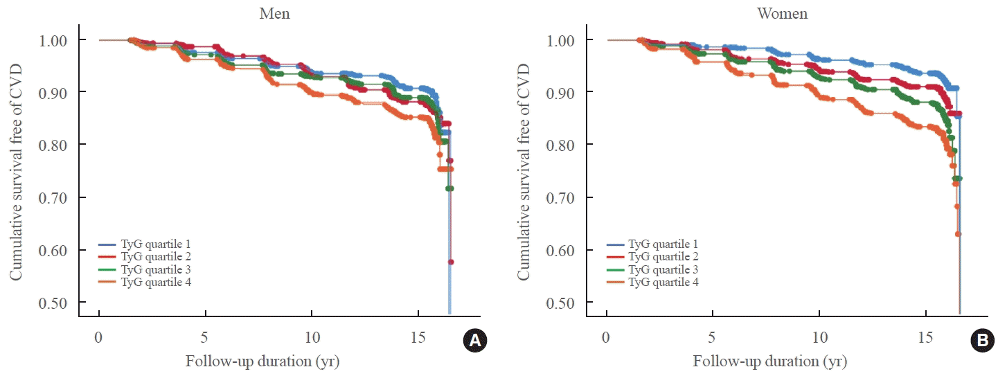
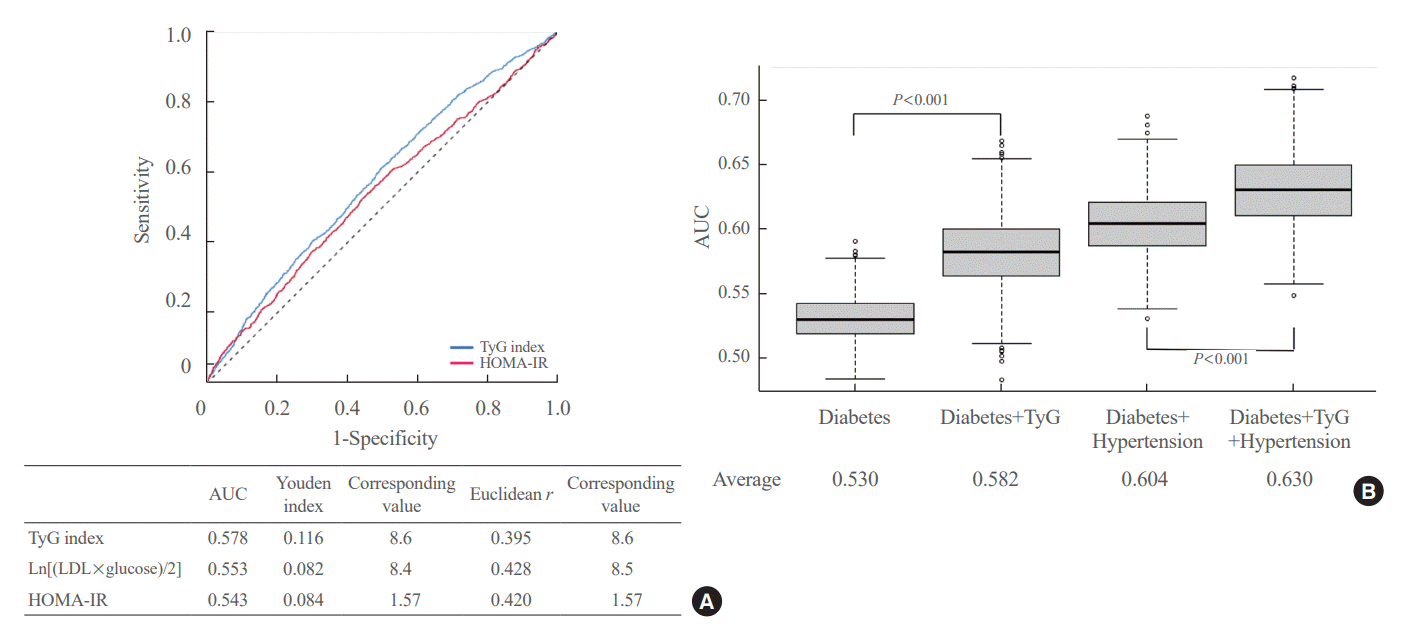
Fig. 1.
Cumulative survival for incident cardiovascular disease according to triglyceride-glucose (TyG) index. (A, B) Cumulative survivals for incident cardiovascular disease (n=8,551; 931 [10.9%] developed cardiovascular disease [CVD]) according to quartiles of TyG index in (A) men and (B) women during 16 years of follow-up are depicted using Kaplan-Meier analysis.
enm-2023-1703f1.tif
Fig. 2.
Receiver operating characteristic (ROC) analysis of triglyceride-glucose (TyG) and other indices for predicting incident cardiovascular disease. (A) The ROC curves are plotted to compare the predictive power for incident cardiovascular disease of indices including TyG index. The estimates of the ROC curves including the area under the curve (AUC), Youden index, Euclidean r, and their corresponding values of each index are presented. (B) The box plots show the distributions of AUC values from 100 iterations of 10-fold cross-validation. First box plot indicates analysis of diabetes only model, second box plot indicates analysis of multiple logistic regression model with diabetes and TyG index, third box plot is with diabetes and hypertension, and fourth box plot is from the multiple regression model with diabetes and hypertension, and TyG index. HOMA-IR, homeostasis model assessment of insulin resistance; LDL, low-density lipoprotein.
enm-2023-1703f2.tif
Table 1.
Clinical Characteristics and Incidence of Cardiovascular Disease according to Triglyceride-Glucose Index
Characteristic TyG index quartile (male)
TyG index quartile (female)
1 2 3 4 Total P value 1 2 3 4 Total P value
Number 1,009 1,012 1,011 1,010 4,042 1,117 1,117 1,118 1,117 4,469
TyG index 8.1±0.0 8.6±0.0 8.9±0.0 9.6±0.0 8.8±0.0 <0.001 8.0±0.0 8.4±0.0 8.7±0.0 9.3±0.0 8.6±0.0 <0.001
CVD 92 (9.1) 113 (11.2) 107 (10.6) 141 (14.0) 453 (11.2) 0.002 73 (6.5) 97 (8.7) 131 (11.7) 177 (15.8) 478 (10.7) <0.001
Age, yr 52.2±8.9 52.0±9.0 50.9±8.4 50.8±8.1 51.4±8.6 <0.001 49.2±8.2 51.1±8.7 53.9±8.9 54.9±8.7 52.3±8.9 <0.001
BMI, kg/m2 22.7±2.8 23.8±2.8 24.8±2.7 25.5±2.7 24.2±2.9 <0.001 23.6±3.1 24.6±3.1 25.1±3.2 26.2±3.2 24.9±3.3 <0.001
Total cholesterol, mg/dL 181.3±31.9 189.9±33.2 196.4±35.3 206.7±37.5 193.6±35.7 <0.001 178.6±31.4 188±33.7 199±32.8 206.6±37.5 193.1±35.6 <0.001
HDL-C, mg/dL 50.5±11.7 46.3±10.2 43.1±9.1 40.6±8.9 45.1±10.7 <0.001 53.5±10.7 49.1±10.5 46.3±10.1 41.2±8.5 47.5±10.9 <0.001
Triglyceride, mg/dL 82.6±17.8 124.8±17.7 173.4±29.6 311.4±146.2 173±114.6 <0.001 74.1±13.1 105.5±13.6 143.4±20.9 242.7±99.8 141.4±81.9 <0.001
LDL-C, mg/dL 114.3±30.3 118.7±32 118.5±34.3 122.7±47.7 118.5±36.8 <0.001 110.4±28.4 117.8±30.1 123.9±30.2 123.3±39.3 118.9±32.7 <0.001
Glucose, mg/dL 83.6±0.3 86.4±0.3 90.2±0.5 104.7±1.2 91.2±0.4 <0.001 80.8±0.2 82.8±0.3 84.7±0.3 97.0±1.0 86.3±0.3 <0.001
HbA1c, % 5.3±0.4 5.4±0.5 5.5±0.6 6±1.3 5.6±0.8 <0.001 5.2±0.3 5.3±0.4 5.5±0.5 6±1.3 5.5±0.8 <0.001
HOMA-β 131.7±152.4 130±204 119.2±95.7 108.3±87.2 122.3±143.2 <0.001 173.7±172.3 166.6±139.8 174.7±183.3 147.1±149.7 165.5±162.5 <0.001
HOMA-IR 1.3±0.7 1.5±1.0 1.6±1.0 2.1±1.5 1.6±1.2 <0.001 1.4±0.7 1.6±1.0 1.8±1.3 2.3±2.2 1.8±1.4 <0.001
SBP, mm Hg 113.2±15.7 116.9±16.6 118.2±15.9 120±16.2 117.1±16.3 <0.001 110.5±16.9 114.5±18.3 119.1±19.3 123.4±19.3 116.9±19.1 <0.001
DBP, mm Hg 73.6±10.6 76.0±10.6 76.8±10.9 78.5±11.5 76.2±11.1 <0.001 70.3±11 72.5±11.2 74.8±12.1 77.6±11.6 73.8±11.8 <0.001
hsCRP, mg/dL 0.3±0.6 0.2±0.4 0.2±0.3 0.3±0.6 0.2±0.5 0.079 0.2±0.5 0.2±0.3 0.2±0.4 0.3±0.8 0.2±0.5 0.001
Diabetes mellitus 27 (2.7) 52 (5.1) 89 (8.8) 257 (25.4) 425 (10.5) <0.001 20 (1.8) 30 (2.7) 63 (5.6) 241 (21.6) 354 (7.9) <0.001
Hypertension 116 (11.5) 189 (18.7) 221 (21.9) 286 (28.3) 812 (20.1) <0.001 120 (10.7) 191 (17.1) 285 (25.5) 381 (34.1) 977 (21.9) <0.001
Alcohol 359 (35.6) 417 (41.2) 489 (48.4) 543 (53.8) 1,808 (44.7) <0.001 32 (2.9) 37 (3.3) 38 (3.4) 40 (3.6) 147 (3.3) 0.349
Smoking 473 (46.9) 475 (46.9) 478 (47.3) 537 (53.2) 1,963 (48.6) 0.006 24 (2.1) 30 (2.7) 41 (3.7) 50 (4.5) 145 (3.2) 0.001
Exercise 364 (36.1) 369 (36.5) 400 (39.6) 417 (41.3) 1,550 (38.3) 0.006 391 (35.0) 373 (33.4) 348 (31.1) 330 (29.5) 1,442 (32.3) 0.003

Values are expressed as mean±standard error or number (%).

TyG, triglyceride-glucose; CVD, cardiovascular disease; BMI, body mass index; HDL-C, high-density lipoprotein cholesterol; LDL-C, low-density lipoprotein cholesterol; HbA1c, glycated hemoglobin; HOMA-β, homeostatic model assessment value of β cell function; HOMA-IR, homeostatic model assessment value of insulin resistance; SBP, systolic blood pressure; DBP, diastolic blood pressure; hsCRP, high-sensitivity C-reactive protein.

Table 2.
Hazard Ratios for Cardiovascular Disease according to Triglyceride-Glucose Index
Variable All participants
Non-diabetics
Univariate
Multivariate
Univariate
Multivariate
HR 95% CI P value HR 95% CI P value HR 95% CI P value HR 95% CI P value
TyG index
 Q1 Reference Reference Reference Reference
 Q2 1.30 1.06–1.60 0.011 1.12 0.91–1.38 0.287 1.26 1.02–1.56 0.029 1.09 0.88–1.35 0.449
 Q3 1.55 1.27–1.88 <0.001 1.17 0.95–1.44 0.154 1.48 1.21–1.82 <0.001 1.13 0.91–1.41 0.264
 Q4 2.10 1.74–2.53 <0.001 1.36 1.10–1.68 0.004 1.91 1.56–2.34 <0.001 1.35 1.08–1.68 0.009
Age 1.07 1.06–1.07 <0.001 1.06 1.05–1.07 <0.001 1.07 1.06–1.07 <0.001 1.06 1.05–1.07 <0.001
Sex 0.89 0.79–1.02 0.086 0.84 0.71–0.99 0.042 0.91 0.79–1.05 0.200 0.90 0.74–1.08 0.244
Body mass index 1.04 1.02–1.06 <0.001 1.02 1.00–1.05 0.062 1.03 1.01–1.06 0.006 1.02 0.99–1.04 0.185
Diabetes mellitus 1.99 1.66–2.39 <0.001 1.40 1.15–1.70 0.001 - -
Hypertension 2.47 2.16–2.82 <0.001 1.74 1.50–2.01 <0.001 2.50 2.16–2.89 <0.001 1.80 1.53–2.11 <0.001
Total cholesterol 1.005 1.003–1.007 <0.001 1.003 1.001–1.005 0.006 1.005 1.003–1.007 <0.001 1.003 1.001–1.005 0.004
Smoking 1.28 1.11–1.48 0.001 1.39 1.16–1.66 <0.001 1.31 1.12–1.53 0.001 1.48 1.22–1.80 <0.001
Alcohol 0.95 0.82–1.12 0.558 0.84 0.70–1.01 0.070 0.91 0.76–1.08 0.288 0.83 0.68–1.02 0.073
Exercise 0.76 0.66–0.87 <0.001 0.86 0.75–1.00 0.050 0.73 0.62–0.85 <0.001 0.87 0.74–1.02 0.092
hsCRP 1.11 1.05–1.18 0.001 1.08 1.01–1.16 0.035 1.11 1.05–1.19 0.001 1.08 1.01–1.17 0.035

HRs of cardiovascular disease (n=8,551; 931 [10.9%] developed cardiovascular disease [CVD]) for median 15.6 years of follow-up were evaluated with Cox proportional hazard model. Each variable including quartiles of TyG index was evaluated for the risk of incident CVD in univariate analysis. Multivariate analysis was adjusted for age, sex, body mass index, diabetes mellitus, hypertension, total cholesterol, smoking, alcohol, exercise, and hsCRP.

HR, hazard ratio; CI, confidence interval; TyG, triglyceride-glucose; hsCRP, high-sensitivity C-reactive protein.

Table 3.
Presence of Carotid Plaque and Carotid Intima-Media Thickness according to Triglyceride-Glucose Index among Non-Diabetic Subjects
Variable TyG index quartile
P value
1 2 3 4
Male (n=973)
Plaque (–) 180 (66.7) 146 (57.5) 136 (61.8) 129 (56.3) 0.049
Plaque (+) 90 (33.3) 108 (42.5) 84 (38.2) 100 (43.7)
Left IMT 0.73±0.01 0.74±0.01 0.72±0.01 0.73±0.01 0.814
Right IMT 0.74±0.01 0.76±0.01 0.77±0.02 0.74±0.01 0.220
Female (n=1,403)
Plaque (–) 306 (87.9) 261 (75.2) 261 (77.0) 259 (70.2) <0.001
Plaque (+) 42 (12.1) 86 (24.8) 78 (23.0) 110 (29.8)
Left IMT 0.66±0.01 0.68±0.01 0.69±0.01 0.70±0.01 0.010
Right IMT 0.67±0.01 0.69±0.01 0.72±0.01 0.70±0.01 0.005

Values are expressed as number (%) or mean±standard error. The presence of carotid plaque and carotid IMT were evaluated in the bilateral proximal to distal common carotid artery and the bifurcations of the external and internal carotid artery using carotid ultrasonography.

TyG, triglyceride-glucose; IMT, intima-media thickness.

TOOLS
Similar articles