Journal List > Korean J Sports Med > v.33(2) > 1054544

Korean J Sports Med. 2015 Dec;33(2):143-146. Korean.
Published online December 07, 2015.  https://doi.org/10.5763/kjsm.2015.33.2.143
Copyright © 2015 The Korean Society of Sports Medicine
Evaluation and Application of Muscle Injuries Using Tensiomyography
Sang Won Bae,1,* Jung Hoon Chai,2,* Bo Kyeong Kim,3 Chul Hyun Kim,2 and Chan Kim4
1Daejeon Teun Teun Hospital, Daejeon, Korea.
2Department of Sports Medicine, Soonchunyang University, Asan, Korea.
3Department of Social Physical Education, Korea Unversity, Seoul, Korea.
4Department of Physiology and Biophysics, Eulji University School of Medicine, Daejeon, Korea.

Correspondence: Chan Kim. Department of Physiology and Biophysics, Eulji University School of Medicine, 77 Gyeryong-ro 771beon-gil, Jung-gu, Daejeon 34824, Korea. Tel: +82-42-259-1631, Email: ckim@eulji.ac.kr

*These authors contribute equally to this study.

Received November 13, 2015; Revised November 27, 2015; Accepted November 29, 2015.

This is an Open Access article distributed under the terms of the Creative Commons Attribution Non-Commercial License (http://creativecommons.org/licenses/by-nc/4.0) which permits unrestricted non-commercial use, distribution, and reproduction in any medium, provided the original work is properly cited.


Abstract

Tensiomyography (TMG) is known as non-invasive method which assesses the muscular characteristics such as contraction velocity or maximal displacement of the belly. The aim of this study was to evaluate muscular responses by TMG after muscle injury and to introduce using TMG first in Korea. This study was performed with a subject who was diagnosed with muscular injury and consent measuring and following up TMG analyses. A female patient, who was diagnosed left hip adductor muscle strain, underwent magnetic resonance imaging (MRI) and TMG at intervals of two weeks. We obtained decreased in displace maximum (Dm, 4.14 vs. 5.69) and altered curve shape in the injured muscle at the initial TMG assessment in comparison to the non-injured side. After two weeks, MRI findings and symptom were improved and Dm in the injured side increased as in the non-injured muscle. These findings suggest that a decrease in Dm indicate increased in muscle stiffness after muscle injury, and an increase in Dm as in the non-injured side after two weeks indicate recovered status. TMG may be useful as a simple and non-invasive device for monitoring muscle function after muscle injury and during the recovery.

Keywords: Muscle injury; Tensiomyography; Muscle belly; Noninvasive

Figures


Fig. 1
TMG parameters. Td: delay time, Tc: contraction time, Ts: sustain time, Tr: relaxation time, Dm: displace maximum, TMG: tensiomyography.
Click for larger image


Fig. 2
Magnetic resonance imaging follow up (diagnosis: left adductor strain) yellow arrow means injury area.
Click for larger image


Fig. 3
Tensiomyography results (injured and recovered). Lt: left, Dm: displace maximum.
Click for larger image


Fig. 4
Comparison of Dm (maximal displacement of the left hip adductor belly) between injured and non-injured during recovery.
Click for larger image

Tables


Table 1
Patient information
Click for larger image

Notes

Conflict of Interest:No potential conflict of interest relevant to this article was reported.

References
1. Dahmane R, Valen i V, Knez N, Er en I. Evaluation of the ability to make non-invasive estimation of muscle contractile properties on the basis of the muscle belly response. Med Biol Eng Comput 2001;39:51–55.
2. Valencic V, Knez N. Measuring of skeletal muscles' dynamic properties. Artif Organs 1997;21:240–242.
3. Rey E, Lago-Penas C, Lago-Ballesteros J. Tensiomyography of selected lower-limb muscles in professional soccer players. J Electromyogr Kinesiol 2012;22:866–872.
4. Dahmane R, Djordjevic S, Simunic B, Valencic V. Spatial fiber type distribution in normal human muscle Histochemical and tensiomyographical evaluation. J Biomech 2005;38:2451–2459.
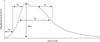
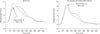
5. Krizaj D, Simunic B, Zagar T. Short-term repeatability of parameters extracted from radial displacement of muscle belly. J Electromyogr Kinesiol 2008;18:645–651.
6. Pisot R, Narici MV, Simunic B, et al. Whole muscle contractile parameters and thickness loss during 35-day bed rest. Eur J Appl Physiol 2008;104:409–414.
7. Garcia-Manso JM, Rodriguez-Ruiz D, Rodriguez-Matoso D, de Saa Y, Sarmiento S, Quiroga M. Assessment of muscle fatigue after an ultra-endurance triathlon using tensiomyography (TMG). J Sports Sci 2011;29:619–625.
8. Rodriguez Ruiz D, Quiroga Escudero ME, Rodriguez Matoso D, et al. Tensiomiografia utilizada para a avaliacao de jogadores de volei de praia de alto nivel. Rev Bras Med Esporte 2012;18:95–99.
9. Rusu LD, Cosma GG, Cernaianu SM, et al. Tensiomyography method used for neuromuscular assessment of muscle training. J Neuroeng Rehabil 2013;10:67.
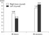
10. Alentorn-Geli E, Alvarez-Diaz P, Ramon S, et al. Assessment of neuromuscular risk factors for anterior cruciate ligament injury through tensiomyography in male soccer players. Knee Surg Sports Traumatol Arthrosc 2015;23:2508–2513.
11. Alentorn-Geli E, Alvarez-Diaz P, Ramon S, et al. Assessment of gastrocnemius tensiomyographic neuromuscular characteristics as risk factors for anterior cruciate ligament injury in male soccer players. Knee Surg Sports Traumatol Arthrosc 2015;23:2502–2507.
12. Alvarez-Diaz P, Alentorn-Geli E, Ramon S, et al. Effects of anterior cruciate ligament injury on neuromuscular tensiomyographic characteristics of the lower extremity in competitive male soccer players. Knee Surg Sports Traumatol Arthrosc. 2014 Sep 25; [doi: 10.1007/s00167-014-3319-4]
[Epub].
13. Alvarez-Diaz P, Alentorn-Geli E, Ramon S, et al. Comparison of tensiomyographic neuromuscular characteristics between muscles of the dominant and non-dominant lower extremity in male soccer players. Knee Surg Sports Traumatol Arthrosc. 2014 Sep 19; [doi: 10.1007/s00167-014-3298-5]
[Epub].
14. Alvarez-Diaz P, Alentorn-Geli E, Ramon S, et al. Effects of anterior cruciate ligament reconstruction on neuromuscular tensiomyographic characteristics of the lower extremity in competitive male soccer players. Knee Surg Sports Traumatol Arthrosc 2015;23:3407–3413.
TOOLS
Similar articles

A Novel Method for the Assessment of Muscle Injuries

Evaluation of Muscle Damage by Central Fatigue Using Tensiomyography

Analysis of Bodybuilder's Skeletal Muscle Characteristics Using Tensiomyography

Analysis of Lower Extremity Contraction According to Gender Using Tensiomyography

Evaluation of Reliability of Tensiomyography Measurement