Abstract
Purpose
This study is designed to evaluate the reliability for studies of tensiomyography (TMG). TMG can evaluate muscle function noninvasively and selectively.
Methods
We measured 12 male volunteers (age, 26.5±7.6 years; height, 175.3±4.7 cm; weight, 78.8±13.3 kg) in this study and measured TMG during three occasions over 3 consecutive days. None of the participants has had any history of neuromuscular disorders or muscle diseases. Vastus lateralis, vastus medialis (VM), rectus femoris (RF) in quadriceps and biceps femoris, semitendinosus in hamstrings muscles were measured. Coefficient of variation (CV%) and intraclass correlation coefficient (ICC) have been calculated about maximal displacement (Dm, mm) and contraction time (Tc, ms) which are main parameters.
Results
Most of the ICC of Dm were over 0.8 and the highest among the muscles except both VM. And, most ICC of Tc was lower than Dm except both BF (right, 18.31; left, 15.03). But, the ICC of Tc was lower than Dm except left RF (0.890) and VM (0.859).
Conclusion
This study has shown that the Dm is high levels of the ICC and CV(%) in thigh muscle except VM. In the future, we plan to establish the method of measurement more clearly for reducing the errors of measurements. The technique of correct palpation of measurable muscles using TMG devices is also necessary.
Go to : 
REFERENCES
1. Valencic V, Knez N. Measuring of skeletal muscles' dynamic properties. Artif Organs. 1997; 21:240–2.
2. Dahmane R, Valen i V, Knez N, Er en I. Evaluation of the ability to make non-invasive estimation of muscle contractile properties on the basis of the muscle belly response. Med Biol Eng Comput. 2001; 39:51–5.

3. Kim C, Chai JH, Kim BK, Kim CH, Bae SW. A novel method for the assessment of muscle injuries. Korean J Sports Med. 2015; 33:59–66.

4. Rey E, Lago-Penas C, Lago-Ballesteros J. Tensiomyography of selected lower-limb muscles in professional soccer players. J Electromyogr Kinesiol. 2012; 22:866–72.

5. Alvarez-Diaz P, Alentorn-Geli E, Ramon S, et al. Effects of anterior cruciate ligament injury on neuromuscular tensiomyographic characteristics of the lower extremity in competitive male soccer players. Knee Surg Sports Traumatol Arthrosc. 2016; 24:2264–70.

6. Alvarez-Diaz P, Alentorn-Geli E, Ramon S, et al. Effects of anterior cruciate ligament reconstruction on neuromuscular tensiomyographic characteristics of the lower extremity in competitive male soccer players. Knee Surg Sports Traumatol Arthrosc. 2015; 23:3407–13.

7. Neamtu MC, Rusu L, Neamtu OM, et al. Analysis of neuromuscular parameters in patients with multiple sclerosis and gait disorders. Rom J Morphol Embryol. 2014; 55:1423–8.
8. Chai JH, Kim C, Kim CH. TMG(tensiomyography): noninvasive method of evaluation of muscle function. Korean J Phys Educ. 2017; 56:519–26.

9. Pisot R, Narici MV, Simunic B, et al. Whole muscle contractile parameters and thickness loss during 35-day bed rest. Eur J Appl Physiol. 2008; 104:409–14.

10. Neamtu MC, Rusu L, Rusu PF, Neamtu OM, Georgescu D, Iancau M. Neuromuscular assessment in the study of structural changes of striated muscle in multiple sclerosis. Rom J Morphol Embryol. 2011; 52:1299–303.
11. Rodriguez-Ruiz D, Diez-Vega I, Rodriguez-Matoso D, Fernandez-del-Valle M, Sagastume R, Molina JJ. Analysis of the response speed of musculature of the knee in professional male and female volleyball players. Biomed Res Int. 2014; 2014:239708.
12. Garcia-Manso JM, Rodriguez-Matoso D, Sarmiento S, et al. Effect of high-load and high-volume resistance exercise on the tensiomyographic twitch response of biceps brachii. J Electromyogr Kinesiol. 2012; 22:612–9.
13. Macgregor LJ, Ditroilo M, Smith IJ, Fairweather MM, Hunter AM. Reduced radial displacement of the gastrocnemius medialis muscle after electrically elicited fatigue. J Sport Rehabil. 2016; 25:241–7.

14. Simunic B, Degens H, Rittweger J, Narici M, Mekjavic IB, Pisot R. Noninvasive estimation of myosin heavy chain composition in human skeletal muscle. Med Sci Sports Exerc. 2011; 43:1619–25.
15. Martin-Rodriguez S, Alentorn-Geli E, Tous-Fajardo J, et al. Is tensiomyography a useful assessment tool in sports medicine? Knee Surg Sports Traumatol Arthrosc. 2017; 25:3980–1.

16. Tous-Fajardo J, Moras G, Rodriguez-Jimenez S, Usach R, Doutres DM, Maffiuletti NA. Inter-rater reliability of muscle contractile property measurements using non-invasive tensiomyography. J Electromyogr Kinesiol. 2010; 20:761–6.

17. Simunic B. Between-day reliability of a method for noninvasive estimation of muscle composition. J Electromyogr Kinesiol. 2012; 22:527–30.
18. Wilson HV, Johnson MI, Francis P. Repeated stimulation, inter-stimulus interval and inter-electrode distance alters muscle contractile properties as measured by Tensiomyography. PLoS One. 2018; 13:e0191965.

19. Chai JH, Kim BK, Kim C, Kim CH, Bae SW. Analysis of bodybuilder's skeletal muscle characteristics using tensiomyography. Korean J Sports Med. 2016; 34:146–52.

20. Bae SW, Chai JH, Kim BK, Kim CH, Kim C. Evaluation and application of muscle injuries using tensiomyography. Korean J Sports Med. 2015; 33:143–6.

21. Krizaj D, Simunic B, Zagar T. Short-term repeatability of parameters extracted from radial displacement of muscle belly. J Electromyogr Kinesiol. 2008; 18:645–51.
22. Delagi EF, Perotto AO, Iazzetti J, Morrison D. Anatomical guide for the electromyographer: the limbs and trunk. 4th ed.Springfield: Charles. C. Thomas;2005.
23. Lohr C, Braumann KM, Reer R, Schroeder J, Schmidt T. Reliability of tensiomyography and myotonometry in detecting mechanical and contractile characteristics of the lumbar erector spinae in healthy volunteers. Eur J Appl Physiol. 2018; 118:1349–59.

24. Garcia-Garcia O, Serrano-Gomez V, Hernandez-Mendo A, Morales-Sanchez V. Baseline mechanical and neuromuscular profile of knee extensor and flexor muscles in professional soccer players at the start of the pre-season. J Hum Kinet. 2017; 58:23–34.
Go to : 
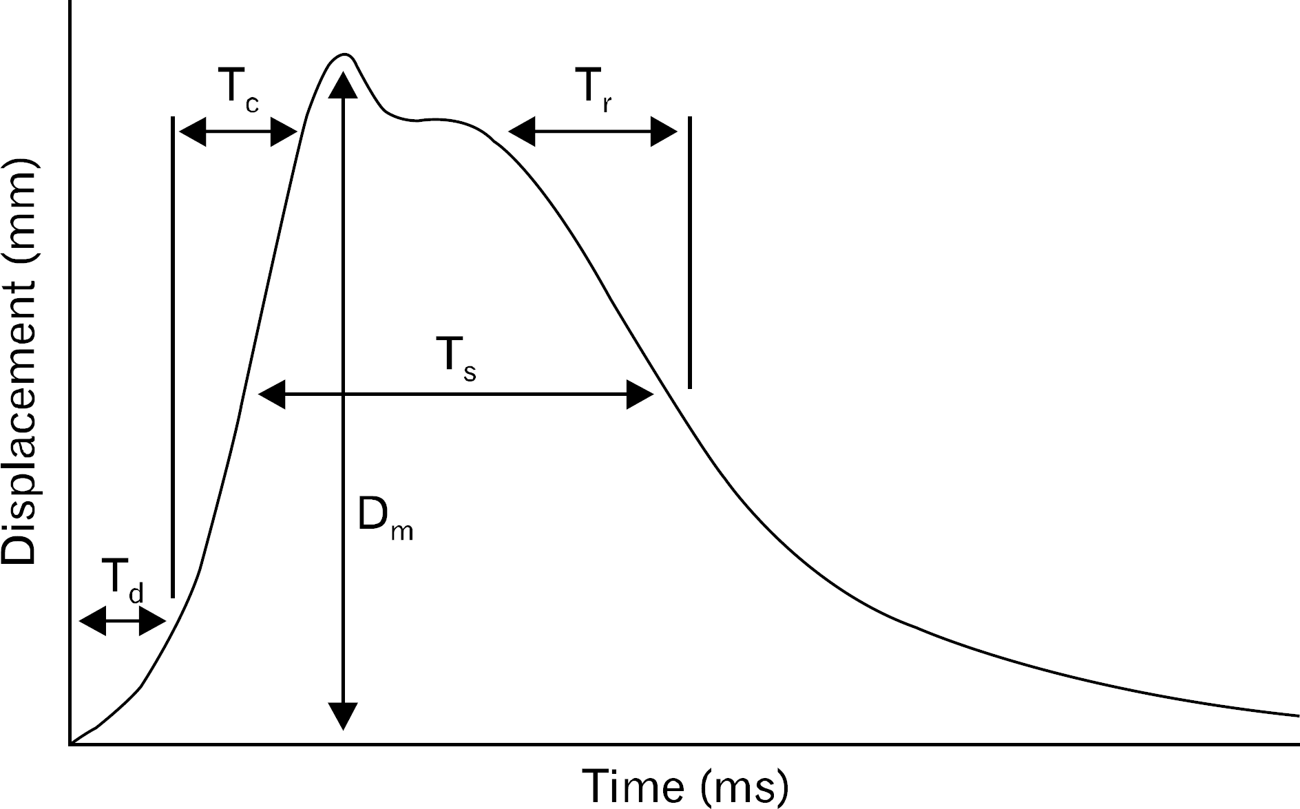
 | Fig. 2.Tensiomyography parameters. Tc: contraction time, Tr: relaxation time, Ts: sustain time, Td: delay time, Dm: maximal displacement. |
Table 1.
Physical characteristics (n=12)
| Sex | Age (yr) | Height (cm) | Weight (kg) |
|---|---|---|---|
| Male | 26.5±7.6 | 175.3±4.7 | 78.8±13.3 |
Table 2.
Maximal displacement (mm)
Table 3.
Contraction time (ms)


 PDF
PDF ePub
ePub Citation
 Print
Print


 XML Download