Journal List > J Korean Acad Prosthodont > v.55(1) > 1034961

J Korean Acad Prosthodont. 2017 Jan;55(1):79-87. Korean.
Published online Jan 25, 2017.  https://doi.org/10.4047/jkap.2017.55.1.79
© 2017 The Korean Academy of Prosthodontics
Full-mouth rehabilitation of skeletal anterior open bite with severely decayed dentition: A case report
Seong-A Kim, Kwantae Noh, Ahran Pae and Yi-Hyung Woo
Department of Prosthodontics, School of Dentistry, Kyung Hee University, Seoul, Republic of Korea.

Corresponding Author: Yi-Hyung Woo. Department of Prosthodontics, School of Dentistry, Kyung Hee University, 26, Kyungheedae-ro, Dongdaemun-gu, Seoul 02447, Republic of Korea. +82 (0)2 958 9340, Email: yhwoo@khu.ac.kr
Received July 25, 2016; Revised August 25, 2016; Accepted September 12, 2016.

This is an Open Access article distributed under the terms of the Creative Commons Attribution Non-Commercial License (http://creativecommons.org/licenses/by-nc/3.0/) which permits unrestricted non-commercial use, distribution, and reproduction in any medium, provided the original work is properly cited.


Abstract

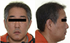
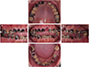
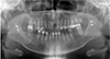
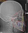
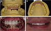
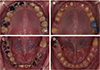
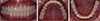
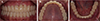
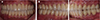
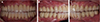
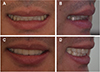
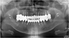
The open bite malocclusion is a common clinical entity and has multifactorial causes. Development of effective treatment plan and management is dependent on proper diagnosis. The skeletal open bite patient requires a coordinated orthodontic and orthognathic surgical approach to achieve stable occlusion, acceptable esthetics, and improved function. But in case of open bite with severely decayed dentition, restoration in the entire dentition is necessary. Using the facial analysis and diagnostic wax-up, the most effective treatment was prosthetic rehabilitation. The provisional restorations were fabricated to satisfy esthetic and functional requirements, which result in the uniformly distributed occlusal force, anterior and canine guidance. The inter-arch relationship, labio-dental harmony, and the soft tissue aspect, which is important to estimate the longevity were evaluated. Definitive restorations of monolithic zirconia were made by replicating provisional restorations by using the latest CAD/CAM technology. They were delivered to the patient and clinical follow-up observation was satisfactory.

Keywords: Skeletal anterior open bite; Rampant caries; CAD/CAM; Maintenance

Figures


Fig. 1
Preoperative extraoral view; long and narrow face with no lip-incompetence at rest posture; not a typical facial appearance of the skeletal anterior open bite.
Click for larger image


Fig. 2
Preoperative intraoral view showing severely decayed dentition and skeletal anterior open bite. (A) Occlusal view of maxilla, (B) Lateral view (right side), (C) Frontal view, (D) Lateral view (left side), (E) Occlusal view of mandible.
Click for larger image


Fig. 3
Preoperative panoramic view.
Click for larger image


Fig. 4
Preoperative cephalometric view.
Click for larger image


Fig. 5
Diagnostic wax-up for anterior programming device. (A) Frontal view, (B) Occlusal view, (C) Intraoral view of centric relation, (D) Evaluation of maxillary incisal crown length.
Click for larger image


Fig. 6
Diagnostic wax-up. (A) Frontal view, (B, C) Occlusal view of maxilla and mandible.
Click for larger image


Fig. 7
Intraoral view. (A, B) Before preliminary treatment, (C, D) After preliminary treatment.
Click for larger image


Fig. 8
1st provisional restoration. (A) Frontal view, (B, C) Occlusal view of maxilla and mandible.
Click for larger image


Fig. 9
2nd provisional restoration. (A) Frontal view, (B, C) Occlusal view of maxilla and mandible.
Click for larger image


Fig. 10
Assessment of anterior teeth. (A) Rest position, (B) Smile view, (C) Lateral view.
Click for larger image


Fig. 11
Definitive restoration. (A) Frontal view, (B, C) Lateral view.
Click for larger image


Fig. 12
Definitive restoration. (A) Frontal view (protrusive position), (B, C) Lateral view (eccentric position).
Click for larger image


Fig. 13
Postoperative view with dento-labial harmony. (A) Frontal view at rest position, (B) Lateral view at rest position, (C) Frontal view at smile, (D) Lateral view at smile.
Click for larger image


Fig. 14
Postoperative panoramic view.
Click for larger image

References
1. Beane RA Jr. Nonsurgical management of the anterior open bite: a review of the options. Semin Orthod 1999;5:275–283.
2. Huang GJ. Long-term stability of anterior open-bitetherapy: A review. Semin Orthod 2002;8:162–172.
3. Finkelstein T, Shapira Y, Shpack N. Nonsurgical treatment of severe open bite associated with amelogenesis imperfecta. J Clin Orthod 2012;46:427–433.
4. Dawson PE. In: Functional occlusion: from TMJ to smile design. Mosby; 2007. pp. 480-492.
5. Turner KA, Missirlian DM. Restoration of the extremely worn dentition. J Prosthet Dent 1984;52:467–474.
6. Dawson PE. Determining the determinants of occlusion. Int J Periodontics Restorative Dent 1983;3:8–21.
7. Akin E, Sayin MO, Karaçay S, Bulakbaşi N. Real-time balanced turbo field echo cine-magnetic resonance imaging evaluation of tongue movements during deglutition in subjects with anterior open bite. Am J Orthod Dentofacial Orthop 2006;129:24–28.
8. Lopez-Gavito G, Wallen TR, Little RM, Joondeph DR. Anterior open-bite malocclusion: a longitudinal 10-year postretention evaluation of orthodontically treated patients. Am J Orthod 1985;87:175–186.
9. Denison TF, Kokich VG, Shapiro PA. Stability of maxillary surgery in openbite versus nonopenbite malocclusions. Angle Orthod 1989;59:5–10.
TOOLS
ORCID iDs

Seong-A Kim
https://orcid.org/http://orcid.org/0000-0002-5980-7875

Kwantae Noh
https://orcid.org/http://orcid.org/0000-0003-3480-7737

Ahran Pae
https://orcid.org/http://orcid.org/0000-0001-8758-0754

Similar articles

Full-mouth rehabilitation in an amelogenesis imperfecta patient with anterior open bite using CAD/CAM system

Full mouth rehabilitation with extremely worn dentition

Full mouth rehabilitation using zirconia crown in severe worn dentition: a case report

Full mouth rehabilitation of a patient with worn dentition and loss of posterior support by vertical dimension reestablishment: a clinical report

Full mouth rehabilitation in a patient with loss of vertical dimension caused by severe tooth loss: a case report