

 PDF
PDF ePub
ePub Citation
 Print
Print


Jung-Hyun Ryu , Won-Sup Lee, Cheol-Won Lee, Su-Young Lee
, Won-Sup Lee, Cheol-Won Lee, Su-Young Lee
Abstract
Excessive teeth wear may result in the complications such as esthetic problems, hypersensitivity, and loss of vertical dimension. This clinical report focuses on the causes of severely worn dentition and the full-mouth rehabilitation of a patient with rheumatoid arthritis for 20 years. An interview, clinical and radiological examinations were performed to analyze the causes and decide the treatment plan. After delivery of the final prostheses, a night guard was used to protect the restorations and temporomandibular joints. The patient was satisfied esthetically and functionally.
Figures and Tables
Fig. 1
(A) Panoramic radiograph before treatment, (B) Temporomandibular joint radiograph before treatment. No evidence of pathologic change.
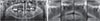
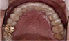
Fig. 2
Pretreatment state. (A) Maxillary occlusal view, (B) Right lateral view, (C) Frontal view, (D) Left lateral view, (E) Mandibular occlusal view.
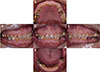
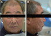
Fig. 4
(A) Facebow transfer, (B) Occlusal registration, (C) Length of anterior teeth, (D) Distance between upper and lower vestibule, (E) Diagnostic wax up model with new occlusal vertical dimension, (F) 1st Provisional restorations, (G) Stent, (H) Try-in of the provisional restorations.
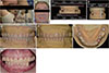
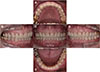
Fig. 5
(A) Lower provisional restoration with metal coping, (B) Radiograph of lower provisional restoration, (C) Provisional restoration before final impression.
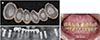
Fig. 6
(A) Customized incisal guide table, (B) Upper teeth preparation, (C) Lower teeth preparation, (D) Final impression of the upper teeth, (E) Final impression of the lower teeth, (F) Bite registration for cross mounting, (G) Mounting of the definitive cast.
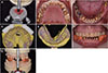
Fig. 7
Computer-aided design. (A) Scan of upper teeth, (B) Scan of the lower teeth, (C) Full contour of upper teeth, (D) Full contour of lower teeth.
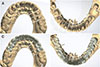
Fig. 8
(A) Panoramic radiograph after treatment, (B) Temporomandibular joint radiograph after treatment. No evidence of pathologic change.

References
1. Grippo JO, Simring M, Schreiner S. Attrition, abrasion, corrosion and abfraction revisited: a new perspective on tooth surface lesions. J Am Dent Assoc. 2004; 135:1109–1118.
2. Crothers AJ. Tooth wear and facial morphology. J Dent. 1992; 20:333–341.

3. Verrett RG. Analyzing the etiology of an extremely worn dentition. J Prosthodont. 2001; 10:224–233.
4. Smith BG, Knight JK. A comparison of patterns of tooth wear with aetiological factors. Br Dent J. 1984; 157:16–19.
5. Davies SJ, Gray RJ, Qualtrough AJ. Management of tooth surface loss. Br Dent J. 2002; 192:11–16. 19–23.
6. Muts EJ, van Pelt H, Edelhoff D, Krejci I, Cune M. Tooth wear: a systematic review of treatment options. J Prosthet Dent. 2014; 112:752–759.
7. Dawson PE. Functional occlusion: From TMJ to smile design. St. Louis, MO: Elsevier Health Sciences;2006.
8. Turner KA, Missirlian DM. Restoration of the extremely worn dentition. J Prosthet Dent. 1984; 52:467–474.
9. Abduo J, Lyons K. Clinical considerations for increasing occlusal vertical dimension: a review. Aust Dent J. 2012; 57:2–10.
10. Korean Academy of Orofacial Pain and Oral Medicine. Orofacial pains and temporomandibular disorders. 4th ed. Seoul, Korea: 2012. p. 155–156.
11. Nelson SJ. Wheeler's dental anatomy, physiology, and occlusion. 10th ed. St. Louis, MO: Elsevier, Saunders;2015. p. 13.
12. Goiato MC, Pellizzer EP, da Silva EV, Bonatto Lda R, dos Santos DM. Is the internal connection more efficient than external connection in mechanical, biological, and esthetical point of views? A systematic review. Oral Maxillofac Surg. 2015; 19:229–242.
13. Kim KS, Lim YJ, Kim MJ, Kwon HB, Yang JH, Lee JB, Yim SH. Variation in the total lengths of abutment/implant assemblies generated with a function of applied tightening torque in external and internal implant-abutment connection. Clin Oral Implants Res. 2011; 22:834–839.
14. Ozkurt Z, Kazazoğlu E. Clinical success of zirconia in dental applications. J Prosthodont. 2010; 19:64–68.
15. Sato S, Hotta TH, Pedrazzi V. Removable occlusal overlay splint in the management of tooth wear: a clinical report. J Prosthet Dent. 2000; 83:392–395.
16. Ehrlich J, Hochman N, Yaffe A. Contribution of oral habits to dental disorders. Cranio. 1992; 10:144–147.
17. Gregory-Head B, Curtis DA. Erosion caused by gastroesophageal reflux: diagnostic considerations. J Prosthodont. 1997; 6:278–285.
18. Chronopoulos V, Maroulakos G, Tsoutis K, Stathopoulou P, Nagy WW. Complete mouth rehabilitation and gastroesophageal reflux disease: Conventional and contemporary treatment approaches. J Prosthet Dent. 2017; 117:1–7.
19. Kuybulu FI, Gemalmaz D, Pameijer CH, Yarat A, Alcan T. Erosion of luting cements exposed to acidic buffer solutions. Int J Prosthodont. 2007; 20:494–495.
20. Rivera-Morales WC, Mohl ND. Relationship of occlusal vertical dimension to the health of the masticatory system. J Prosthet Dent. 1991; 65:547–553.
 XML Download