Journal List > J Korean Acad Prosthodont > v.54(1) > 1034858

J Korean Acad Prosthodont. 2016 Jan;54(1):1-7. Korean.
Published online Jan 29, 2016.  https://doi.org/10.4047/jkap.2016.54.1.1
Copyright © 2016 The Korean Academy of Prosthodontics
A study of lower facial change according to facial type when virtually vertical dimension increases
Nam-Woo Kim,1 Gung-Chol Lee,2 Cheol-Hyun Moon,2 Jung-Yoon Bae,1 and Ji-Yeon Kim1
1Department of Prosthodontics, Gil Medical Center, Gachon University, Inchon, Republic of Korea.
2Department of Orthodontics, Gil Medical Center, Gachon University, Inchon, Republic of Korea.

Corresponding Author: Ji-Yeon Kim. Department of Prosthodontics, Gil Medical Center, Gachon University, 774-34, Namdongdae-ro, Namdong-gu, Inchon 21565, Republic of Korea. +82 32 460 3376: Email: prosthetic02@hanmail.net
Received August 05, 2015; Revised August 25, 2015; Accepted September 02, 2015.

This is an Open Access article distributed under the terms of the Creative Commons Attribution Non-Commercial License (http://creativecommons.org/licenses/by-nc/3.0/) which permits unrestricted non-commercial use, distribution, and reproduction in any medium, provided the original work is properly cited.


Abstract

Purpose

The aim of this study was to evaluate the effect of increased vertical dimension of occlusion on lower facial changes by facial type.

Materials and methods

Lateral cephalograms from 261 patients were obtained and classified by sagittal (Class I, II, and III) and vertical (hypodivergent, normodivergent, and hyperdivergent) facial patterns. Retrusive displacement of soft tissue Pogonion and downward displacement of soft tissue Menton were measured in each group after 2 mm of vertical dimension of occlusion was increased at the lower central incisor using a virtual simulation program. The ratio of both displacements was calculated in all groups. The statistical analysis was done by 2-way ANOVA and Post hoc was done by Tukey test (5% level of significance).

Results

Retrusive displacement of soft tissue Pogonion in Class III group was statistically different compared to Class I and II, and in vertical facial groups all 3 groups were significantly different (P<.05). Downward displacement of soft tissue Menton showed statistically significant difference between all sagittal groups and vertical groups (P<.05). The ratio of both displacements showed statistically significant difference in all sagittal groups and vertical groups (P<.05), and Class II hyperdivergent group had the highest value.

Conclusion

Lower facial change was statically significant according to the facial type when vertical dimension of occlusion increased. Class II hyperdivergent facial type showed the highest ratio after increase in vertical dimension of occlusion.

Keywords: Vertical dimension; Vertical dimension of occlusion

Figures


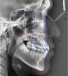
Fig. 1
Digital tracing of Lateral Cephalometric Radiography.
Click for larger image

Tables


Table 1
Definition of cephalometric landmarks
Click for larger image


Table 2
Definition of reference plane
Click for larger image


Table 3
Mean Value and Standard Deviation of measurements
Click for larger image


Table 4
Numbers in each group
Click for larger image


Table 5
Mean and standard deviation (SD) of the soft tissue Pogonion displacement on X axis (mm)
Click for larger image


Table 6
Mean and standard deviation (SD) of the soft tissue Menton displacement on Y axis (mm)
Click for larger image


Table 7
Mean and standard deviation (SD) of the ratio of the soft tissue Pogonion displacement on X axis over the soft tissue Menton displacement on Y axis
Click for larger image

References
1. Orthlieb JD, Laurent M, Laplanche O. Cephalometric estimation of vertical dimension of occlusion. J Oral Rehabil 2000;27:802–807.
2. Turner KA, Missirlian DM. Restoration of the extremely worn dentition. J Prosthet Dent 1984;52:467–474.
3. Burstone CR. Deep overbite correction by intrusion. Am J Orthod 1977;72:1–22.
4. Park YC, Lee SY, Kim DH, Jee SH. Intrusion of posterior teeth using mini-screw implants. Am J Orthod Dentofacial Orthop 2003;123:690–694.
5. Ramfjord SP, Blankenship JR. Increased occlusal vertical dimension in adult monkeys. J Prosthet Dent 1981;45:74–83.
6. Berry DC, Poole DF. Attrition: possible mechanisms of compensation. J Oral Rehabil 1976;3:201–206.
7. Crothers A, Sandham A. Vertical height differences in subjects with severe dental wear. Eur J Orthod 1993;15:519–525.
8. Varrela TM, Paunio K, Wouters FR, Tiekso J, Söder PO. The relation between tooth eruption and alveolar crest height in a human skeletal sample. Arch Oral Biol 1995;40:175–180.
9. Dahl BL, Krogstad O. The effect of a partial bite-raising splint on the inclination of upper and lower front teeth. Acta Odontol Scand 1983;41:311–314.
10. Mohindra NK, Bulman JS. The effect of increasing vertical dimension of occlusion on facial aesthetics. Br Dent J 2002;192:164–168.
11. Albarakati SF, Kula KS, Ghoneima AA. The reliability and reproducibility of cephalometric measurements: a comparison of conventional and digital methods. Dentomaxillofac Radiol 2012;41:11–17.
12. Chung KR. In: The result report about measurement of lateral cephalogram in Korean adult who have normal occlusion. Seoul, Rep. of Korea: Korean Assoc Orthodont; 1997. pp. 13-17.
13. Broadbent BH. A new x-ray technique and its application to orthodontia. Angle Orthod 1931;1:45–66.
14. Ricketts RM. The role of cephalometrics in prosthetic diagnosis. J Prosthet Dent 1956;6:488–503.
15. DiPietro GJ, Moergeli JR. Significance of the Frankfortmandibular plane angle to prosthodontics. J Prosthet Dent 1976;36:624–635.
16. McGEE GF. Use of facial measurements in determining vertical dimension. J Am Dent Assoc 1947;35:342–350.
17. Ellinger CW. Radiographic study of oral structures and their relation to anterior tooth position. J Prosthet Dent 1968;19:36–45.
18. Fayz F, Eslami A. Determination of occlusal vertical dimension: a literature review. J Prosthet Dent 1988;59:321–323.
19. Carlsson GE, Ingervall B, Kocak G. Effect of increasing vertical dimension on the masticatory system in subjects with natural teeth. J Prosthet Dent 1979;41:284–289.
20. Rebibo M, Darmouni L, Jouvin J, Orthlieb JD. Vertical dimension of occlusion: the keys to decision. Int J stomatol occlu med 2009;2:147–159.
21. Scheideman GB, Bell WH, Legan HL, Finn RA, Reisch JS. Cephalometric analysis of dentofacial normals. Am J Orthod 1980;78:404–420.
22. Mack MR. Vertical dimension: A dynamic concept based on facial form and oropharyngeal function. J Prosthet Dent 1991;66:478–485.
23. Parker CD, Nanda RS, Currier GF. Skeletal and dental changes associated with the treatment of deep bite malocclusion. Am J Orthod Dentofacial Orthop 1995;107:382–393.
24. Geelen W, Wenzel A, Gotfredsen E, Kruger M, Hansson LG. Reproducibility of cephalometric landmarks on conventional film, hardcopy, and monitor-displayed images obtained by the storage phosphor technique. Eur J Orthod 1998;20:331–340.
25. Yoon YJ, Kim KS, Hwang MS, Kim HJ, Choi EH, Kim KW. Effect of head rotation on lateral cephalometric radiographs. Angle Orthod 2001;71:396–403.
26. Lauritzen AG, Bodner GH. Variations in location of arbitrary and true hinge axis points. J Prosthet Dent 1961;11:224–229.
TOOLS
ORCID iDs

Ji-Yeon Kim
https://orcid.org/http://orcid.org/0000-0002-9609-8529

Similar articles

Full mouth rehabilitation of the patient with severely worn dentition: a case report

Full mouth rehabilitation accompanied by phonetic analysis of a patient with reduction of vertical dimension of occlusion, and inaccurate pronunciation due to numerous tooth loss: a case report

Mouth rehabilitation of a patient with severely worn dentition with vertical dimension increase

Prediction of frontal soft tissue changes after mandibular surgery in facial asymmetry individuals

Full mouth rehabilitation using removable partial denture in patient with loss of vertical dimension due to worn dentition