

 PDF
PDF ePub
ePub Citation
 Print
Print


Eun-Young Shim , Na-Young Lee, Jeong-Kyung Kang
, Na-Young Lee, Jeong-Kyung Kang
Abstract
Gradual attrition is a normal process of aging, but severe attrition causes occlusal disharmony, functional disorder and esthetic problems. The collapse of posterior support may cause attrition of anterior teeth, and loss of occlusal vertical dimension (OVD). And it induces the pathologic change of the TMJ, unaesthetic facial appearance and decreased masticatory function. In this case, 70 year-old male presented with decreased vertical dimension and esthetic problems due to worn dentition. Based on assessment of intraoral findings, diagnostic cast and radiographic examination, full-mouth rehabilitation with increase of OVD was planned. After 10 month follow-up, occlusal stability is maintained and through this procedure, satisfactory outcomes were achieved in esthetic and functional aspects.
Figures and Tables
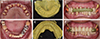
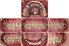
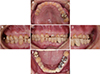 | Fig. 1Intraoral photograph before treatment. (A) Upper view, (B) Right side view, (C) Frontal view, (D) Left side view, (E) Lower view.
|
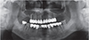
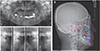 | Fig. 2(A) Panoramic radiograph before treatment, (B) Cephalometric radiograph before treatment, (C) TMJ series before treatment.
|
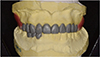
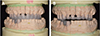
 | Fig. 5(A, D) Tooth preparation, (B, E) Final impression, (C, F) interocclusal relationship registrations using provisional restorations.
|

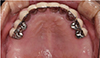
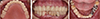
 | Fig. 9Maxillary removable partial denture fabrication. (A) impression, (B) framework, (C) wax denture.
|

References
1. Ibbetson RJ, Setchell DJ. Treatment of the worn dentition: 2. Dent Update. 1989; 16:300–302. 305–307.
2. Hemmings KW, Howlett JA, Woodley NJ, Griffiths BM. Partial dentures for patients with advanced tooth wear. Dent Update. 1995; 22:52–59.
3. Turner KA, Missirlian DM. Restoration of the extremely worn dentition. J Prosthet Dent. 1984; 52:467–474.

4. Briggs P, Bishop K. Fixed prostheses in the treatment of tooth wear. Eur J Prosthodont Restor Dent. 1997; 5:175–180.
5. Willis FM. Features of the face involved in full denture prosthesis. Dental Cosmos. 1935; 77:851–854.
6. Dawson PE. Functional occlusion: From TMJ to smile design. St. Louis; MO: Elsevier Health Sciences;2006.
7. Tench RW. Dangers in dental reconstruction in-volving increase of the vertical dimension of the lower third of the human face. J Am Dent Assoc. 1938; 25:566–570.
8. Monteith B. The role of the free-way space in the generation of muscle pain among denture-wearers. J Oral Rehabil. 1984; 11:483–498.
9. Carlsson GE, Ingervall B, Kocak G. Effect of increasing vertical dimension on the masticatory system in subjects with natural teeth. J Prosthet Dent. 1979; 41:284–289.
10. Abduo J. Safety of increasing vertical dimension of occlusion: a systematic review. Quintessence Int. 2012; 43:369–380.
11. Abduo J, Lyons K. Clinical considerations for increasing occlusal vertical dimension: a review. Aust Dent J. 2012; 57:2–10.
12. DiPietro GJ. A study of occlusion as related to the Frankfortmandibular plane angle. J Prosthet Dent. 1977; 38:452–458.
 XML Download