

 PDF
PDF ePub
ePub Citation
 Print
Print


Abstract
Objective
To aid the development of a frontal image simulating program, we evaluated the soft tissue frontal changes in relationship to movement of hard tissue with orthognathic surgery of facial asymmetry patients.
Methods
Preoperative and postoperative frontal cephalograms and frontal view photographs of 45 mandibular surgery patients with facial asymmetry were obtained in a standardized manner. Vertical and horizontal changes of hard tissue and soft tissue were measured from cephalograms and photographs, respectively. Soft tissue change in result to hard tissue change was then analyzed.
Results
Both vertical and horizontal correlation analysis showed a weak relationship between the changes. Hard tissue points that were picked for 1:1 mean ratio with soft tissue points did not show any significant relevance. For each soft tissue change, regressive equation was formulated by stepwise multiple regression analysis, and the equation for soft tissue Menton was most reliable in predicting changes. Both vertical and horizontal hard tissue changes were used together in prediction of vertical or horizontal soft tissue change.
Figures and Tables
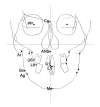 | Fig 1Cephalometric landmarks to represent hard tissue change. Detailed description of landmarks is in Table 2.
|
 | Fig 2Photometric landmarks to represent soft tissue change. Detailed description of landmarks is in Table 3.
|
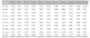
Table 1
Description of the subjects used in this study and summary of orthognathic surgery performed
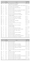
![]()
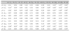
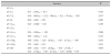
Table 4
Correlation coefficient between hard tissue change and soft tissue change in the horizontal direction
![]()
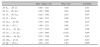
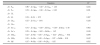
Table 5
Correlation coefficient between hard tissue change and soft tissue change in the vertical direction
![]()
References
1. Robinson SW, Speidel TM, Isaacson RJ, Worms FW. Soft tissue profile change produced by reduction of mandibular prognathism. Angle Orthod. 1972. 42:227–235.
2. Hershey HG, Smith LH. Soft-tissue profile change associated with surgical correction of the prognathic mandible. Am J Orthod. 1974. 65:483–502.

3. Lines PA, Steinhauser EW. Soft tissue changes in relationship to movement of hard structures in orthognathic surgery: a preliminary report. J Oral Surg. 1974. 32:891–896.
4. Mansour S, Burstone C, Legan H. An evaluation of soft tissue changes resulting from Le Fort I maxillary surgery. Am J Orthod. 1983. 84:37–47.
5. Jensen AC, Sinclair PM, Wolford LM. Soft tissue changes associated with double jaw surgery. Am J Orthod Dentofacial Orthop. 1992. 101:266–275.
6. Lin SS, Kerr WJ. Soft and hard tissue changes in Class III patients treated by bimaxillary surgery. Eur J Orthod. 1998. 20:25–33.
7. Enacar A, Taner T, Toroğlu S. Analysis of soft tissue profile changes associated with mandibular setback and double-jaw surgeries. Int J Adult Orthodon Orthognath Surg. 1999. 14:27–35.
8. Chew MT. Soft and hard tissue changes after bimaxillary surgery in Chinese Class III patients. Angle Orthod. 2005. 75:959–963.
9. McNeill RW, Proffit WR, White RP. Cephalometric prediction for orthodontic surgery. Angle Orthod. 1972. 42:154–164.
10. Kinnebrew MC, Hoffman DR, Carlton DM. Projecting the soft-tissue outcome of surgical and orthodontic manipulation of the maxillofacial skeleton. Am J Orthod. 1983. 84:508–519.
11. Bhatia SN, Sowray JH. A computer-aided design for orthognathic surgery. Br J Oral Maxillofac Surg. 1984. 22:237–253.
12. Harradine NW, Birnie DJ. Computerized prediction of the results of orthognathic surgery. J Maxillofac Surg. 1985. 13:245–249.
13. Sarver DM, Johnston MW, Matukas VJ. Video imaging for planning and counseling in orthognathic surgery. J Oral Maxillofac Surg. 1988. 46:939–945.
14. Sarver DM, Johnston MW. Video imaging: techniques for superimposition of cephalometric radiography and profile images. Int J Adult Orthodon Orthognath Surg. 1990. 5:241–248.
15. Hwang HS, Lee KH, Park JY, Kang BC, Park JW, Lee JS. Development of posteroranterior cephalometric analysis for the diagnosis of facial asymmetry. J Korean Dent Assoc. 2004. 42:219–231.
16. Kim EH, Hwang HS. The validity of head posture aligner in posteroanterior cephalometry. Korean J Orthod. 2000. 30:543–552.
17. Kim KM, Lee KH, Kim WS, Hwang HS. A comparative study on the construction of the reference line in posteroanterior cephalometry. J Korean Dent Assoc. 2001. 39:676–683.
18. Hwang HS, Eun CS, Hwang CH, Lim HJ. Three-dimensional CT image study on the correction of gonial angle width enlarged on frontal cephalogram. Korean J Orthod. 2005. 35:251–261.
19. Major PW, Johnson DE, Hesse KL, Glover KE. Landmark identification error in posterior anterior cephalometrics. Angle Orthod. 1994. 64:447–454.
20. Legrell PE, Nyquist H, Isberg A. Validity of identification of gonion and antegonion in frontal cephalograms. Angle Orthod. 2000. 70:157–164.
 XML Download