Journal List > Cancer Res Treat > v.56(4) > 1516088705

Choi, Chung, Choi, Kim, Kim, Yoo, Ryoo, Lee, Choi, and Choi: Clinical and Radiologic Predictors of Response to Atezolizumab-Bevacizumab in Advanced Hepatocellular Carcinoma

Abstract

Purpose

This study aimed to identify clinical and radiologic characteristics that could predict response to atezolizumab-bevacizumab combination therapy in patients with advanced hepatocellular carcinoma (HCC).

Materials and Methods

This single-center retrospective study included 108 advanced HCC patients with intrahepatic lesions who were treated with atezolizumab-bevacizumab. Two radiologists independently analyzed imaging characteristics of the index tumor on pretreatment computed tomography. Predictive factors associated with progressive disease (PD) at the best response based on Response Evaluation Criteria in Solid Tumors, ver. 1.1 were evaluated using logistic regression analysis. Progression-free survival (PFS) was estimated by the Kaplan-Meier method and compared with the log-rank test.

Results

Of 108 patients with a median PFS of 15 weeks, 40 (37.0%) had PD during treatment. Factors associated with PD included the presence of extrahepatic metastases (adjusted odds ratio [aOR], 4.13; 95% confidence interval [CI], 1.19 to 14.35; p=0.03), the infiltrative appearance of the tumor (aOR, 3.07; 95% CI, 1.05 to 8.93; p=0.04), and the absence of arterial-phase hyperenhancement (APHE) (aOR, 6.34; 95% CI, 2.18 to 18.47; p < 0.001). Patients with two or more of these factors had a PD of 66.7% and a median PFS of 8 weeks, indicating a significantly worse outcome compared to the patients with one or no of these factors.

Conclusion

In patients with advanced HCC treated with atezolizumab-bevacizumab treatment, the absence of APHE, infiltrative appearance of the intrahepatic tumor, and presence of extrahepatic metastases were associated with poor response and survival. Evaluation of early response may be necessary in patients with these factors.

Introduction

Hepatocellular carcinoma (HCC) is among the most prevalent cancers worldwide and causes approximately 800,000 deaths annually, making it the third leading cause of cancer-related deaths [1]. Despite routine surveillance of populations at risk, many patients are still diagnosed with HCC at an advanced stage [2,3]. Numerous trials are being conducted on the treatment of advanced HCC due to its poor prognosis. The combination of the immune checkpoint inhibitor (ICI) atezolizumab and the vascular endothelial growth factor (VEGF) inhibitor bevacizumab has shown superior efficacy when compared with the tyrosine kinase inhibitor sorafenib [4], leading to the use of this combination as first-line therapy in patients with advanced HCC.
Trials in multiple malignancies have shown that only about 20%-40% of patients experience an objective tumor response to ICIs, with a smaller percentage achieving long-term remission [5-7]. Studies attempting to identify pretreatment predictors of response to ICIs have found that the level of expression of programmed death-ligand 1 (PD-L1) in tumor cells was associated with responses to treatment with ICIs, with high PD-L1 expression being associated with a favorable response to programmed death-1 (PD-1) or PD-L1 inhibitors [8]. Because the environment of HCC includes the interaction of hepatitis and/or cirrhosis with the host immune system, the HCC microenvironment is more complex than that of other solid tumors [9]. Indeed, it has been found that intrahepatic HCCs respond less favorably to ICIs than extrahepatic tumors [10], highlighting the importance of predicting the responses of intrahepatic HCCs to treatment with ICIs. Moreover, because HCC is primarily diagnosed based on imaging findings at high-risk setting (such as the presence of the hepatitis B or C virus or cirrhosis) [2,3,11], biopsies are not routinely performed, and pathologic prognostic factors such as PD-L1 expression may not be available in most HCC patients. Rather, because several imaging features of HCCs have been shown to correlate with their pathological characteristics, the potential role of radiologic findings as surrogate markers of pathologic data is currently under investigation [12].
Identifying pretreatment predictors based on clinical and radiologic findings of response to atezolizumab-bevacizumab could be keys to optimizing systemic therapy of HCC. The present study therefore sought to identify clinical and radiologic characteristics that could predict response to atezolizumab-bevacizumab combination therapy in patients with advanced HCC.

Materials and Methods

1. Study population

This retrospective study included patients who received atezolizumab-bevacizumab as first-line systemic treatment at a single tertiary referral hospital between July 1, 2016, and December 31, 2021. Patients previously enrolled in phase I trials or those who received atezolizumab-bevacizumab after the drugs were approved for treating unresectable HCC were evaluated for study eligibility. Patients were included according to the following criteria: (1) age ≥ 18 years; (2) HCC diagnosed by radiographic characteristics on computed tomography (CT) and/or magnetic resonance imaging (MRI) or histological findings, in accordance with the HCC guidelines of the American Association for the Study of Liver Diseases (AASLD) [3]; (3) advanced and unresectable HCC; (4) atezolizumab-bevacizumab treatment as first-line systemic treatment; and (5) available pretreatment multiphase liver CT within 1 month before atezolizumab-bevacizumab treatment. Patients were excluded if they (1) had been treated with either atezolizumab or bevacizumab alone; (2) had poor performance status, defined as an Eastern Cooperative Oncology Group (ECOG) performance status > 2; (3) had no evidence of HCC in the liver; or (4) had intrahepatic but nonmeasurable HCC (Fig. 1). Atezolizumab (fixed dose of 1,200 mg) plus bevacizumab (15 mg/kg) were administered intravenously every 3 weeks [4] until disease progression, severe adverse events, or death. According to patient tolerance, dosage delays were granted. Clinical information, including demographic characteristics, laboratory results, imaging characteristics, and clinical outcomes, was obtained from patients’ electronic medical records.

2. Image acquisition

All patients underwent pretreatment multiphase liver CT (Siemens Healthineers, Erlangen, Germany) within 1 month before atezolizumab-bevacizumab treatment. All imaging examinations met the technical acquisition standards of the Liver Imaging Reporting and Data System (LI-RADS) [13]. CT examinations were performed on 64 channel multidetector CT scanners. After unenhanced images were acquired, iodinated contrast medium (Ultravist 370, Bayer Schering Pharma, Berlin, Germany) was injected intravenously using a power injector at a rate of 3 mL/sec, followed by the acquisition of late-arterial phase (determined using a bolus triggering method), portal venous phase (70-90 seconds), and delayed phase (about 180 seconds) images. Details of the CT parameters are summarized in S1 Table.

3. Image analysis

All images were anonymized, randomized, and independently reviewed by two board-certified abdominal radiologists (with 8 and 4 years of experience in hepatic imaging, respectively). Because this study was mainly focused on the characteristics of index tumors on pretreatment CT, not on the diagnostic performance in lesion detection, the readers were informed that all patients had HCCs and were told the size and location of the index tumor to be analyzed. However, they were blinded to the clinical data, including imaging evaluations by other readers and treatment outcomes. The list of index tumors was prepared by an investigator who was not involved in the image analysis. When multiple tumors were present, the largest lesion was selected as the index tumor. Any discrepancies between the readers were settled by a consensus review with a third invited reader with 19 years of experience in hepatic imaging.
Imaging characteristics of the index tumor on pretreatment CT were evaluated, including tumor size, tumor location, the number of involved liver segments, tumor margins (smooth vs. nonsmooth), infiltrative appearance (yes vs. no), arterial-phase hyperenhancement (APHE; yes vs. no), rim enhancement (yes vs. no), washout appearance (yes vs. no), intratumoral necrosis (yes vs. no), peritumoral bile duct dilatation (yes vs. no), tumor-in-vein (yes vs. no), and multifocality (yes vs. no), as determined by LI-RADS lexicon [14]. Detailed definitions of the analyzed imaging characteristics are summarized in S2 Table.

4. Outcome assessment

Tumor response was assessed based on Response Evaluation Criteria in Solid Tumors, ver. 1.1 (RECIST 1.1). Responses were evaluated by serial multiphasic CT or MRI, performed after every two to three cycles of treatment (6-9 weeks), with additional evaluation as warranted. The primary outcome of interest was progressive disease (PD) at best response. The secondary outcome was progression-free survival (PFS).

5. Statistical analysis

Continuous variables were expressed as median and interquartile range or mean and standard deviation and were compared using unpaired two-tailed t-tests. Categorical variables were expressed as frequencies and percentages and were compared using Fisher’s exact test or the chi-square test. Inter-reader agreement on imaging characteristics was evaluated using intraclass correlation coefficients and kappa statistics.
To determine factors significantly predictive of PD, patients were classified into two groups (i.e., PD vs. non-PD), and univariable logistic regression analysis was performed using various clinical and imaging characteristics. Factors found to differ significantly on univariable analyses were included in a multivariable logistic regression analysis to determine factors independently predictive of PD after atezolizumab-bevacizumab treatment. Based on a multivariable logistic regression analysis, the risk for PD was presented graphically as a nomogram. The predictive accuracy of the nomogram was assessed using the concordance index (c-index), and a calibration plot was generated to compare the predicted and observed probability of PD by the nomogram. PFS was analyzed by the Kaplan-Meier method according to the presence or absence of a predictive factor and compared with the log-rank test. Hazard ratios (HRs) for survival outcomes were calculated using a Cox proportional hazards model. In addition, tumor response and PFS were compared according to the number of significant predictive factors (i.e., N=0, N=1, or N ≥ 2).
Statistical analyses were performed using SPSS ver. 21 (IBM Corp., Armonk, NY) or R statistics ver. 4.0.3 (R Foundation for Statistical Computing, Vienna, Austria), with p-values < 0.05 defined as statistically significant.

Results

1. Patient characteristics

Of the 160 eligible patients, 52 were excluded, including six who received atezolizumab or bevacizumab monotherapy, 14 who were not followed up for assessment of treatment response, two with ECOG performance status > 2, 26 without no HCC in the liver, and four with nonmeasurable intrahepatic lesions. The study therefore included 108 patients, 95 men and 13 women, of mean age 58±12 years (Fig. 1). Their clinical characteristics are presented in Table 1. Of the 108 patients, 77 (71.3%) had hepatitis B, making it the most common risk factor for HCC; 55 (50.9%) had Child-Pugh class A disease; and 101 (93.5%) had advanced Barcelona Clinic Liver Cancer stage disease. Seventy-two patients (66.7%) received locoregional treatments, including ablation, radiation therapy, and transarterial chemoembolization, prior to treatment with atezolizumab-bevacizumab. Eighty patients (74.1%) had extrahepatic metastases, with the lungs being the most frequent sites of extrahepatic metastases.
During treatment, two patients (1.9%) achieved a complete response; 13 (12.0%) achieved a partial response; 53 (49.1%) had stable disease, making the disease control rate 63.0%; and 40 (37.0%) had PD at the best response. Over a median follow-up period of 8 months (maximum, 27 months), 87 patients (80.6%) died, and 88 (81.5%) experienced disease progression.

2. Pretreatment imaging characteristics

Pretreatment imaging characteristics are summarized in Table 2. Mean tumor sizes were 9.9±5.7 cm in the PD group and 8.3±5.4 cm in the non-PD group. The numbers of tumor-involved liver segments were similar (4.7±2.5 vs. 4.4±2.8, p=0.69), although infiltrative appearance was significantly more frequent in the PD than in the non-PD group (45.0% vs. 19.1%, p=0.005). In addition, absence of APHE (45.0% vs. 11.8%, p < 0.001) and presence of tumor-in-vein (70.0% vs. 38.2%, p=0.002) were significantly more frequent in the PD than in the non-PD group (Fig. 2).
The kappa value of inter-reader agreement for the infiltrative appearance, the absence of APHE, and tumor-in-vein was 0.60, 0.72, and 0.83, respectively (S3 Table).

3. Determination of factors predictive of progressive disease

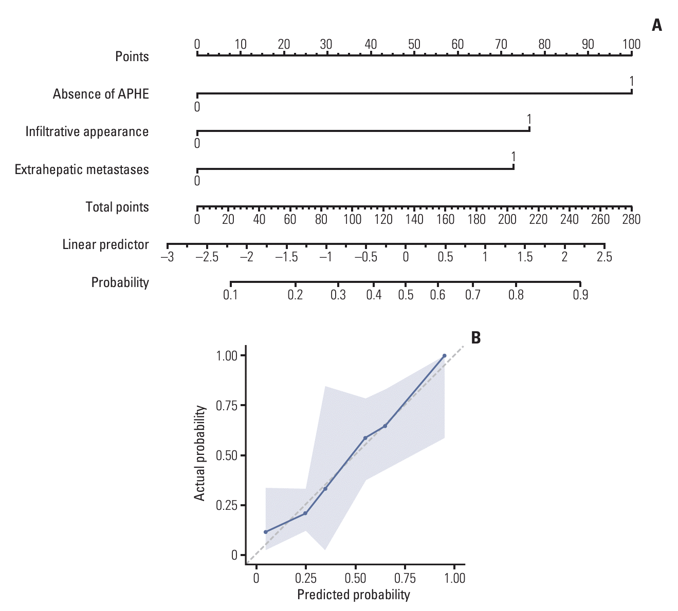
Univariable analysis identified five clinical and imaging characteristics associated with PD: the presence of extrahepatic metastases, high α-fetoprotein (AFP; ≥ 400 ng/mL), the absence of APHE in the tumor, the infiltrative appearance of the tumor, and tumor-in-vein (Table 3). Multivariable analysis showed that the presence of extrahepatic metastases (adjusted odds ratio [aOR], 4.13; 95% confidence interval [CI], 1.19 to 14.35; p=0.03), the absence of APHE in the tumor (aOR, 6.34; 95% CI, 2.18 to 18.47; p < 0.001), and the infiltrative appearance of the tumor (aOR, 3.07; 95% CI, 1.05 to 8.93; p=0.04) were independent predictive factors of PD. Based on the results of this model, a nomogram was displayed showing the probability of PD (Fig. 3A). The model had a c-index of 0.78 (95% CI, 0.68 to 0.87), and the calibration plot showed a good calibration (Fig. 3B).
When comparing tumor response according to the number of significant predictive factors, the disease control rate was significantly lower in the group with two or more predictive factors compared to the group without predictive factors (33.3% vs. 83.3%, p < 0.001) or compared to the group with one predictive factor (33.3% vs. 78.4%, p < 0.001) (Table 4).

4. PFS stratified by significant predictive factors

Accordingly, median PFS was significantly shorter in patients with than without extrahepatic metastases (11.3 vs. 30.0 weeks; HR, 2.10; 95% CI, 1.29 to 3.42; p=0.03) (S4 Fig.), in patients without than with APHE (8.9 vs. 17.6 weeks; HR, 1.76; 95% CI, 1.11 to 2.79; p=0.02), and in patients with than without infiltrative appearance (8.3 vs. 17.1 weeks; HR, 1.65; 95% CI, 1.07 to 2.56; p=0.02).
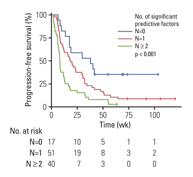
Furthermore, when the patients were stratified by the number of significant predictive factors, the median PFS in the group with two or more predictive factors was significantly shorter than that both the group with one predictive factor (8.1 vs. 18.4 weeks; HR, 1.99; 95% CI, 1.03 to 3.86; p=0.04) and the group without predictive factors (8.1 vs. 37.9 weeks; HR, 3.80; 95% CI, 1.93 to 7.49; p < 0.001) (Fig. 4).

Discussion

The present study analyzed baseline predictors of response to the combination of atezolizumab and bevacizumab in patients with advanced HCC. Although tumor markers did not differ significantly between the PD and non-PD groups, clinical and imaging characteristics, such as APHE in the tumor, the infiltrative appearance of the tumor, and the presence of extrahepatic metastases, were predictive of treatment response. In particular, when patients were stratified based on the number of predictive factors, those with two or more predictive factors had a disease control rate of only 33.3% and a median PFS of only 8 weeks, indicating a dismal outcome compared to those with one or no predictive factors.
Multitargeted tyrosine kinase inhibitors, including sorafenib, lenvatinib, regorafenib, and cabozantinib, often result in disease control with a modest response and have become the backbone of systemic therapy for HCC [15-18]. Most patients, however, develop resistance to these tyrosine kinase inhibitors after a certain period of time. The introduction of ICIs markedly altered the treatment landscape of HCC with a radiological response rate of 10%-20% with a durable response by anti–PD-1 monotherapy in patients with HCC [19,20]. The approval of the combination of atezolizumab-bevacizumab as first-line treatment for HCC was a significant milestone, leading to an increased response rate of up to 30% [4]. Although it remains unclear whether the combination of atezolizumab-bevacizumab elicits various organ-specific immune responses, tumor responses to ICIs have been reported to depend on the specific organ [21]. For example, intrahepatic HCCs were found to be less responsive to ICIs than extrahepatic metastases [10]. The progression of intrahepatic HCC is often accompanied by increased hepatic dysfunction, thus precluding the use of subsequent therapy and worsening prognosis. Therefore, predicting the response of intrahepatic HCC is crucial in managing the advanced HCC patients with intrahepatic lesions. The significance of this study lies in distinguishing between advanced HCC patients with a poor response and those with a favorable response based on pretreatment clinical and radiological characteristics, such as the absence of APHE, the infiltrative appearance of the tumor, and the presence of extrahepatic metastases.
Evaluating the presence of APHE in tumors is crucial for patients undergoing atezolizumab-bevacizumab treatment. In hepatocarcinogenesis, APHE in HCC can vary depending on the hepatic arterial blood supply. For instance, the absence of APHE (hypovascular HCC) may be observed in both poorly differentiated HCC and early HCC [22]. The VEGF inhibitor bevacizumab can block angiogenesis and cut off blood supply to the tumor, leading to severe hypoxia, deprivation of critical nutrients, and eventually necrosis [23]. Moreover, anti-VEGF therapy can enhance the efficacy of anti–PD-1 and anti–PD-L1 agents by reversing VEGF-mediated immunosuppression and promoting T-cell infiltration in tumors [24]. Considering that patients with more angiogenic tumors generally respond better to angiogenesis inhibitors [25,26], the absence of APHE in tumors could be an important and specific imaging feature for predicting PD after atezolizumab-bevacizumab treatment. A recent study indicated that patients with rim APHE demonstrated better response rates and PFS compared to those without rim APHE, with a higher proportion of CD8+ tumor-infiltrating lymphocytes [27]. However, due to the small number of patients with rim APHE in our study (8/108, 7.4%), our study had a limitation in evaluating whether rim APHE could be a significant predictor of better prognosis.
Furthermore, the presence of extrahepatic metastases indicates tumor spread and a high tumor burden. Generally, the likelihood of extrahepatic metastatic lesions increases with more advanced intrahepatic tumor stages. For example, one study found that 87% of patients with extrahepatic HCC had intrahepatic stages III (10%) and IVA (76%) tumors [28]. Moreover, characteristics associated with higher tumor burden, including the presence of extrahepatic metastases, were significant negative predictors of disease control in patients receiving nivolumab for advanced HCC [29].
Infiltrative HCC is very well known to have a poorer prognosis than nodular HCC as the former is characterized by a high tumor burden, the presence of vascular invasion and extrahepatic metastases, a high blood AFP level, and symptoms at presentation [30]. The growth rate of infiltrative HCC is generally very rapid, such that angiogenesis cannot catch up with tumor growth, reducing immune cell recruitment within the tumor and preventing a sufficient response to atezolizumab-bevacizumab treatment. While infiltrative appearance is well-known as an imaging characteristic indicative of a rapid growth pattern [12], the pathogenesis explaining its specific association with a poor response to atezolizumab-bevacizumab treatment is still lacking.
A previous study identified that hepatic vein thrombi and pleural effusion as factors independent prognostic of poor PFS [31], but these factors were not statistically significant in the present study. This discrepancy may be due to infiltrative HCC frequently involving tumor-in-vein, with the previous study not analyzing the presence of infiltrative appearance alone and patients with infiltrative tumors or irregular tumor margins grouped into a single category [31]. In the present study, however, 25 (80.6%) of 31 infiltrative HCCs had tumor-in-vein, with multivariable analysis finding that infiltrative appearance rather than tumor-in-vein was independently predictive of outcome. This previous study reported ascites, pleural effusion, and low density on CT as factors independent prognostic of poor overall survival [31]. Ascites and pleural effusion may be predictor of worse long-term survival in cirrhotic patients, suggesting decompensated liver disease or the presence of coexisting cardiac disease [32,33]. Also, the low density of HCC may be a predictor of poor overall survival, as it has been associated with poorly differentiated HCC [34]. However, given the conflicting results for PFS and the lack of evaluation of overall survival in our study, further research is necessary to confirm these findings.
This study had several limitations. First, the retrospective design of this study and its inclusion of patients at a single center indicated that selection bias could not be ruled out completely. Most patients were registered before atezolizumab-bevacizumab combination therapy was approved for national insurance coverage; thus, some patients may not have been treated with these agents for financial reasons, introducing a selection bias. Second, this study included patients who received atezolizumab-bevacizumab between January 2016 and December 2021. Because the purpose of this study was to analyze the response after atezolizumab-bevacizumab treatment, it was necessary to include eligible patients who have a sufficient follow-up period. Third, due to the small number of patients and the analysis limited to CT (i.e., not included MRI), it may limit the general applicability of these study results. Particularly, this study was able to identify predictors of poor response to treatment but could not identify the clinical and imaging characteristics associated with favorable responses in patients with long-lasting CR and PR. Additional studies with larger populations are required to determine the clinical and imaging characteristics of patients with good responses to atezolizumab-bevacizumab treatment. Fourth, the lack of histopathological correlations can hinder a comprehensive understanding of the significant imaging features observed. Last, the results of the present study cannot be extrapolated to estimate whether patients who are predicted to have a poor response to atezolizumab-bevacizumab combination therapy will benefit from alternative treatment with multi-tyrosine kinase inhibitors such as sorafenib or lenvatinib, and further research is needed to address this issue.
In conclusion, the present study identified several pretreatment clinical and imaging characteristics that were associated with poor response in advanced HCC patients with intrahepatic lesions who were treated with a combination of atezolizumab and bevacizumab. These factors included the absence of APHE, the infiltrative appearance of the tumor, and the presence of extrahepatic metastases. These pretreatment factors may guide the selection of patients who would benefit from atezolizumab-bevacizumab treatment. Further studies are warranted to validate the findings of the present study.

Electronic Supplementary Material

Supplementary materials are available at Cancer Research and Treatment website (https://www.e-crt.org).

Notes

Ethical Statement

The study protocol was in accordance with the guidelines of the Declaration of Helsinki and was approved by the Institutional Review Board of Asan Medical Center (approval no.: 2022-0101), which waived the requirement for informed patient consent due to the retrospective nature of this study.

Author Contributions

Conceived and designed the analysis: Choi SH, Choi WM.

Collected the data: Choi J, Kim KM, Kim HF, Yoo C, Ryoo BY.

Contributed data or analysis tools: Choi SJ, Chung SW.

Performed the analysis: Choi SJ, Chung SW.

Wrote the paper: Choi SJ, Chung SW, Choi J, Kim KM, Kim HD, Yoo C, Ryoo BY, Lee SS, Choi SH, Choi WM.

Visualization: Lee SS.

Conflict of Interest

Conflict of interest relevant to this article was not reported.

References

1. Sung H, Ferlay J, Siegel RL, Laversanne M, Soerjomataram I, Jemal A, et al. Global cancer statistics 2020: GLOBOCAN estimates of incidence and mortality worldwide for 36 cancers in 185 countries. CA Cancer J Clin. 2021; 71:209–49.
crossref
2. European Association for the Study of the Liver. EASL clinical practice guidelines: management of hepatocellular carcinoma. J Hepatol. 2018; 69:182–236.
3. Heimbach JK, Kulik LM, Finn RS, Sirlin CB, Abecassis MM, Roberts LR, et al. AASLD guidelines for the treatment of hepatocellular carcinoma. Hepatology. 2018; 67:358–80.
crossref
4. Finn RS, Qin S, Ikeda M, Galle PR, Ducreux M, Kim TY, et al. Atezolizumab plus bevacizumab in unresectable hepatocellular carcinoma. N Engl J Med. 2020; 382:1894–905.
crossref
5. Hamid O, Robert C, Daud A, Hodi FS, Hwu WJ, Kefford R, et al. Five-year survival outcomes for patients with advanced melanoma treated with pembrolizumab in KEYNOTE-001. Ann Oncol. 2019; 30:582–8.
crossref
6. Garon EB, Hellmann MD, Rizvi NA, Carcereny E, Leighl NB, Ahn MJ, et al. Five-year overall survival for patients with advanced non‒small-cell lung cancer treated with pembrolizumab: results from the phase I KEYNOTE-001 study. J Clin Oncol. 2019; 37:2518–27.
crossref
7. Brahmer JR, Govindan R, Anders RA, Antonia SJ, Sagorsky S, Davies MJ, et al. The Society for Immunotherapy of Cancer consensus statement on immunotherapy for the treatment of non-small cell lung cancer (NSCLC). J Immunother Cancer. 2018; 6:75.
crossref
8. Reck M, Rodriguez-Abreu D, Robinson AG, Hui R, Csoszi T, Fulop A, et al. Pembrolizumab versus chemotherapy for PD-L1-positive non-small-cell lung cancer. N Engl J Med. 2016; 375:1823–33.
crossref
9. Sangro B, Sarobe P, Hervas-Stubbs S, Melero I. Advances in immunotherapy for hepatocellular carcinoma. Nat Rev Gastroenterol Hepatol. 2021; 18:525–43.
crossref
10. Lu LC, Hsu C, Shao YY, Chao Y, Yen CJ, Shih IL, et al. Differential organ-specific tumor response to immune checkpoint inhibitors in hepatocellular carcinoma. Liver Cancer. 2019; 8:480–90.
crossref
11. Korean Liver Cancer Association; National Cancer Center Korea. 2022 KLCA-NCC Korea practice guidelines for the management of hepatocellular carcinoma. Clin Mol Hepatol. 2022; 28:583–705.
12. Fowler KJ, Burgoyne A, Fraum TJ, Hosseini M, Ichikawa S, Kim S, et al. Pathologic, molecular, and prognostic radiologic features of hepatocellular carcinoma. Radiographics. 2021; 41:1611–31.
crossref
13. CT/MRI LI-RADS v2018 CORE [Internet]. Reston, VA: American College of Radiology;2018. [cited 2024 May 1]. Available from: https://www.acr.org/-/media/ACR/Files/RADS/LIRADS/LI-RADS-2018-Core.pdf.
14. LI-RADS lexicon table [Internet]. Reston, VA: American College of Radiology;2020. [cited 2024 May 1]. Available from: https://www.acr.org/-/media/ACR/Files/RADS/LI-RADS/LIRADS-Lexicon-Table.pdf.
15. Cheng AL, Kang YK, Chen Z, Tsao CJ, Qin S, Kim JS, et al. Efficacy and safety of sorafenib in patients in the Asia-Pacific region with advanced hepatocellular carcinoma: a phase III randomised, double-blind, placebo-controlled trial. Lancet Oncol. 2009; 10:25–34.
crossref
16. Kudo M, Finn RS, Qin S, Han KH, Ikeda K, Piscaglia F, et al. Lenvatinib versus sorafenib in first-line treatment of patients with unresectable hepatocellular carcinoma: a randomised phase 3 non-inferiority trial. Lancet. 2018; 391:1163–73.
crossref
17. Bruix J, Qin S, Merle P, Granito A, Huang YH, Bodoky G, et al. Regorafenib for patients with hepatocellular carcinoma who progressed on sorafenib treatment (RESORCE): a randomised, double-blind, placebo-controlled, phase 3 trial. Lancet. 2017; 389:56–66.
crossref
18. Abou-Alfa GK, Meyer T, Cheng AL, El-Khoueiry AB, Rimassa L, Ryoo BY, et al. Cabozantinib in patients with advanced and progressing hepatocellular carcinoma. N Engl J Med. 2018; 379:54–63.
crossref
19. El-Khoueiry AB, Sangro B, Yau T, Crocenzi TS, Kudo M, Hsu C, et al. Nivolumab in patients with advanced hepatocellular carcinoma (CheckMate 040): an open-label, non-comparative, phase 1/2 dose escalation and expansion trial. Lancet. 2017; 389:2492–502.
crossref
20. Zhu AX, Finn RS, Edeline J, Cattan S, Ogasawara S, Palmer D, et al. Pembrolizumab in patients with advanced hepatocellular carcinoma previously treated with sorafenib (KEYNOTE-224): a non-randomised, open-label phase 2 trial. Lancet Oncol. 2018; 19:940–52.
21. Osorio JC, Arbour KC, Le DT, Durham JN, Plodkowski AJ, Halpenny DF, et al. Lesion-level response dynamics to programmed cell death protein (PD-1) blockade. J Clin Oncol. 2019; 37:3546–55.
crossref
22. Yoneda N, Matsui O, Kobayashi S, Kitao A, Kozaka K, Inoue D, et al. Current status of imaging biomarkers predicting the biological nature of hepatocellular carcinoma. Jpn J Radiol. 2019; 37:191–208.
crossref
23. Brackenier C, Kinget L, Cappuyns S, Verslype C, Beuselinck B, Dekervel J. Unraveling the synergy between atezolizumab and bevacizumab for the treatment of hepatocellular carcinoma. Cancers (Basel). 2023; 15:348.
crossref
24. Hegde PS, Wallin JJ, Mancao C. Predictive markers of antiVEGF and emerging role of angiogenesis inhibitors as immunotherapeutics. Semin Cancer Biol. 2018; 52:117–24.
crossref
25. Verbiest A, Renders I, Caruso S, Couchy G, Job S, Laenen A, et al. Clear-cell renal cell carcinoma: molecular characterization of IMDC Risk Groups and Sarcomatoid Tumors. Clin Genitourin Cancer. 2019; 17:e981–94.
crossref
26. Roussel E, Kinget L, Verbiest A, Boeckx B, Zucman-Rossi J, Couchy G, et al. Molecular underpinnings of glandular tropism in metastatic clear cell renal cell carcinoma: therapeutic implications. Acta Oncol. 2021; 60:1499–506.
crossref
27. Kuwano A, Yada M, Miyazaki Y, Tanaka K, Koga Y, Ohishi Y, et al. An imaging feature predicts efficacy of atezolizumab plus bevacizumab in unresectable hepatocellular carcinoma. Cancer Diagn Progn. 2023; 3:468–74.
crossref
28. Katyal S, Oliver JH 3rd, Peterson MS, Ferris JV, Carr BS, Baron RL. Extrahepatic metastases of hepatocellular carcinoma. Radiology. 2000; 216:698–703.
crossref
29. Choi WM, Lee D, Shim JH, Kim KM, Lim YS, Lee HC, et al. Effectiveness and safety of nivolumab in Child-Pugh B patients with hepatocellular carcinoma: a real-world cohort study. Cancers (Basel). 2020; 12:1968.
crossref
30. Yopp AC, Mokdad A, Zhu H, Mansour JC, Balch GC, Choti MA, et al. Infiltrative hepatocellular carcinoma: natural history and comparison with multifocal, nodular hepatocellular carcinoma. Ann Surg Oncol. 2015; 22 Suppl 3:S1075–82.
crossref
31. Awiwi MO, Elsayes KM, Mohamed YI, Altameemi L, Gjoni M, Irshad OM, et al. The prognostic value of baseline clinical and radiologic imaging features in patients with unresectable hepatocellular carcinoma treated with atezolizumab plus bevacizumab. J Hepatocell Carcinoma. 2022; 9:913–27.
crossref
32. Mansour D, McPherson S. Management of decompensated cirrhosis. Clin Med (Lond). 2018; 18:s60–5.
crossref
33. Hung TH, Tseng CW, Tsai CC, Tsai CC, Tseng KC, Hsieh YH. The long-term outcomes of cirrhotic patients with pleural effusion. Saudi J Gastroenterol. 2018; 24:46–51.
crossref
34. Lee JH, Lee JM, Kim SJ, Baek JH, Yun SH, Kim KW, et al. Enhancement patterns of hepatocellular carcinomas on multiphasicmultidetector row CT: comparison with pathological differentiation. Br J Radiol. 2012; 85:e573–83.
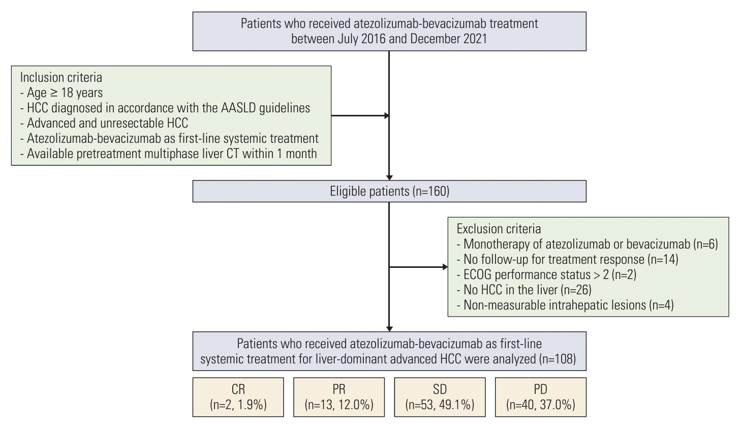
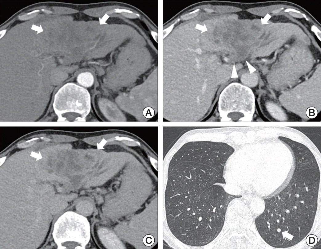
Fig. 1.
Flow diagram for patient enrollment. AASLD, American Association for the Study of Liver Diseases; CR, complete response; CT, computed tomography; ECOG, Eastern Cooperative Oncology Group; HCC, hepatocellular carcinoma; PD, progressive disease; PR, partial response; SD, stable disease.
crt-2024-283f1.tif
Fig. 2.
A 57-year-old male with biopsy proven hepatocellular carcinoma in the left hepatic lobe. (A-C) Pretreatment dynamic contrast-enhanced computed tomography showing a 9.8 cm infiltrative mass (arrow) without arterial-phase hyperenhancement (A). Nonsmooth tumor margins (arrows) on portal venous (B) and delayed-phase (C) images with tumor-in-vein in the left portal vein (arrowheads). (D) Metastatic nodules in the left lower lobe (arrow). This patient experienced disease progression after 12.5 weeks.
crt-2024-283f2.tif
Fig. 3.
Nomogram for predicting progressive disease. (A) The nomogram depicts the predicted probability of progressive disease on a scale of 0 to 280, derived from a multivariable logistic regression model. (B) Calibration plot for predicted probability vs. actual probability for progressive disease. The dotted line indicates predicted probability, while the black line represents the actual probability, and the gray zone indicates the 95% confidence intervals. APHE, arterial-phase hyperenhancement.
crt-2024-283f3.tif
Fig. 4.
Kaplan-Meier analyses of progression-free survival outcomes according to the number of significant predictive factors (N=0, N=1, or N ≥ 2). Significant predictive factors include the absence of APHE, the infiltrative appearance of the tumor, and the presence of extrahepatic metastases.
crt-2024-283f4.tif
Table 1.
Clinical characteristics of the 108 patients included in this study
Variable Value
Age (yr) 58±12
Sex
 Men 95 (88.0)
 Women 13 (12.0)
Etiology
 HBV 77 (71.3)
 HCV 6 (5.6)
 Others 25 (23.1)
Child-Pugh class
 A 55 (50.9)
 B 53 (49.1)
ECOG performance status
 0 11 (10.2)
 1 94 (87.0)
 2 3 (2.8)
Ascites 10 (9.3)
Pleural effusion 10 (9.3)
Prior treatment
 Surgery 28 (25.9)
 Ablation 13 (12.0)
 TACE 69 (63.9)
 RT 41 (38.0)
BCLC stage
 Intermediate 7 (6.5)
 Advanced 101 (93.5)
Extrahepatic metastasis 80 (74.1)
Involved organ
 Lung 54 (50.0)
 Bone 17 (15.7)
 Lymph node 26 (24.1)
Multiorgan metastases 25 (23.2)
Laboratory data
 Albumin (g/dL) 3.5 (3.2, 3.9)
 Bilirubin (g/dL) 0.8 (0.5, 1.1)
 INR 1.1 (1.0, 1.2)
ALBI grade
 Grade 1 21 (19.4)
 Grade 2 83 (76.9)
 Grade 3 4 (3.7)
Tumor markers
 AFP (ng/mL) 359 (15-4,463)
 PIVKA-II (mAU/mL) 784 (121-7,587)

Values are resented as mean±SD, median (IQR), or number (%). AFP, α-fetoprotein; ALBI, albumin-bilirubin; BCLC, Barcelona Clinic Liver Cancer; ECOG, Eastern Cooperative Oncology Group; HBV, hepatitis B virus; HCV, hepatitis C virus; INR, international normalized ratio; IQR, interquartile range; PIVKAII, protein induced by vitamin K absence or antagonist-II; RT, radiation therapy; SD, standard deviation; TACE, transarterial chemoembolization.

Table 2.
Pretreatment imaging characteristics of the index tumor
Variable PD (n=40) Non-PD (n=68) p-value
Tumor size (cm) 9.9±5.7 8.3±5.4 0.13
No. of involved segments 4.7±2.5 4.4±2.8 0.69
Tumor margin
 Smooth 13 (32.5) 31 (45.6) 0.18
 Nonsmooth 27 (67.5) 37 (54.4)
Infiltrative appearance
 Present 18 (45.0) 13 (19.1) 0.005
 Absent 22 (55.0) 55 (80.9)
APHE
 Present 22 (55.0) 60 (88.2) < 0.001
 Absent 18 (45.0) 8 (11.8)
Rim enhancement
 Present 4 (10.0) 4 (5.9) 0.43
 Absent 36 (90.0) 64 (94.1)
Washout appearance
 Present 36 (90.0) 53 (77.9) 0.12
 Absent 4 (10.0) 15 (22.1)
Intratumoral necrosis
 Present 17 (42.5) 28 (41.2) 0.89
 Absent 23 (57.5) 40 (58.8)
Peritumoral bile duct dilatation
 Present 3 (7.5) 7 (10.3) 0.63
 Absent 37 (92.5) 61 (89.7)
Tumor-in-vein
 Present 28 (70.0) 26 (38.2) 0.002
 Absent 12 (30.0) 42 (61.7)
Multifocality in liver
 Present 29 (72.5) 46 (67.6) 0.60
 Absent 11 (27.5) 22 (32.4)

Values are resented as mean±SD or number (%). APHE, arterial-phase hyperenhancement; PD, progressive disease; SD, standard deviation.

Table 3.
Univariable and multivariable analyses of factors predictive of progressive disease
Variable Univariable analysis
Multivariable analysis
OR (95% CI) p-value OR (95% CI) p-value
Age, per 1-year increase 0.99 (0.96-1.03) 0.72
Sex
 Women 1 (reference) 0.28
 Men 2.13 (0.55-8.24)
Etiology
 Non-HBV 1 (reference) 0.83
 HBV 1.10 (0.46-2.62)
Child-Pugh class
 A 1 (reference) 0.52
 B 0.77 (0.35-1.69)
ECOG performance
 0 or 1 1 (reference) 0.19
 2 2.87 (0.59-14.03)
Ascites
 Present 1.15 (0.30-4.34) 0.84
Pleural effusion
 Present 1.80 (0.49-6.65) 0.38
Prior treatment
 Surgery 1.39 (0.58-3.35) 0.46
 Ablation 1.54 (0.48-4.95) 0.47
 TACE 1.08 (0.48-2.44) 0.85
 RT 1.60 (0.72-3.56) 0.25
BCLC stage
 B 1 (reference) 0.23
 C 3.77 (0.44-32.55)
Extrahepatic metastasis
 Present 3.58 (1.24-10.36) 0.02 4.13 (1.19-14.35) 0.03
Involved organ
 Lung 2.65 (1.18-5.96) 0.02
 Bone 2.18 (0.77-6.20) 0.15
 Lymph node 1.08 (0.44-2.69) 0.86
Multiorgan metastases
 Present 1.65 (0.68-4.05) 0.27
ALBI grade
 Grade 1 1 (reference) 0.54
 Grade 2 or 3 0.74 (0.28-1.95)
AFP (ng/mL)
 < 400 1 (reference) 0.03
 ≥ 400 2.38 (1.07-5.31) 1.55 (0.59-4.07) 0.38
PIVKA-II (mAU/mL)
 < 2,000 1 (reference) 0.10
 ≥ 2,000 1.93 (0.88-4.26)
Tumor size, per 1 cm increase 1.06 (0.98-1.13) 0.13
No. of involved segments 1.03 (0.89-1.20) 0.69
Tumor margin
 Smooth 1 (reference) 0.18
 Nonsmooth 1.74 (0.77-3.93)
Infiltrative appearance
 Present 3.46 (1.45-8.25) 0.005 3.07 (1.05-8.93) 0.04
APHE
 Absent 6.14 (2.34-16.12) < 0.001 6.34 (2.18-18.47) < 0.001
Rim enhancement
 Present 1.88 (0.44-8.00) 0.43
Washout appearance
 Present 2.55 (0.78-8.30) 0.12
Intratumoral necrosis
 Present 1.06 (0.48-2.33) 0.89
Peritumoral bile duct dilatation
 Present 0.71 (0.17-2.90) 0.63
Tumor-in-vein
 Present 3.77 (1.64-8.68) 0.002 2.07 (0.74-5.77) 0.16
Multifocality in liver
 Present 1.26 (0.53-2.98) 0.60

AFP, α-fetoprotein; ALBI, albumin-bilirubin; APHE, arterial-phase hyperenhancement; BCLC, Barcelona Clinic Liver Cancer; CI, confidence interval; ECOG, Eastern Cooperative Oncology Group; HBV, hepatitis B virus; OR, odds ratio; PIVKA-II, protein induced by vitamin K absence or antagonist-II; RT, radiation therapy; TACE, transarterial chemoembolization.

Table 4.
Treatment response and disease control rate according to the number of significant predictive factors
Treatment response No. of significant predictive factors
p-value
N=0 (n=18) N=1 (n=51) N ≥ 2 (n=39)
Complete response 1 0 1
Partial response 5 6 2
Stable disease 9 34 10
Progressive disease 3 11 26
Disease control rate (%) 83.3 78.4 33.3 < 0.001
TOOLS
Similar articles