Journal List > J Stroke > v.26(3) > 1516088614

Salim, Yedavalli, Musmar, Adeeb, Essibayi, Naamani, Henninger, Sundararajan, Kühn, Khalife, Ghozy, Scarcia, Tan, Pulli, Heit, Regenhardt, Cancelliere, Bernstock, Rouchaud, Fiehler, Sheth, Puri, Dyzmann, Colasurdo, Barreau, Renieri, Filipe, Harker, Radu, Marotta, Spears, Ota, Mowla, Jabbour, Biswas, Clarençon, Siegler, Nguyen, Varela, Baker, Altschul, Gonzalez, Möhlenbruch, Costalat, Gory, Stracke, Aziz-Sultan, Hecker, Shaikh, Liebeskind, Pedicelli, Alexandre, Tancredi, Faizy, Kalsoum, Lubicz, Patel, Pereira, Guenego, Dmytriw, and the MAD MT Investigators: Mechanical Thrombectomy Versus Intravenous Thrombolysis in Distal Medium Vessel Acute Ischemic Stroke: A Multinational Multicenter Propensity Score-Matched Study

Abstract

Background and Purpose

The management of acute ischemic stroke (AIS) due to distal medium vessel occlusion (DMVO) remains uncertain, particularly in comparing the effectiveness of intravenous thrombolysis (IVT) plus mechanical thrombectomy (MT) versus IVT alone. This study aimed to evaluate the safety and efficacy in DMVO patients treated with either MT-IVT or IVT alone.

Methods

This multinational study analyzed data from 37 centers across North America, Asia, and Europe. Patients with AIS due to DMVO were included, with data collected from September 2017 to July 2023. The primary outcome was functional independence, with secondary outcomes including mortality and safety measures such as types of intracerebral hemorrhage.

Results

The study involved 1,057 patients before matching, and 640 patients post-matching. Functional outcomes at 90 days showed no significant difference between groups in achieving good functional recovery (modified Rankin Scale 0–1 and 0–2), with adjusted odds ratios (OR) of 1.21 (95% confidence interval [CI] 0.81 to 1.79; P=0.35) and 1.00 (95% CI 0.66 to 1.51; P>0.99), respectively. Mortality rates at 90 days were similar between the two groups (OR 0.75, 95% CI 0.44 to 1.29; P=0.30). The incidence of symptomatic intracerebral hemorrhage was comparable, but any type of intracranial hemorrhage was significantly higher in the MT-IVT group (OR 0.43, 95% CI 0.29 to 0.63; P<0.001).

Conclusion

The results of this study indicate that while MT-IVT and IVT alone show similar functional and mortality outcomes in DMVO patients, MT-IVT presents a higher risk of hemorrhagic complications, thus MT-IVT may not routinely offer additional benefits over IVT alone for all DMVO stroke patients. Further prospective randomized trials are needed to identify patient subgroups most likely to benefit from MT-IVT treatment in DMVO.

Introduction

The management of acute ischemic stroke (AIS) attributable to distal medium vessel occlusion (DMVO) presents a significant challenge in the field of neurointerventional therapy. Mechanical thrombectomy (MT), while established as the standard of care for large vessel occlusion (LVO) strokes [1-5], exhibits uncertain efficacy in DMVO cases [6]. This uncertainty is noteworthy as DMVOs are estimated to account for 25%–40% of AIS instances with discernible arterial blockage.
Traditionally, DMVOs have been scarcely represented in clinical trials and practice, leading to a significant gap in evidence-based guidelines for managing these patients. However, a number of studies are now investigating the safety and effectiveness of MT for DMVO strokes, including DISTAL (EnDovascular Therapy Plus Best Medical Treatment [BMT] vs. BMT Alone for MedIum VeSsel Occlusion sTroke, ClinicalTrials.gov Identifier: NCT05029414), DISTALS (Distal Ischemic Stroke Treatment With Adjustable Low-profile Stentriever, NCT05152524), DISCOUNT (Evaluation of Mechanical Thrombectomy in Acute Ischemic Stroke Related to a Distal Arterial Occlusion, NCT05030142), ESCAPE-MeVO (EndovaSCular TreAtment to imProve outcomEs for Medium Vessel Occlusions, NCT05151172), and FRONTIER-AP (Randomized controlled trial of the clinical outcome and safety of endovascular vs. standard medical therapy for stroke with medium sized vessel occlusion) [7,8], which explore various aspects of endovascular therapy in comparison to or alongside best medical treatment. Despite the potential benefits of MT, concerns arise over its application in DMVO due to the smaller size and complex structure of distal cerebral arteries, which may complicate the reperfusion process. Consequently, this raises significant safety and efficacy concerns regarding the use of MT in treating DMVOs [6,9].
A central point of uncertainty is whether MT can improve clinical outcomes compared to those attainable with intravenous thrombolysis (IVT) [10]. However, advancements in endovascular technologies, including retriever and aspiration devices, have expanded the application of MT in treating distal vessel occlusions [9].
In response to this clinical gap, our multicenter cohort study aimed to evaluate the safety and efficacy in DMVO patients undergoing treatment with either IVT alone or a combination of MT and IVT (MT-IVT).

Methods

Study design

This study represents a detailed analysis within the Multicenter Analysis of primary Distal medium vessel occlusions: effect of Mechanical Thrombectomy (MAD-MT) registry [8,11-14]. This research adheres to the Strengthening the Reporting of Observational Studies in Epidemiology (STROBE) guidelines [15,16].

Ethical approval and patient consent

Ethical clearance for this retrospective study was obtained from the institutional review board or local ethical standards committee at each of the 37 participating centers across North America, Asia, and Europe. Considering the retrospective nature of our analysis, the requirement for informed consent was waived. The dataset supporting the conclusions of this article is available from the corresponding author upon reasonable request.

Study population and selection criteria

Our target population comprised patients who experienced AIS due to DMVO. Inclusion criteria were: (1) complete data for 3-month modified Rankin Scale (mRS) score, intervention type, and hemorrhagic complications including symptomatic intracerebral hemorrhage (sICH); and (2) patients treated with either MT-IVT or IVT alone. We excluded patients who received MT alone or did not receive either MT or intravenous tissue plasminogen activator (IV-tPA) treatment.

Data collection and review process

Data were collected from September 2017 to July 2023, encompassing consecutive patients treated with MT or IVT for primary medium proximal vessel occlusion (M2, A1, P1) or primary medium distal vessel occlusion (M3/M4, A2/A3, P2/P3) as defined by Saver et al. [9]. This included both prospective data collection and retrospective analysis. Local neurointerventionalists initially evaluated angiographic treatment success, subsequently contributing their data to the MAD-MT consortium. The data were self-reported by each participating center.

Variables and data recording

We meticulously documented a comprehensive set of baseline clinical and demographic characteristics, including sex, age, medical history (hypertension, hypercholesterolemia, diabetes mellitus, atrial fibrillation), smoking status, pre-stroke mRS score, location of the occluded vessel, National Institutes of Health Stroke Scale (NIHSS) score upon presentation, and baseline Alberta Stroke Program Early Computed Tomography Score (ASPECTS). Patients were also categorized based on their occlusion site during initial angiography into medium proximal (M2, A1, P1) and medium distal vessels (M3/M4, A2/A3, P2/P3).
We additionally recorded information on antiplatelet and anticoagulant medication use, treatment approach (mothership vs. drip-and-ship), timing from symptom onset to arterial puncture and recanalization, vital signs, glycemic levels, anesthesia type, access site, and post-MT imaging modalities.

Outcome measures

The primary outcome focused on functional independence (mRS 0–2), with secondary outcomes including an excellent outcome (mRS 0–1) and mortality (mRS 6). Safety outcomes centered on types of intracerebral hemorrhage (ICH) and sICH, defined in accordance with “The Heidelberg Bleeding Classification.” [17]

Procedural and technical aspects

Treatment modalities included MT-IVT and IVT alone. The choice of treatment, procedural techniques (aspiration, stent retriever, combined techniques like Solumbra), access site (femoral or radial artery), and number of passes were at the discretion of the treating clinicians, aligned with institutional protocols and guidelines.

Statistical analysis

Statistical analysis was conducted using R (version 4.2.2; R Foundation for Statistical Computing, Vienna, Austria). For categorical variables, we calculated frequencies and percentages and employed the χ2 test for comparison. In instances where cell counts were fewer than five, Fisher’s exact test was utilized. Continuous variables were summarized using median values and interquartile ranges (IQRs) and were compared using the Mann-Whitney U test to assess differences between groups.
Propensity score matching (PSM) controlled for potential confounders [18,19]. Propensity scores were estimated using logistic regression, and an optimal matching algorithm was employed with a 1:1 ratio. Variables likely to affect the outcomes according to previous studies [20-22] were included in the PSM, which included age, sex, baseline NIHSS, pre-admission mRS scores, history of antiplatelet and anticoagulant medication use, pertinent comorbidities (diabetes mellitus, hypertension, hypercholesterolemia, atrial fibrillation), and occlusion location. Covariate balance postPSM was assessed using the standardized mean difference, with <0.1 indicating effective balance. A P-value of 0.05 or less was considered statistically significant.

Missing data

In this study, we encountered a proportion of missing data, as documented in Supplementary Figure 1. Missing data were assumed to be either completely missing at random or randomly missing. To avoid bias and reduction in statistical power, we included cases with missing values by employing multiple imputation using chained equations for missing data handling [23], producing fifty imputed datasets with five iterations each. Imputations were performed on matched data only, with no imputation on cases with missing outcomes (90-day mRS, mortality, hemorrhagic complications).
Post-imputation, logistic regression models were applied to determine the relationship of IVT alone versus MT-IVT and observed outcomes. Multivariable models were adjusted to include age, sex, comorbidities (diabetes, hypertension, hypercholesterolemia, atrial fibrillation), baseline NIHSS, occlusion location, ASPECTS, and pre-stroke use of antiplatelet or anticoagulant agents.

Results

Patient characteristics

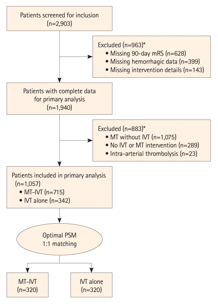
Before propensity score matching, our study involved 1,057 patients, with 715 undergoing MT-IVT and 342 receiving IVT alone (Figure 1). Prior to matching, the MT-IVT group displayed a median age of 74 years, similar to the IVT alone group (P=0.54). Notably, there was a lower prevalence of hypercholesterolemia in the MT-IVT group (34%) compared to the IVT alone group (51%; P<0.001). Similarly, the rate of atrial fibrillation was higher in the IVT alone group (37% vs. 30% in MT-IVT; P=0.02). Rates of hypertension were comparable, though the IVT alone group had a higher prevalence of diabetes (27% vs. 19% in MT-IVT; P=0.009). Smoking history and previous use of antiplatelet or anticoagulant drugs were also similar between the groups. Significant differences were noted in baseline mRS scores and site of initial occlusion, with more medium vessel (M2, P1, A1) occlusions in the MT-IVT group (81% vs. 74% in IVT alone; P=0.011). The median ASPECTS score was 9.00 in both groups, but the baseline NIHSS score was higher in the MT-IVT group (median 12 vs. 6 in IVT alone; P<0.001) (Supplementary Table 1).
After propensity score matching, a total of 640 patients were analyzed, and the cohorts were well balanced across all measured variables. Sex distribution, median age, and prevalence of hypercholesterolemia, hypertension, diabetes, atrial fibrillation, smoking history, and previous medication use showed no significant differences between the two groups. Baseline mRS scores, site of initial occlusion, ASPECTS scores, and baseline NIHSS scores were also similar (Table 1).

Periprocedural details

Table 2 provides comprehensive periprocedural data for the study cohort post-matching. In the MT-IVT group, the first-line procedural techniques were distributed as follows: aspiration in 23%, a combination of techniques in 64%, and stentretriever in 13%. The median times from onset to IVT needle were similar in both groups, recorded as 155 minutes and 164 minutes, respectively. Additionally, in the MT-IVT group the median time from symptom onset to recanalization was 283 minutes. Periprocedural data before PSM are documented Supplementary Table 2.

Outcomes after matching

Following the matching process (Table 3), the median number of passes required for thrombectomy in the MT-IVT group was 1.00. On the first day post-admission, the median NIHSS score was 3 (IQR 1–8) across the entire cohort (IQR: 1–11 in MT-IVT vs. 1–6 in IVT alone; P=0.061). In terms of angiographic outcomes, a high rate of successful recanalization (modified Thrombolysis in Cerebral Infarction [mTICI] 2b–3) was achieved in 91% of the MT-IVT group. Complete reperfusion (mTICI 2c–3) was accomplished in 61% of this group.
Functional outcomes at 90 days, assessed by mRS scores, showed no significant difference between the two groups for achieving mRS 0–1 (45% in MT-IVT vs. 50% in IVT alone; P=0.21) or mRS 0–2 (62% in MT-IVT vs. 63% in IVT alone; P=0.74). Mortality rates at 90 days were 14% in the MT-IVT group and 11% in the IVT alone group, with no statistically significant difference (P=0.4) (Figure 2A).
The incidence of sICH was similar between the two groups (8.3% in MT-IVT vs. 6.9% in IVT alone; P=0.59). However, any type of ICH was significantly higher in the MT-IVT group (32% vs. 17% in IVT alone; P<0.001). Notably, hemorrhagic infarction type 1 (HI1) and subarachnoid hemorrhage (SAH) were significantly more common in the MT-IVT group (P<0.001 for both). Other ICH types, including HI2, PH1, and PH2, did not significantly differ between the groups (Figure 2B). Perforations were observed solely in the MT-IVT group, with a total of 14 events. These perforations were associated with 12 instances of any type of ICH, including three cases of sICH and five cases of SAH. Other complications, such as embolization in new territories and artery dissection, were infrequent and did not differ significantly between the groups. Outcomes before matching were documented in Supplementary Table 3.

Logistic regression analysis

The unadjusted and adjusted logistic regression models (Table 4) reinforced these findings. In the adjusted model, the odds ratios for 90-day mRS 0–1 and mRS 0–2 were 1.21 (95% CI 0.81 to 1.79; P=0.35) and 1.00 (95% CI 0.66 to 1.51; P>0.99), respectively, indicating no significant difference in functional outcomes between the two treatment modalities. Similarly, the adjusted model showed no significant differences in mortality rates (OR 0.75, 95% CI 0.44 to 1.29; P=0.3) and rates of sICH (OR 0.78, 95% CI 0.42 to 1.45; P=0.44).
However, the risk of any type of ICH was significantly lower in the IVT alone group, with an OR of 0.43 (95% CI 0.29 to 0.63; P<0.001). Specifically, hemorrhagic infarction type 1 (HI1) showed a significantly reduced risk in the IVT alone group (OR 0.17, 95% CI 0.08 to 0.36; P<0.001), as did subarachnoid hemorrhage (SAH) (OR 0.17, 95% CI 0.07 to 0.41; P<0.001). Conversely, hemorrhagic infarction type 2 (HI2) was more often in the IVT alone group (OR 4.37, 95% CI 1.42 to 13.5; P=0.01), whereas parenchymal hematoma types 1 and 2 (PH1 and PH2) showed no significant difference in odds between the groups.

Sensitivity analysis

Sensitivity analysis using pre-imputation and imputed models showed similar trends, confirming the robustness of the findings (Supplementary Table 4).

Subgroup analysis

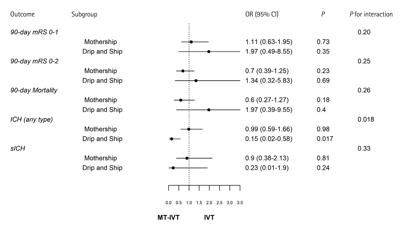
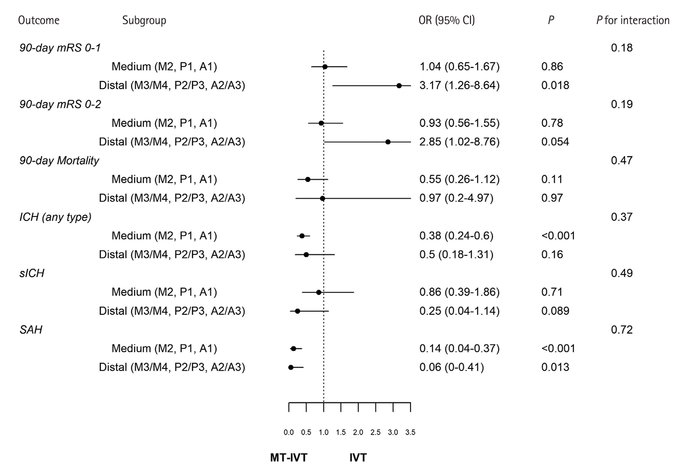
Additional subgroup analyses comparing treatment outcomes between the “mothership” scenario (n=366) and “drip-and-ship” scenario (n=166) showed no significant differences in achieving mRS 0–1, mRS 0–2, mortality, and sICH. However, there was a significant difference in the rate of ICH between these subgroups (P=0.018), with the “drip-and-ship” group having an increased risk of ICH in the MT-IVT treatment (Figure 3). Subgroup analysis based on occlusion location medium (n=492) and distal (n=148) showed no differences in outcomes between the subgroups (Figure 4).

Discussion

In this study, we sought to determine the safety and efficacy of MT-IVT compared to IVT alone in patients with AIS due to DMVO. We found similar rates of 90-day good and excellent functional recovery and mortality between the two treatment strategies. In addition, there was an increased incidence of any ICH in the MT-IVT group, despite no significant difference in sICH.
DMVOs represent a challenging subset of AIS, with existing recommendations primarily targeting larger vessel occlusions [24]. Current recommendations from the American Stroke Association predominantly address large vessel occlusion strokes. They suggest that MT for MCA M3 occlusions may be reasonable, while no specific recommendations are provided for M4 occlusions or DMVOs [16,25]. This is echoed in European guidelines, which do not provide specific MT recommendations for DMVO [26].
Notably, our findings align with previous research. A recent meta-analysis of 2,469 patients, comparing endovascular therapy with best medical therapy in medium vessel occlusion AIS, found no significant difference in functional independence, mortality rates, and incidence of sICH between the two groups [27]. The observational study by Saber et al. [28] indicated a slightly higher rate of excellent mRS outcomes in the MT-treated group for primary DMVO stroke, without notable differences in complications. This is consistent with our findings, which suggest comparable efficacy of the two treatment strategies but without significant difference in sICH. Notably, the older age (median: 74, IQR: 63–83) in our study cohort compared to that in the study by Saber et al. [28] might have contributed to the higher mortality rate observed in our cohort, despite lower baseline NIHSS score. Consequently, the odds of mortality were similar in both studies.
Recent observational studies have also explored the bridging treatment approach (IVT followed by MT) and noted an enhanced reperfusion rate in MT without a significant difference in complication rates [29,30]. Consistent with these studies, we observed high successful (mTICI 2b–3) and excellent (mTICI 2c–3) recanalization rates with MT-IVT. Nevertheless, since we lack comparable data for the IVT alone group, further study is required to determine whether combined treatment is superior to IVT alone.
The similar outcomes in function and mortality observed across the treatment modalities may be attributed to the generally less severe clinical presentations of DMVO strokes in comparison with LVOs. This distinction is reflected in our PSM model, which demonstrated a tendency to select patients presenting with lower NIHSS scores at admission for the MT-IVT group. This selection bias suggests that our findings may primarily apply to patients experiencing mild to moderate DMVO strokes and may not be generalizable to those with severe strokes characterized by higher NIHSS scores. Furthermore, the applicability of the commonly utilized mRS scores in evaluating outcomes after DMVO strokes warrants further discussion, raising questions about the adequacy of these measures in capturing the full spectrum of patient experiences and outcomes following such interventions [28,31]. In a previous study, we demonstrated that there is no significant difference in the ICH rate (OR, 1.10; 95% CI 0.96–1.25), successful recanalization rate (OR, 2.22; 95% CI 0.61–8.03), or 90-day functional mRS outcomes between aspiration and stent-retriever thrombectomy in DMVO stroke patients [12], indicating that the choice of MT technique likely did not influence our findings.
Our findings indicate that while the rate of ICH in the MT-IVT group is elevated, it remains comparable to bleeding rates observed in prior studies focused on MT for acute M2 occlusion [32]. These hemorrhagic incidents are predominantly mild and asymptomatic. This observation is supported by the lack of significant difference in the occurrence of sICH between the treatment cohorts. The observed increase in any ICH in DMVO treated with MT may be attributed to the fragility of smaller-sized vessels. This risk is particularly notable in patients eligible for IVT, aligning with a meta-analysis that identified an elevated risk of ICH in patients treated with MT for distal MCA occlusions [31-34]. Moreover, certain MT-IVT subpopulations, such as patients treated in a “drip-and-ship” scenario, might be at increased risk of ICH [35,36]. Our subgroup analysis revealed a significant interaction (P=0.018), indicating that the MT-IVT group in the “drip-and-ship” scenario had a significantly increased risk of any ICH (OR 0.15, 95% CI 0.02–0.58; P=0.017). In contrast, there was no significant difference in the “mothership” scenario (OR 0.99, 95% CI 0.59–1.66; P=0.98). This increased risk is, in part, likely due to the longer time interval between IVT administration and MT. The subgroup analysis based on occlusion site, as shown in Figure 4, revealed no significant interaction differences between medium and distal occlusions across various outcomes, including 90-day mRS scores, mortality, and ICH complications. A notable finding was the significance of ICH in medium occlusions (OR 0.38, 95% CI 0.24–0.6, P<0.001) but not in distal vessel occlusions (OR 0.5, 95% CI 0.18–1.31, P=0.16). This discrepancy is likely attributable to the smaller sample size of the distal occlusion subgroup (n=148 vs. n=492 in medium occlusions), which may have limited the statistical power to detect significant differences. Consequently, the P-value for interaction was not significant (P=0.37). A similar observation applies to the 90-day mRS 0–1 outcome, where there was a nonsignificant interaction (P=0.18).
This study’s strengths include its extensive scope, incorporating large-scale data from multiple nations and centers, thereby enhancing the generalizability of our findings to real-world settings. However, there are limitations inherent to its retrospective design. Treatment decisions based on physician discretion and institutional protocols may introduce biases. Even with the application of propensity score matching, the potential for unmeasured confounders to influence outcomes remains. The variability in procedural techniques and the range of operator experience across participating centers might also limit the universality of our results. A notable limitation is the absence of long-term outcome data beyond the 90-day mark, which constrains our ability to comprehensively understand the sustained effects of MT on patients’ functional recovery and quality of life. Additionally, this study did not assess reperfusion rates in the IVT alone group, an aspect that could offer further insight into treatment effectiveness. Moreover, lack of perfusion scans and lack of data on the location of the ICH and perforations are other limitations. It is important also to highlight that the baseline NIHSS scores in our matched cohorts were relatively low in both groups. Specifically, the 25th percentile for the MT-IVT group was 4, indicating that some patients with NIHSS score of <4 were included. Clinical trials largely excluded these patients. According to current evidence including PRISMS (The Potential of rtPA for Ischemic Strokes With Mild Symptoms) and ARAMIS (Antiplatelet vs. R-tPA for Acute Mild Ischemic Stroke) trials, the standard of care for patients with low NIHSS scores, particularly up to an NIHSS of 5 is not clear yet, and might be better managed with dual-antiplatelet therapy rather than IVT [37,38]. Given these limitations, there is a clear need for prospective randomized clinical trials to better identify DMVO patient subgroups that are most likely to benefit from MT.

Conclusions

In conclusion, this comprehensive retrospective study provides valuable insights into the efficacy and safety of MT-IVT compared to IVT alone in DMVO acute ischemic stroke patients. The analysis indicated comparable 90-day mortality and functional outcomes (mRS 0–1 and 0–2) between the two treatment groups. However, a higher rate of any hemorrhagic transformation was noted in the MT-IVT group. These findings suggest that MT in combination with IVT for all DMVO stroke patients may not always be beneficial. Further randomized clinical trials are warranted to identify DMVO patients most likely to benefit from MT-IVT treatment.

Supplementary materials

Supplementary materials related to this article can be found online at https://doi.org/10.5853/jos.2024.01389.
Supplementary Table 1.
Baseline characteristics of the included patients before matching
jos-2024-01389-Table-1.pdf
Supplementary Table 2.
Periprocedural details of the included patients before matching
jos-2024-01389-Table-2.pdf
Supplementary Table 3.
Outcome data of the included patients before matching
jos-2024-01389-Table-3.pdf
Supplementary Table 4.
Pre-imputation multivariable logistic regression model (complete case analysis) compared with the imputed model
jos-2024-01389-Table-4.pdf
Supplementary Figure 1.
Proportion of missing data per variable. (A) Missing variables after PSM. (B) Missing variables before PSM. PSM, propensity score matching; mRS, modified Rankin Scale; NIHSS, National Institutes of Health Stroke Scale; IV-tPA, intravenous tissue plasminogen activator.
jos-2024-01389-Fig-1.pdf

Notes

Funding statement

None

Conflicts of interest

Dr. Regenhardt serves on a DSMB for a trial sponsored by Rapid Medical, serves as site PI for studies sponsored by Penumbra and Microvention, and receives stroke research grant funding from the National Institutes of Health, Society of Vascular and Interventional Neurology, and Heitman Stroke Foundation. Dr. Guenego reports consultancy for Rapid Medical and Phenox, not directly related to the present work. Dr. Clarençon reports conflicts of interest with Medtronic, Balt Extrusion (consultant), ClinSearch (core lab), Penumbra, Stryker (payment for reading) and Artedrone (Board); all not directly related to the present work. Dr. Henninger received support from CDMRP/DoD W81XWH-19-PRARP-RPA and NINDS NS131756, during the conduct of the study. Dr. Liebeskind is consultant as Imaging Core Lab to Cerenovus, Genentech, Medtronic, Stryker, Rapid Medical. Dr. Yeo reports Advisory work for AstraZeneca, Substantial support from NMRC Singapore and is a medical advisor for See-mode, Cortiro and Sunbird Bio, with equity in Ceroflo. All unrelated to the present work. Dr. Griessenauer reports a proctoring agreement with Medtronic and research funding by Penumbra. Dr. Marnat reports conflicts of interest with Microvention Europe, Stryker Neurovascular, Balt (consulting), Medtronic, Johnson & Johnson and Phenox (paid lectures), all not directly related to the present work. Dr. Puri is a consultant for Medtronic Neurovascular, Stryker NeurovascularBalt, Q’Apel Medical, Cerenovus, Microvention, Imperative Care, Agile, Merit, CereVasc and Arsenal Medical, he received research grants from NIH, Microvention, Cerenovus, Medtronic Neurovascular and Stryker Neurovascular, and holds stocks in InNeuroCo, Agile, Perfuze, Galaxy and NTI. Dr. Tjoumakaris is a consultant for Medtronic and Microvention (funds paid to institution, not personally). Dr. Jabbour is a consultant for Medtronic, Microvention and Cerus. All remaining authors have declared no conflicts of interest.

Author contribution

Conceptualization: HAS. Study design: AD, AG. Methodology: HAS, AD, AG. Data collection: HAS, VY, BM, NA, KN, NH, SHS, AK, JK, SG, LS, BT, JH, RR, NC, JB, AR, JF, SS, ME, AP, CD, MC, XB, LR, JF, PH, RR, TM, JS, TO, AM, PJ, AB (Arundhati Biswas), FC, JS, TN, RV, AB (Amanda Baker), DA, NG, MM, VC, BG, CS, MA, CH, HS, DL, AP, AA, IT, TF, EK, BL, AP, VP, AG, AD. Investigation: HAS, VY, BM, NA, KN, NH, SHS, AK, JK, SG, LS, BT, JH, RR, NC, JB, AR, JF, SS, ME, AP, CD, MC, XB, LR, JF, PH, RR, TM, JS, TO, AM, PJ, AB (Arundhati Biswas), FC, JS, TN, RV, AB (Amanda Baker), DA, NG, MM, VC, BG, CS, MA, CH, HS, DL, AP, AA, IT, TF, EK, BL, AP, VP, AG, AD. Statistical analysis: HAS. Writing—original draft: HAS, VY. Writing—review & editing: HAS, VY, BM, NA, KN, NH, SHS, AK, JK, SG, LS, BT, JH, RR, NC, JB, AR, JF, SS, ME, AP, CD, MC, XB, LR, JF, PH, RR, TM, JS, TO, AM, PJ, AB (Arundhati Biswas), FC, JS, TN, RV, AB (Amanda Baker), DA, NG, MM, VC, BG, CS, MA, CH, HS, DL, AP, AA, IT, TF, EK, BL, AP, VP, AG, AD. Approval of final manuscript: all authors.

ACKNOWLEDGMENTS

We acknowledge the radiographers, paramedics, and nurses, without whom this work would be impossible.

References

1. Berkhemer OA, Fransen PS, Beumer D, van den Berg LA, Lingsma HF, Yoo AJ, et al. A randomized trial of intraarterial treatment for acute ischemic stroke. N Engl J Med. 2015; 372:11–20.
2. Jovin TG, Chamorro A, Cobo E, de Miquel MA, Molina CA, Rovira A, et al. Thrombectomy within 8 hours after symptom onset in ischemic stroke. N Engl J Med. 2015; 372:2296–2306.
crossref
3. Saver JL, Goyal M, Bonafe A, Diener HC, Levy EI, Pereira VM, et al. Stent-retriever thrombectomy after intravenous t-PA vs. t-PA alone in stroke. N Engl J Med. 2015; 372:2285–2295.
crossref
4. Goyal M, Demchuk AM, Menon BK, Eesa M, Rempel JL, Thornton J, et al. Randomized assessment of rapid endovascular treatment of ischemic stroke. N Engl J Med. 2015; 372:1019–1030.
crossref
5. Goyal M, Menon BK, van Zwam WH, Dippel DW, Mitchell PJ, Demchuk AM, et al. Endovascular thrombectomy after large-vessel ischaemic stroke: a meta-analysis of individual patient data from five randomised trials. Lancet. 2016; 387:1723–1731.
6. Ospel JM, Goyal M. A review of endovascular treatment for medium vessel occlusion stroke. J Neurointerv Surg. 2021; 13:623–630.
crossref
7. Australian New Zealand Clinical Trials Registry. Trial review: trial registered on ANZCTR [Internet]. Camperdown: ANZCTR;2021. [accessed March 23, 2024]. Available from: https://www.anzctr.org.au/Trial/Registration/TrialReview.aspx?ACTRN=12621001746820p.
8. Dmytriw AA, Musmar B, Salim H, Ghozy S, Siegler JE, Kobeissi H, et al. Incidence and clinical outcomes of perforations during mechanical thrombectomy for medium vessel occlusion in acute ischemic stroke: a retrospective, multicenter, and multinational study. Eur Stroke J. 2024; 9:328–337.
9. Saver JL, Chapot R, Agid R, Hassan A, Jadhav AP, Liebeskind DS, et al. Thrombectomy for distal, medium vessel occlusions: a consensus statement on present knowledge and promising directions. Stroke. 2020; 51:2872–2884.
10. Sarraj A, Sangha N, Hussain MS, Wisco D, Vora N, Elijovich L, et al. Endovascular therapy for acute ischemic stroke with occlusion of the middle cerebral artery M2 segment. JAMA Neurol. 2016; 73:1291–1296.
crossref
11. Dmytriw AA, Ghozy S, Salim HA, Musmar B, Siegler JE, Kobeissi H, et al. Assessment of thrombectomy versus combined thrombolysis and thrombectomy in patients with acute ischemic stroke and medium vessel occlusion. Radiology. 2024; 312:e233041.
12. Siegler JE, Shaikh H, Khalife J, Oak S, Zhang L, Abdalkader M, et al. Aspiration versus stent-retriever as first-line endovascular therapy technique for primary medium and distal intracranial occlusions: a propensity-score matched multicenter analysis. Stroke Vasc Interv Neurol. 2023; 3:e000931.
13. Salim H, Musmar B, Adeeb N, Yedavalli V, Lakhani D, Grewal SS, et al. Outcomes of mechanical thrombectomy in anticoagulated patients with acute distal and medium vessel stroke. Eur Stroke J. 2024; May. 10. [Epub]. Available from: https://doi.org/10.1177/23969873241249295.
crossref
14. Radu RA, Costalat V, Romoli M, Musmar B, Siegler JE, Ghozy S, et al. Outcomes with general anesthesia compared to conscious sedation for endovascular treatment of medium vessel occlusions: results of an international multicentric study. Clin Neuroradiol. 2024; Apr. 30. [Epub]. Available from: https://doi.org/10.1007/s00062-024-01415-1.
crossref
15. Cuschieri S. The STROBE guidelines. Saudi J Anaesth. 2019; 13(Suppl 1):S31–S34.
crossref
16. von Elm E, Altman DG, Egger M, Pocock SJ, Gøtzsche PC, Vandenbroucke JP. The strengthening the reporting of observational studies in epidemiology (STROBE) statement: guidelines for reporting observational studies. PLoS Med. 2007; 4:e296.
17. von Kummer R, Broderick JP, Campbell BC, Demchuk A, Goyal M, Hill MD, et al. The Heidelberg bleeding classification: classification of bleeding events after ischemic stroke and reperfusion therapy. Stroke. 2015; 46:2981–2986.
18. Stuart EA. Matching methods for causal inference: a review and a look forward. Stat Sci. 2010; 25:1–21.
crossref
19. Rosenbaum PR, Rubin DB. The central role of the propensity score in observational studies for causal effects. Biometrika. 1983; 70:41–55.
crossref
20. Scheitz JF, Seiffge DJ, Tütüncü S, Gensicke H, Audebert HJ, Bonati LH, et al. Dose-related effects of statins on symptomatic intracerebral hemorrhage and outcome after thrombolysis for ischemic stroke. Stroke. 2014; 45:509–514.
crossref
21. Zinkstok SM, Engelter ST, Gensicke H, Lyrer PA, Ringleb PA, Artto V, et al. Safety of thrombolysis in stroke mimics: results from a multicenter cohort study. Stroke. 2013; 44:1080–1084.
22. Engelter ST, Soinne L, Ringleb P, Sarikaya H, Bordet R, Berrouschot J, et al. IV thrombolysis and statins. Neurology. 2011; 77:888–895.
crossref
23. Rubin DB. Multiple imputation. In : van Buuren S, editor. Flexible imputation of missing data. 2nd ed. New York: Chapman and Hall/CRC;2018. p. 29–62.
24. Goyal M, Ospel JM, Menon BK, Hill MD. MeVO: the next frontier? J Neurointerv Surg. 2020; 12:545–547.
crossref
25. Powers WJ, Rabinstein AA, Ackerson T, Adeoye OM, Bambakidis NC, Becker K, et al. Guidelines for the early management of patients with acute ischemic stroke: 2019 update to the 2018 guidelines for the early management of acute ischemic stroke: a guideline for healthcare professionals from the American Heart Association/American Stroke Association. Stroke. 2019; 50:e344–e418.
crossref
26. Turc G, Bhogal P, Fischer U, Khatri P, Lobotesis K, Mazighi M, et al. European Stroke Organisation (ESO) - European Society for Minimally Invasive Neurological Therapy (ESMINT) guidelines on mechanical thrombectomy in acute ischemic Stroke. J Neurointerv Surg. 2023; 15:e8.
27. Loh EW, Toh KZX, Kwok GYR, Teo YH, Teo YN, Goh C, et al. Endovascular therapy for acute ischemic stroke with distal medium vessel occlusion: a systematic review and meta-analysis. J Neurointerv Surg. 2023; 15:e452–e459.
crossref
28. Saber H, Desai SM, Haussen D, Al-Bayati A, Majidi S, Mocco J, et al. Endovascular therapy vs medical management for patients with acute stroke with medium vessel occlusion in the anterior circulation. JAMA Netw Open. 2022; 5:e2238154.
crossref
29. Grossberg JA, Rebello LC, Haussen DC, Bouslama M, Bowen M, Barreira CM, et al. Beyond large vessel occlusion strokes: distal occlusion thrombectomy. Stroke. 2018; 49:1662–1668.
30. Rizzo F, Romoli M, Simonetti L, Gentile M, Forlivesi S, Piccolo L, et al. Reperfusion strategies in stroke with medium-to-distal vessel occlusion: a prospective observational study. Neurol Sci. 2024; 45:1129–1134.
crossref
31. Strambo D, Bartolini B, Beaud V, Marto JP, Sirimarco G, Dunet V, et al. Thrombectomy and thrombolysis of isolated posterior cerebral artery occlusion: cognitive, visual, and disability outcomes. Stroke. 2020; 51:254–261.
32. Saber H, Narayanan S, Palla M, Saver JL, Nogueira RG, Yoo AJ, et al. Mechanical thrombectomy for acute ischemic stroke with occlusion of the M2 segment of the middle cerebral artery: a meta-analysis. J Neurointerv Surg. 2018; 10:620–624.
crossref
33. Nogueira RG, Zaidat OO, Castonguay AC, Haussen DC, Martin CO, Holloway WE, et al. Rescue thrombectomy in large vessel occlusion strokes leads to better outcomes than intravenous thrombolysis alone: a ‘real world’ applicability of the recent trials. Interv Neurol. 2016; 5:101–110.
crossref
34. Sarraj A, Hassan A, Savitz SI, Grotta JC, Cai C, Parsha KN, et al. Endovascular thrombectomy for mild strokes: how low should we go? Stroke. 2018; 49:2398–2405.
crossref
35. Sarraj A, Grotta J, Albers GW, Hassan AE, Blackburn S, Day A, et al. Clinical and neuroimaging outcomes of direct thrombectomy vs bridging therapy in large vessel occlusion: analysis of the SELECT cohort study. Neurology. 2021; 96:e2839–e2853.
36. Katsanos AH, Sarraj A, Froehler M, Purrucker J, Goyal N, Regenhardt RW, et al. IV thrombolysis initiated before transfer for endovascular stroke thrombectomy: a systematic review and meta-analysis. Neurology. 2023; 100:e1436–e1443.
37. Khatri P, Kleindorfer DO, Devlin T, Sawyer RN Jr, Starr M, Mejilla J, et al. Effect of alteplase vs aspirin on functional outcome for patients with acute ischemic stroke and minor nondisabling neurologic deficits: the PRISMS randomized clinical trial. JAMA. 2018; 320:156–166.
crossref
38. Chen HS, Cui Y, Zhou ZH, Zhang H, Wang LX, Wang WZ, et al. Dual antiplatelet therapy vs alteplase for patients with minor nondisabling acute ischemic stroke: the ARAMIS randomized clinical trial. JAMA. 2023; 329:2135–2144.
crossref

Figure 1.
Patient selection and propensity score matching flowchart. mRS, modified Rankin Scale; MT, mechanical thrombectomy; IVT, intravenous thrombolysis; PSM, propensity score matching ratio. *Multipl selection of patients possible.
jos-2024-01389f1.tif
Figure 2.
Comparative outcomes of MT-IVT versus IVT alone. (A) The percentage distribution of 90-day mRS scores for patients treated with IVT alone compared to those who received MT in addition to IVT (MT-IVT). The mRS scores range from 0 (no symptoms) to 6 (death), with the number of patients achieving each score level represented by the length of the bars along the X-axis. (B) The proportion of patients experiencing various types of hemorrhagic complications post-treatment. mRS, modified Rankin Scale; IVT, intravenous thrombolysis; MT, mechanical thrombectomy; sICH, symptomatic intracerebral hemorrhage; ICH, intracerebral hemorrhage; SAH, subarachnoid hemorrhage; HI1, hemorrhagic infarction type 1; HI2, hemorrhagic infarction type 2; PH1, parenchymal hematoma type 1; PH2, parenchymal hematoma type 2. ***P<0.001.
jos-2024-01389f2.tif
Figure 3.
Subgroup analysis by scenario “Drip and Ship” versus “Mothership.” mRS, modified Rankin Scale; MT, mechanical thrombectomy; IVT, intravenous thrombolysis; ICH, intracerebral hemorrhage; sICH, symptomatic intracerebral hemorrhage; OR, odds ratio; CI, confidence interval.
jos-2024-01389f3.tif
Figure 4.
Subgroup analysis by location medium versus distal. mRS, modified Rankin Scale; MT, mechanical thrombectomy; IVT, intravenous thrombolysis; ICH, intracerebral hemorrhage; sICH, symptomatic intracerebral hemorrhage; SAH, subarachnoid hemorrhage; OR, odds ratio; CI, confidence interval.
jos-2024-01389f4.tif
Table 1.
Baseline characteristics of the included patients after matching
Variable Overall (n=640) MT-IVT (n=320) IVT alone (n=320) P
Male sex 335 (52) 170 (53) 165 (52) 0.75
Age (yr) 74 (63–83) 74 (64–83) 74 (63–83) 0.86
Hypercholesterolemia 296 (47) 145 (47) 151 (47) >0.99
Hypertension 428 (67) 219 (68) 209 (65) 0.45
Diabetes 158 (25) 74 (23) 84 (26) 0.41
Atrial fibrillation 213 (33) 102 (32) 111 (35) 0.50
Current smokers 112 (18) 52 (16) 60 (19) 0.47
Previous use of antiplatelet drugs 119 (29) 88 (30) 31 (27) 0.61
Previous use of anticoagulant drugs 64 (15) 45 (16) 19 (15) 0.95
Baseline mRS 0.55
 0 431 (71) 226 (72) 205 (71)
 1 65 (11) 32 (10) 33 (11)
 2 33 (5.5) 20 (6.3) 13 (4.5)
 3 46 (7.6) 25 (7.9) 21 (7.2)
 4 30 (5.0) 12 (3.8) 18 (6.2)
Site of initial occlusion 0.40
 Medium (M2, P1, A1) 492 (77) 251 (78) 241 (75)
 Distal (M3/M4, P2/P3, A2/A3) 148 (23) 69 (22) 79 (25)
ASPECTS 9.00 (8.00–10.00) 9.00 (8.00–10.00) 9.00 (8.00–10.00) 0.49
Baseline NIHSS 6 (4–13) 6 (4–14) 6 (3–13) 0.16

Values are presented as n (%) or median (interquartile range).

MT, mechanical thrombectomy; IVT, intravenous thrombolysis; mRS, modified Rankin Scale; ASPECTS, Alberta Stroke Program Early Computed Tomography Score; NIHSS, National Institutes of Health Stroke Scale.

Table 2.
Periprocedural details of the included patients after matching
Variable Overall (n=640) MT-IVT (n=320) IVT alone (n=320) P
First line technique
 Aspiration - 72 (23) NA
 Both - 195 (64) NA
 Stentretriever - 40 (13) NA
Side >0.99
 Right 272 (45) 142 (45) 130 (45)
 Left 331 (55) 174 (55) 157 (55)
Onset to IVT needle time (min) 160 (110–240) 155 (105–230) 164 (115–247) 0.31
Onset to arterial puncture (min) - 227 (165–323) NA
Puncture to recanalization time (min) - 39 (26–62) NA
Onset to recanalization (min) - 283 (213–381) NA
Anesthesia
 CS/LA - 208 (66) NA
 GA - 108 (34) NA
Puncture site
 Femoral - 202 (94) NA
 Radial - 14 (6.5) NA
Imaging after MT
 CT - 197 (63) NA
 Both - 45 (14) NA
 MRI - 69 (22) NA
 No imaging - 1 (0.3) NA

Values are presented as n (%) or median (interquartile range).

MT, mechanical thrombectomy; IVT, intravenous thrombolysis; CS/LA, conscious sedation/local anesthesia; GA, general anesthesia; CT, computed tomography; MRI, magnetic resonance imaging; NA, not applicable.

Table 3.
Outcomes after the matching
Variable Overall (n=640) MT-IVT (n=320) IVT alone (n=320) P
Total number of passes - 1.00 (1.00–3.00) NA
Day one NIHSS 3 (1–8) 3 (1–11) 3 (1–6) 0.061
NIHSS shift -2 (-5–1) -2 (-5–2) -2 (-6–0) 0.046*
mTICI 2c–3 - 187 (61) NA
mTICI 2b–3 - 279 (91) NA
FPE - 110 (38) NA
90-day mRS 0–1 303 (47) 143 (45) 160 (50) 0.21
90-day mRS 0–2 399 (62) 197 (62) 202 (63) 0.74
90-day mortality 80 (13) 44 (14) 36 (11) 0.40
sICH 48 (7.6) 26 (8.3) 22 (6.9) 0.59
ICH (any type) 155 (24) 102 (32) 53 (17) <0.001***
ICH (by type)
 HI1 54 (8.6) 46 (1.4) 8 (2.5) <0.001***
 HI2 19 (3.0) 4 (1.3) 15 (4.8) 0.01*
 PH1 22 (3.5) 10 (3.2) 12 (3.8) 0.82
 PH2 11 (1.7) 4 (1.3) 7 (2.2) 0.54
 SAH 40 (6.3) 34 (1.1) 6 (1.9) <0.001***
Embolization in new territories 12 (3.5) 12 (3.8) 0 (0) 0.57
Perforation 14 (4.1) 14 (4.5) 0 (0) 0.48
Artery dissection 3 (0.9) 3 (1.0) 0 (0) >0.99

Values are presented as median (interquartile range) or n (%).

MT, mechanical thrombectomy; IVT, intravenous thrombolysis; NIHSS, National Institutes of Health Stroke Scale; mTICI, modified Thrombolysis in Cerebral Infarction; FPE, first-pass effect; mRS, modified Rankin Scale; sICH, symptomatic intracerebral hemorrhage; ICH, intracerebral hemorrhage; HI1, hemorrhagic infarction type 1; HI2, hemorrhagic infarction type 2; PH1, parenchymal hemorrhage type 1; PH2, parenchymal hemorrhage type 2; SAH, subarachnoid hemorrhage; NA, not applicable.

* P<0.05;

*** P<0.001.

Table 4.
Univariable and multivariable logistic regression models of IVT alone (vs. MT-IVT) after the matching
Outcome Unadjusted model
Adjusted model
OR (95% CI) P OR (95% CI) P
90-day mRS 0–1 1.24 (0.91–1.69) 0.18 1.21 (0.81–1.79) 0.35
90-day mRS 0–2 1.07 (0.78–1.47) 0.68 1.00 (0.66–1.51) >0.99
90-day mortality 0.80 (0.50–1.27) 0.34 0.75 (0.44–1.29) 0.30
sICH 0.81 (0.45–1.46) 0.48 0.78 (0.42–1.45) 0.44
ICH (any type) 0.42 (0.29–0.62) <0.001 0.43 (0.29–0.63) <0.001
HI1 0.17 (0.08–0.35) <0.001 0.17 (0.08–0.36) <0.001
HI2 4.05 (1.33–12.3) 0.014 4.37 (1.42–13.5) 0.01
PH1 1.29 (0.55–3.02) 0.56 1.30 (0.55–3.07) 0.56
PH2 1.91 (0.56–6.54) 0.30 2.10 (0.59–7.44) 0.25
SAH 0.17 (0.07–0.41) <0.001 0.17 (0.07–0.41) <0.001

IVT, intravenous thrombolysis; MT, mechanical thrombectomy; OR, odds ratio; CI, confidence interval; mRS, modified Rankin Scale; sICH, symptomatic intracerebral hemorrhage; ICH, intracerebral hemorrhage; HI1, hemorrhagic infarction type 1; HI2, hemorrhagic infarction type 2; PH1, parenchymal hemorrhage type 1; PH2, parenchymal hemorrhage type 2; SAH, subarachnoid hemorrhage.

Appendix

Appendix 1.

Collaborators: MAD MT Investigators

Abdelaziz Amllay,1 Achala Vagal,2 Adrien ter Schiphorst,3 Ajith J. Thomas,4 Anil Gopinathan,5 Anne Dusart,6 Carolina Capirossi,7 Charbel Mounayer,8 Charlotte Weyland,9 Cheng-Yang Hsieh,10 Christoph J. Griessenauer,11 Christopher J. Stapleton,12 Erwah Kalsoum,13 Flavio Bellante,6 Gaultier Marnat,14 Géraud Forestier,8 Hamza Shaikh,15 Hugo H. Cuellar-Saenz,12 Iacopo Valente,16 Igor Sibon,17 James D. Rabinov,12 Jérôme Berge,14 Jessica Jesser,9 Juan Carlos Martinez-Gutierrez,18 Kevin Premat,19 Leonard L.L. Yeo,5 Lina Chervak,2 Lukas Meyer,20 Mahmoud Elhorany,19 Miguel Quintero-Consuegra,21 Mohamad Abdalkader,22 Mohammad Ali Aziz-Sultan,23 Monika Killer-Oberpfalzer,11 Peter T. Kan,24 Piers Klein,22 Priyank Khandelwal,25 Ramanathan Kadirvel,4 Robert Fahed,26 Sergio Salazar-Marioni,18 Shogo Dofuku,27 Simona Nedelcu,28 Stavropoula I. Tjoumakaris,1 Suzana Saleme,8 Yasmin Aziz29
1Department of Neurosurgery, Thomas Jefferson University, Philadelphia, PA, USA
2Department of Neurology and Radiology, University of Cincinnati, Cincinnati, OH, USA
3Department of Neurology, Gui de Chauliac Hospital, Montpellier University Medical Center, Montpellier, France
4Departments of Neurological Surgery & Radiology, Mayo Clinic, Rochester, MN, USA
5Department of Medicine, Yong Loo Lin School of Medicine, National University of Singapore, Singapore
6Department of Neurology, Hôpital Civil Marie Curie, Charleroi, Belgium
7Interventistica Neurovascolare, Ospedale Careggi di Firenze, Florence, Italy
8University Hospital of Limoges, Neuroradiology Department, Dupuytren, Université de Limoges, XLIM CNRS, UMR 7252, Limoges, France
9Sektion Vaskuläre und Interventionelle Neuroradiologie, Universitätsklinikum Heidelberg, Heidelberg, Germany
10Neurology Department, Sin-Lau Hospital, Tainan, Taiwan
11Departments of Neurology & Neurosurgery, Christian Doppler Clinic, Paracelsus Medical University, Salzburg, Austria
12Department of Neurosurgery and Interventional Neuroradiology, Louisiana State University, LA, USA
13Department of Neuroradiology, Henri Mondor Hospital, Creteil, France
14Interventional Neuroradiology Department, Bordeaux University Hospital, Bordeaux, France
15Cooper Neurological Institute, Cooper University Hospital, Cooper Medical School of Rowen University, Camden, NJ, USA
16UOSA Neuroradiologia Interventistica, Fondazione Policlinico Universitario A. Gemelli IRCCS, Roma, Italy
17Neurology Department, Bordeaux University Hospital, Bordeaux, France
18Department of Neurology, UTHealth McGovern Medical School, Houston, TX, USA
19Department of Neuroradiology, Pitié-Salpêtrière Hospital, Paris, France; GRC BioFast, Sorbonne University, Paris VI, Paris, France
20Department of Diagnostic and Interventional Neuroradiology, University Medical Center Hamburg-Eppendorf, Hamburg, Germany
21Department of Neurosurgery, Cedars-Sinai Medical Center, Los Angeles, CA, USA
22Departments of Radiology & Neurology, Boston Medical Center, Boston, MA, USA
23Department of Neurosurgery, Brigham and Women’s Hospital, Harvard Medical School, Boston, MA, USA
24Department of Neurosurgery, University of Texas Medical Branch, Galveston, TX, USA
25Department of Endovascular Neurosurgery and Neuroradiology, NJMS, Newark, NJ, USA
26Division of Neurology, Department of Medicine, The Ottawa Hospital, Ottawa Hospital Research Institute and University of Ottawa, Ottawa, ON, Canada
27Department of Neurosurgery, Tokyo Metropolitan Tama Medical Center, Tokyo, Japan
28Department of Neurology, University of Massachusetts Chan Medical School, Worcester, MA, USA
29Department of Neurology, University of Cincinnati Medical Center, Cincinnati, OH, USA
TOOLS
Similar articles