Journal List > Nutr Res Pract > v.18(5) > 1516088429

Ontawong, Aida, Vivithanaporn, Amornlerdpison, and Vaddhanaphuti: Cladophora glomerata Kützing extract exhibits antioxidant, anti-inflammation, and anti-nitrosative stress against impairment of renal organic anion transport in an in vivo study

Abstract

BACKGROUND/OBJECTIVES

Cladophora glomerata extract (CGE), rich in polyphenols, was reported to exhibit antidiabetic and renoprotective effects by modulating the functions of protein kinases-mediated organic anion transporter 1 (Oat1) and 3 (Oat3) in rats with type 2 diabetes mellitus (T2DM). Nevertheless, the antioxidant effects of CGE on such renoprotection have not been investigated. This study examined the mechanisms involved in the antioxidant effects of CGE on renal organic anion transport function in an in vivo study.

MATERIALS/METHODS

Diabetes was induced in the rats through a high-fat diet combined with a single dose of 40 mg/kg body weight (BW) streptozotocin. Subsequently, normal-diet rats were supplemented with a vehicle or 1,000 mg/kg BW of CGE, while T2DM rats were supplemented with a vehicle, CGE, or 200 mg/kg BW of vitamin C for 12 weeks. The study evaluated the general characteristics of T2DM and renal oxidative stress markers. The renal organic transport function was assessed by measuring the para-aminohippurate (PAH) uptake using renal cortical slices and renal inflammatory cytokine expression in the normal diet (ND) and ND + CGE treated groups.

RESULTS

CGE supplementation significantly reduced hyperglycemia, hypertriglyceridemia, insulin resistance, and renal lipid peroxidation in T2DM rats. This was accompanied by the normalization of high expressions of renal glutathione peroxidase and nuclear factor kappa B by CGE and vitamin C. The renal anti-inflammation of CGE was evidenced by the reduction of tumor necrosis factor-1α and interleukin-1β. CGE directly blunted sodium nitroprusside-induced renal oxidative/nitrosative stresses and mediated the PAH uptake in the normally treated CGE in rats was particularly noteworthy. These data also correlated with reduced nitric oxide production, highlighting the potential of CGE as a therapeutic agent for managing T2DM-related renal complications.

CONCLUSION

These findings suggest that CGE has antidiabetic effects and directly prevents diabetic nephropathy through oxidative/nitrosative stress pathways.

INTRODUCTION

Cladophora glomerata (CG) is a freshwater macroalga belonging to the phylum Chlorophyta. CG is grown widely in the Nan River, North Thailand, under the common name “Kai” and is an important human nutrient source. The species contains high quantities of carbohydrates, fat, proteins, vitamins, and minerals [1]. In addition, a CG extract (CGE) has shown anti-inflammatory, anti-hypertensive, and antioxidant activities in vivo and in vitro [23]. A previous study reported that CGE has antidiabetic effects and protects the renal organic anion transport by modulating protein kinases C and ζ [3].
Type 2 diabetes mellitus (T2DM) is a metabolic disorder characterized by hyperglycemia and an insufficiency of endogenous insulin secretion or action [456]. Prolonged hyperglycemia may result in the development of several diabetic complications, including retinopathy, peripheral neuropathy, and nephropathy [7]. On the other hand, elevated blood glucose levels are involved in the overproduction of reactive oxygen species (ROS) and antioxidant depletion in several tissues [8]. Recent studies have indicated that hyperglycemia induces ROS production, leading to oxidative and nitrosative stresses and contributing to diabetic nephropathy (DN) [910]. Nitric oxide (NO) production and function also increase in DN patients and rats [1112].
Several mechanisms are involved in the pathogenesis of DN, including the activation of transcription factors, pro-inflammatory cytokines, chemokines, and adhesion molecules [13]. Among these, nuclear factor kappa B (NF-κB) is the most important therapeutic target in DN [13]. Recent studies have found that deactivating NF-κB by curcumin and thiazolidinediones could improve DN in streptozotocin (STZ)-induced diabetic rats [131415]. In addition, NF-κB can be activated by inflammatory cytokines, tumor necrosis factor-alpha (TNF-α), and interleukin-1β (IL-1β) [71617]. Renal NF-κB are also upregulated and associated with TNF-α, IL-1β, monocyte chemoattractant protein-1, and IL-6 production [18]. The kidney is important in eliminating endogenous and exogenous compounds by various membrane transport proteins [19]. Therefore, organic anion transport mediated by transporters is the critical step in the cellular uptake of organic anions across the basolateral membrane of proximal tubules, resulting in organic anion excretion into the tubular lumen and urine [20].
Among the organic anion transporters (Oats), Oats 1 (SLC22A6) and 3 (SLC22A8) have the highest expression levels and recognize a broad spectrum of substrates and transport with a high-affinity for typical substrate, para-aminohippurate (PAH), and various anionic drugs, such as non-steroidal inflammatory drugs, antivirals and antibiotics in exchange for dicarboxylates inside the cells [2122]. In addition, the changes in organic anion clearances are also involved in the severity and progression of DN. For example, decreased Oat1 and Oat3 gene expression in DN patients resulted in reduced urinary organic acid [11]. Renal PAH transport in T1DM in mice was decreased, corresponding to the downregulation of Oat3 expression, but not Oat1 [23]. Recent studies reported that aqueous extractions of Spirogyra Neglecta and CGE, the major members of fresh macroalgae, exhibited antidiabetic effects and protected the renal organic anion transport function [324]. Nevertheless, it is unknown if the antioxidative effects of CGE have any direct renoprotective effects against T2DM.
Therefore, this study examined the effects of CGE on renal oxidative/nitrosative stresses in T2DM rats and identified the possible mechanisms involved in preventing DN.

MATERIALS AND METHODS

Chemicals

Polyclonal rabbit anti-NF-κB p65 and goat-anti-mouse or rabbit IgG horseradish peroxidase-conjugated secondary antibodies were purchased from Santa Cruz Biotechnology (Santa Cruz, CA, USA). Monoclonal TNF-α and polyclonal IL-1β were procured from R&D systems (Minneapolis, MN, USA). Monoclonal anti-lamin B1 was acquired from Cell Signaling (Danvers, MA, USA), and monoclonal anti-β actin was obtained from Abcam (Waltham, MA, USA). Tritiated para-aminohippurate ([3H]-PAH; specific activity 1 Ci/mmol) was supplied by PerkinElmer Life Sciences (Branford, CT, USA). STZ and CelLytic™ MT cell lysis reagents were purchased from Sigma-Aldrich (St. Louis, MO, USA), and vitamin C (VC) was obtained from Merck (Darmstadt, Germany). All other chemicals of high purity grade were obtained from commercial sources.

CGE preparation, purification, and qualification

CGE was previously identified [3]. A voucher specimen (number AARL G048) was deposited at the herbarium of the Applied Algal Research Laboratory, Department of Biology, Faculty of Science, Chiang Mai University, Chiang Mai, Thailand. The CGE was prepared, purified, and quantitated, as shown in a previous study [3]. In addition, the total phenolic content of CGE was quantified to reach a minimum of 8.36 ± 0.13 mg gallic acid equivalent/g of extract before use in this study.

Animals and induction of diabetes in rats

Male Wistar rats weighing 120–150 g were obtained from the National Laboratory Animal Center, Mahidol University, Salaya, Thailand. The Laboratory of Animal Care and Use Committee at the Faculty of Medicine, Chiang Mai University, Chiang Mai, Thailand, approved the animal facilities and protocols (protocol number 12/2553). All rats were housed in a room maintained at 25 ± 1°C at a 12:12 h dark-light cycle. A previous study reported the anti-hyperglycemic effect of CGE [2]. Therefore, the dose of 1,000 g/kg body weight (BW) of CGE was also used in this study. The animals were divided randomly into 6 groups: normal diet (ND), normal diet supplemented with CGE (ND + CGE), T2DM (DM), T2DM supplemented with CGE at 1,000 mg/kg BW (DM + CGE), T2DM treated with positive control antioxidant, and VC at the dose of 200 mg/kg BW (DM + VC), as described elsewhere [24]. During the initial 2 weeks, the high-fat diet (58% calories as fat) rats were injected intra-peritoneally with a low single dose of 40 mg/kg BW of STZ, as reported elsewhere [24]. Ten days after the injection, the rats in which the fasting blood glucose levels exceeded 250 mg/dL were considered T2DM. All non-diabetic rats were excluded from this study. Each rat received either CGE or VC, daily administered either via a vehicle (distilled water), CGE or VC by oral gavage for 12 weeks until sacrifice.

Determination of plasma glucose, triglyceride, and insulin levels

Hyperglycemia, hyperlipidemia, and insulin resistance were measured to determine the characteristics of T2DM. The total plasma glucose and triglyceride levels were analyzed by commercial enzymatic colorimetric assays (Biotech Reagent, Bangkok, Thailand). In contrast, the plasma insulin concentration was obtained using a Sandwich ELISA assay kit from LINCO Research (Millipore, MA, USA). The homeostasis model assessment of insulin resistance (HOMA-IR) was calculated using the following formula: fasting plasma insulin (µU/mL) × fasting plasma glucose (mmol/L)/(22.5).

Determination of total, reduced, and oxidized plasma glutathione (GSH) levels

The effects of CGE on plasma total, reduced, and oxidized GSH were examined by performing colorimetric assays according to the manufacturer’s protocol (Cayman Chemical, Ann Arbor, MI, USA). The plasma samples were deproteinated by adding 5% metaphosphoric acid (MPA) and centrifuging at 3,500 g for 10 min. The supernatant was added to the assay buffer, and the MPA extract samples were collected for GSH evaluation.

Determination of the total malondialdehyde (MDA) level in renal cortical tissue

The renal oxidative stress condition was examined by measuring the total MDA level according to the manufacturer’s protocol (Cayman Chemical). Briefly, renal cortical tissues were cut and suspended in CelLytic™ MT cell lysis reagent containing protease inhibitors (Roche Applied Science, Indianapolis, IN, USA) according to the manufacturer’s protocol (Sigma-Aldrich). The tissues were then homogenized and centrifuged at 1,600 × g for 10 min at 4°C. The supernatant was then collected for MDA measurement. Each sample was expressed as the total MDA level to total protein concentration (ηmol/mg protein).

Quantitative real-time polymerase chain reaction (qPCR) analysis

The total RNA was extracted from renal cortical tissues using the total RNA extraction kit (Amresco, Solon, OH, USA). First-strand cDNA was obtained using the iScript cDNA synthesis kit (Bio-Rad, Hercules, CA, USA), and qPCR was performed using Bio-Rad iQ SYBR green supermix on Bio-Rad iQ5 (Bio-Rad). The specific primer sets for the antioxidant and β-actin genes were purchased from Integrated DNA Technologies (Coralville, IA, USA), as listed in Table 1. The expression of antioxidant genes was normalized by β-actin and reported as the relative fold changes. The qPCR amplification was performed in duplicate for each cDNA.
Table 1

Primer sequences and expected amplicon sizes for the gene amplification

nrp-18-633-i001
cDNA Genbank Acc. No. Forward primer Reverse primer Amplicon size (bp)
Cu-Zn SOD X05634 5′-GCAGAAGGCAAGCGGTGA AC-3′ 5′-TAGCAGGACAGCAGATGAGT-3′ 387
GPx NM030826 5′-CTCTCCGCGGTGGCACAGT-3′ 5′-CCACCACCGGGTCGGACATAC-3′ 297
CAT AH004967 5′-CCATCGCCAGTGGCAATTAC-3′ 5′-GGCATGTTGCTTGGGTCAA-3′ 670
Actin NM031144 5′-CCTAAGGCCAACCGTGAAAA-3′ 5′-GGAGCGCGTAACCCTCATAG-3′ 181
Cu-Zn SOD, copper zinc superoxide dismutase; GPx, glutathione peroxidase; CAT, catalase.

Subcellular fractions and western blot analysis

Subcellular fractions were extracted from the renal cortical tissues using differential centrifugation to determine the target protein expressions in each cellular compartment, as described in a previous study [24]. Briefly, renal cortical tissues were cut and suspended in CelLytic™ MT cell lysis reagent (Sigma-Aldrich) containing 1% complete protease inhibitor cocktail (Roche Applied Science). The homogenate samples were centrifuged at 5,000 g for 10 min at 4°C, and the supernatant was specified as whole cell lysate. The pellet was re-suspended in the same solution and centrifuged at 10,000 g for 10 min at 4°C. The supernatant from this step was specified as the nuclei-rich fraction. The whole-cell lysate fraction was then centrifuged at 100,000 g for 2 h at 4°C, and the supernatant from the spin was specified as the cytosolic fraction. The commercial Bradford assay (Bio-Rad) was used to measure the total protein concentration of each sample. All samples were stored at −80°C prior to use in further experiments.
For western blotting, the protein samples (50 µg/lane for nuclei samples and 100 µg/lane for whole cell lysate and cytosolic samples) were resolved in 4X Laemmli solution and separated on 10% sodium dodecyl sulfate-polyacrylamide gel. The proteins were then transferred onto a polyvinylidene difluoride (PVDF) membrane (GE Healthcare, Milwaukee, WI, USA) using the Bio-Rad system. The non-specific bindings on the membrane were then eliminated by blocking with 5% non-fat dry milk in 0.05% Tween 20 in Tris-buffered saline (TBS-T) for 1 h at 4°C. The membrane was incubated overnight with the desired specific primary antibodies against NF-κB p65, TNF-α, and IL-1β. An anti-lamin B1 antibody was applied to confirm the enrichment of the nuclei fraction, whereas an anti-β actin antibody was also used as a loading control for all samples. The PVDF membranes were washed with TBS-T and incubated with goat-anti-mouse or rabbit IgG horseradish peroxidase-conjugated secondary antibody (Santa Cruz Biotechnology) for 1 h at 4°C. The target proteins were then detected using an enhanced chemiluminescent kit (GE Healthcare, Buckinghamshire, UK) and quantitatively analyzed by the Image J program from the Research Services Branch of the National Institute of Mental Health (Bethesda, MD, USA).

Renal slice preparation and transport study

The antioxidative effect of CGE on renal transport activity was examined by measuring the PAH uptake in renal cortical slices, as previously described [25]. Briefly, rat kidneys were removed and placed in an oxygenated saline buffer; the renal cortical slices (≤ 0.5 mm; 5–15 mg wet weight) were then cut with a Stadie–Riggs microtome and maintained in ice-cold oxygenated modified Cross and Taggart buffer containing the following (mM): 95 NaCl, 80 mannitol, 5 KCl, 0.74 CaCl2, and 9.5 Na2HPO4, pH 7.4. The renal slices were pre-incubated in modified Cross and Taggart buffer in the absence or presence of 5 mM sodium nitroprusside (SNP) for 30 min and then incubated in a buffer containing 5 μM [3H]-PAH for 30 min at reverse transcription. The uptake was stopped by adding ice-cold buffer. The slices were then washed, blotted, weighed, dissolved in 1 N NaOH, and neutralized with 1 N HCl. A scintillation fluid was added, and radioactivity was measured using a liquid scintillation analyzer (PerkinElmer Life Sciences, Hopkinton, MA, USA). The uptake of [3H]-PAH was calculated as the tissue to medium ratio, i.e., (DPM/g tissue)/(DPM mL medium).

Quantification of total NO level

The production of NO induced by SNP was determined by measuring the nitrite and nitrate levels using chemiluminescence assays. The slices were pre-incubated in 0.5 mL of buffer in the absence or presence of 5 mM SNP for 30 min, and the tissues were homogenized and centrifuged as described above. The nitrite and nitrate levels were measured using a slight modification of the methodologies reported elsewhere [2627]. Briefly, nitrite, iron-nitrosyl, and S-nitrosothiols were reduced to NO gas using a triiodide solution. In contrast, all NO metabolites, including nitrate, were reduced to NO gas using a vanadium III chloride solution at 95°C [2627]. The emitted photons were detected and quantified by a photomultiplier tube of a chemiluminescence-based NO analyzer (Eco Medics AG, Duernten, Switzerland). Sodium nitrite and sodium nitrate were used to produce standard curves for nitrite and nitrate measurement, respectively. The NO levels were calculated from the area under the curve using Origin7 (Origin Lab, Northampton, MA, USA). The value from the vanadium III chloride-based assay was subtracted from the nitrite level from the triiodide-based assay to yield the nitrate level. The nitrite and nitrate levels are reported as the total NO production.

Statistical analysis

The data are reported as mean ± SE of the mean. Statistical differences were assessed using a one-way analysis of variance, followed by a Tukey-Kramer test. The SNP-induced NO production was analyzed using an unpaired, 2-tailed Student’s t-test. Statistical analyses were conducted using the Statistical Package for the Social Sciences (SPSS) version 11.5 (SPSS Inc., Chicago, IL, USA). A P-value of 0.05 was considered significant.

RESULTS

Effects of CGE on general characteristics of T2DM rats

The BW, kidney weight per BW ratio, food consumption, and water intake were similar among the experimental groups (Table 2). The fasting plasma glucose, triglyceride, and HOMA-IR values were significantly higher in the T2DM rats than in the control. In contrast, these parameters were markedly lower in the CGE-treated rats than in the T2DM rats. In addition, the DM + VC group showed significantly reduced triglyceride levels without changes in the glucose levels compared to T2DM rats. Despite no significant difference in the plasma insulin among experimental groups, the HOMA-IR was significantly lower in the CGE-treated rats than in those with T2DM. Moreover, the plasma parameters were not altered in ND + CGE compared to the control. These results suggest that CGE improved the diabetic conditions without affecting the biochemical parameters under normal conditions.
Table 2

Effects of CGE on the general characteristics of T2DM experimental rats

nrp-18-633-i002
Variables ND ND + CGE DM DM + CGE DM + VC
General characteristics
Body weight (g) 428.0 ± 20.5 430.0 ± 23.2 460.3 ± 28.2 430.5 ± 29.6 420.7 ± 60.5
Kidney index 5.4 ± 0.5 5.9 ± 0.6 5.8 ± 0.9 6.1 ± 0.9 6.0 ± 1.0
Food consumption (g/d) 20.8 ± 0.7 21.0 ± 0.5 20.5 ± 1.1 20.0 ± 0.7 22.1 ± 0.3
Energy intake 83.5 ± 2.9 84.3 ± 1.8 105.6 ± 5.4 102.9 ± 3.4 113.9 ± 1.7
Water intake (mL/d) 25.3 ± 7.1 27.2 ± 8.4 36.9 ± 5.3 35.4 ± 9.0 33.6 ± 2.5
Plasma parameters
Glucose (mg/dL) 115.7 ± 5.5 95.5 ± 8.2# 288.6 ± 7.2* 159.2 ± 12.9# 290.9 ± 15.0*‡
Triglyceride (mmol/L) 152.0 ± 15.4 127.5 ± 7.5# 270.9 ± 41.5* 140.3 ± 19.4# 119.4 ± 3.0#
Insulin (ng/mL) 1.8 ± 0.3 2.0 ± 0.3 1.7 ± 0.4 1.5 ± 0.2 2.0 ± 0.6
HOMA-IR 12.2 ± 3.2 12.2 ± 2.6# 31.5 ± 6.5* 14.3 ± 1.9# 25.3 ± 6.3
Data are expressed as the mean ± SE from 6 to 8 animals per group.
CGE, Cladophora glomerata extract; T2DM or DM, type 2 diabetes mellitus; ND, normal diet; VC, vitamin C; HOMA-IR, homeostatic model assessment of insulin resistance.
*P < 0.05 indicates the significant differences from ND; #P < 0.05 indicates the significant differences from DM group; ǂP < 0.05 indicates the significant differences from DM + CGE rats.

Effect of CGE on plasma antioxidant levels and renal cortical oxidative stress

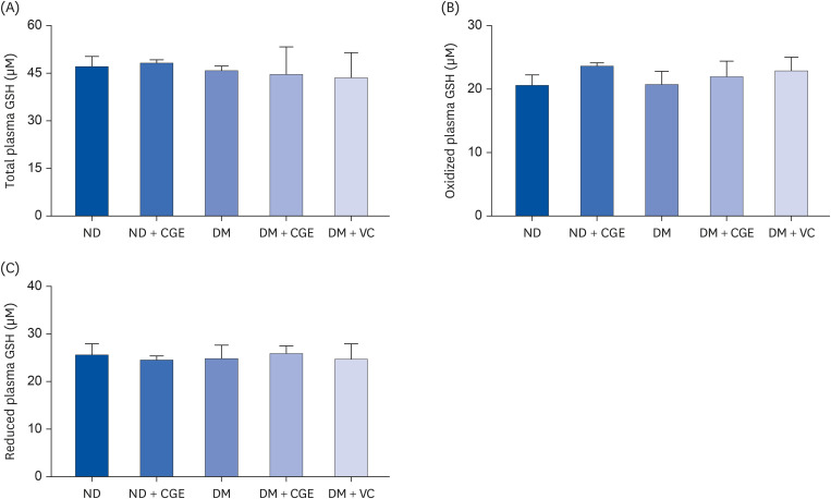
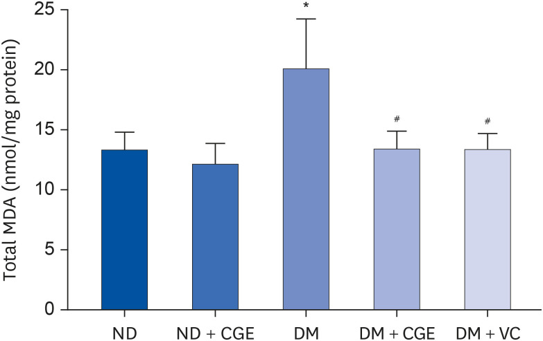
The total, oxidized, and reduced GSH levels were similar in all experimental groups (Fig. 1). In addition, the diabetic rats had significantly higher renal MDA concentrations than the control (Fig. 2), whereas the MDA level was lower in the CGE and VC-treated rats. Furthermore, CGE did not alter the MDA level in normal rats. These results suggest that the CGE could reduce renal oxidative stress in T2DM.
Fig. 1

Effects of CGE on the plasma total, reduced, and oxidized GSH levels, determined in plasma samples from each experimental group using a colorimetric assay kit. The results are expressed as the mean ± SE of the mean (n = 6).

CGE, Cladophora glomerata extract; T2DM or DM, type 2 diabetes mellitus; ND, normal diet; VC, vitamin C; GSH, glutathione.
nrp-18-633-g001
Fig. 2

Effects of CGE on renal cortical MDA level, determined in renal cortical tissue homogenates from each experimental group using a commercial TBARS assay kit. The results are expressed as mean ± SE of the mean (n = 6).

CGE, Cladophora glomerata extract; T2DM or DM, type 2 diabetes mellitus; ND, normal diet; VC, vitamin C; MDA, malondialdehyde.
*P < 0.05 indicates a significant difference from ND; #P < 0.05 indicates a significant difference from DM rats.
nrp-18-633-g002

Effect of CGE on renal antioxidant

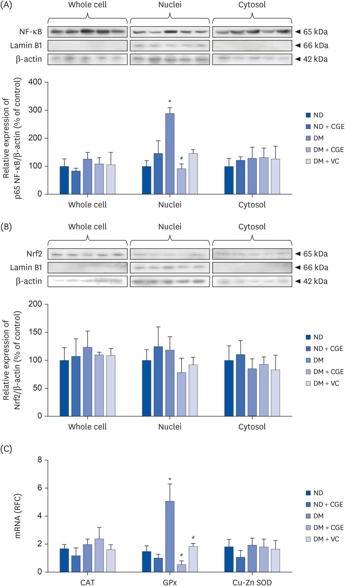
This study further investigated the effects of CGE on oxidative stress markers, including NF-κB, nuclear factor (erythroid-derived 2)-like 2 (Nrf2), catalase (CAT), glutathione peroxidase (GPx) and superoxide dismutase (SOD). Based on a previous study, hyperglycemia, free fatty acid, and oxidative stress were the major stimulators of NF-κB [28]. The levels of NF-κB active subunit (p65 nuclear factor κB [p65NF-κB]) expression in each subcellular fraction extracted from the renal cortical tissues were quantified using western blotting analysis. NF-κB activation (p65NF-κB) was significantly higher in the nuclei fraction of T2DM rat kidneys than in the control (Fig. 3A), whereas CGE and VC markedly reduced its activation. By contrast, there were no significant differences in Nrf2 protein expression in all cell compartments (Fig. 3B). In addition, the T2DM rats showed significantly higher renal GPx gene expression than the control. In contrast, CGE and VC could normalize GPx expression relative to that of T2DM without affecting CAT and SOD expression, suggesting that CGE affected the induction of antioxidant enzyme transcription (Fig. 3C). This result indicates that CGE improved oxidative stress in T2DM by preventing the activation and translocation of NF-κB.
Fig. 3

Effect of CGE on the expression of stress-sensitive markers. (A) p65NF-κB expression in subcellular fractions of rat kidneys. Anti-p65NF-κB antibody was then detected, whereas anti-β-actin and anti-lamin B1 antibodies were used as a loading control and nuclei marker, respectively. The data are expressed as mean ± SE of the mean and repeated for separate sets of animals (n = 3). p65NF-κB protein expression is shown at the top, and the quantification of relative protein expression in each fraction is at the bottom. (B) Nrf2 expression in the subcellular fractions of the rat kidneys. Anti-Nrf2 antibody was subsequently detected, whereas anti-β-actin and anti-lamin B1 antibodies were used as a loading control and nuclei marker, respectively. The data are expressed as the mean ± SE of the mean and repeated for separate sets of animals (n = 3). Nrf2 protein expression is shown at the top and quantification of the relative protein expression in each fraction at the bottom. (C) mRNA expression levels of CAT, GPx, and Cu-Zn SOD from experimental rat kidneys. The values represent mean ± SE of the mean (n = 6).

CGE, Cladophora glomerata extract; T2DM or DM, type 2 diabetes mellitus; ND, normal diet; VC, vitamin C; p65NF-κB, p65 nuclear factor κB; Nrf2, nuclear factor (erythroid-derived 2)-like 2; CAT, catalase; GPx, glutathione peroxidase; Cu-Zn SOD, copper-zinc superoxide dismutase.
*P < 0.05 indicates the significant differences from ND; #P < 0.05 indicates significant differences from DM rats.
nrp-18-633-g003

Direct effect of CGE on renal inflammatory response

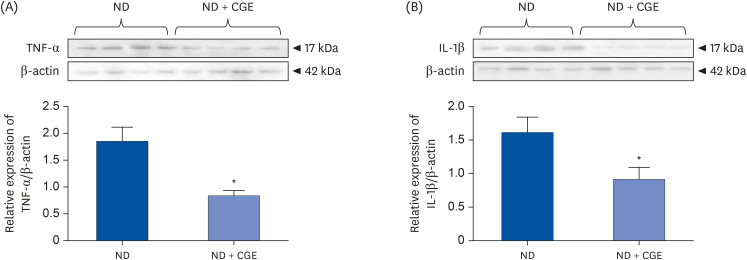
TNF-α and IL-1β protein expression in renal cortical tissues was further determined to confirm whether CGE directly affects NF-κB activation, the downstream of NF-κB. As observed in p65NF-κB, CGE significantly decreased the TNF-α and IL-1β protein expression levels in renal cortical tissues relative to normal kidneys (Fig. 4). Hence, CGE directly improved oxidative stress by reducing pro-inflammatory cytokine production.
Fig. 4

Effect of CGE on the protein expressions of pro-inflammatory cytokines. (A) TNF-α and (B) IL-1β expression in whole-cell lysate from the renal cortical tissues in ND and ND + CGE groups. Anti-TNF-α and anti-IL-1β antibodies were then detected, whereas anti-β-actin antibody was used as a loading control. The values represent the mean ± SE of the mean from separate sets of animals (n = 4).

CGE, Cladophora glomerata extract; ND, normal diet; TNF-α, tumor necrosis factor-alpha; IL-1β, interleukin-1β.
*P < 0.05 indicates significant differences from ND rats.
nrp-18-633-g004

Effect of CGE on SNP-induced renal NO production

The data showed that the pre-incubation with SNP inhibited PAH transport mediated by Oat1 and Oat3 in the renal slices from normal rats (Fig. 5A). Interestingly, the CGE-treated normal rats did not differ in PAH uptake after SNP pre-incubation. This suggests that CGE directly prevented nitrosative stress-induced impairment of the organic anion transport function.
Fig. 5

Effect of CGE on sodium nitroprusside-induced NO production in renal cortical tissue. (A) PAH transport in the renal cortical slices from ND and ND + CGE rats. Rat renal cortical slices were incubated for 30 min in the buffer containing 10 μM of tritiated PAH in the presence or absence of SNP. Data are expressed as mean ± SE of the mean (n = 5). (B) Sodium nitroprusside induced NO production in renal cortical tissues from ND and ND + CGE rats. Rat renal cortical slices were incubated for 30 min in the buffer with or without SNP (n = 5). The data are expressed as mean ± SE.

CGE, Cladophora glomerata extract; ND, normal diet; NO, nitric oxide; PAH, para-aminohippurate; SNP, sodium nitroprusside; T/M, tissue to medium.
*P < 0.05 indicates significant differences from slices incubated with buffer alone.
nrp-18-633-g005
The effects of SNP in renal tissues were confirmed by measuring the total NO levels. The results showed that CGE reduced renal NO production under normal and SNP induction conditions (P < 0.05; Fig. 5B). Furthermore, an increase in NO production induced by SNP was opposed after CGE supplementation (P < 0.05). These data confirm that CGE prevented nitrosative stress, restoring the organic anion transport function.

DISCUSSION

The CGE is widely produced along the Nan River North Thailand. Previous studies have shown its beneficial effects, including antigastric ulcer, analgesic, hypotensive, anti-inflammatory, and antioxidant activities [2]. In addition, a recent study reported that CGE has antidiabetic and renoprotective effects in T2DM rats by restoring the renal organic anion transport function, but the underlying mechanisms are unclear [3]. This question was addressed by evaluating the antioxidant effects of CGE on renal oxidative stress and inflammation in the T2DM rat model. The present work shows that the antioxidant effect of CGE reduced renal cortical oxidative/nitrosative stress by inducing antioxidative gene expression, GPx, inhibiting NKB translocation, and decreasing pro-inflammatory cytokine production. These factors could prevent the impairment of renal organic anion clearance, leading to a significant decrease in the risk of DN. According to a previous study, CGE at 1,000 mg/kg BW in this study can be converted to a human dose at 9,720 mg/60 kg BW/day [28].
Hyperglycemia and hyperlipidemia are prominent causative factors in ROS production [2930]; ROS can directly damage DNA, proteins, and lipids in the cells. Previously, membrane lipids have been shown as one of the targets of ROS [31]. Lipid peroxidation produces several reactive aldehydes, such as MDA, acrolein, and 4-hydroxynonenal [32]. Among these, MDA is a primary biomarker of membrane lipid damage and oxidative stress [33]. In addition, oxidative stress contributes to the progression and severity of DN for 20–30% of types 1 and 2 diabetes patients [3435]. Thus, preventing ROS generation could delay the progression of DN. CGE can decrease renal cortical MDA levels, which could then attenuate renal oxidative stress induced by diabetic conditions. Therefore, the antioxidant effects of CGE are a primary defense mechanism for DN prevention.
According to a previous study, ROS can trigger Nrf2 translocation [36], which could induce the expression of the 3 primary antioxidant enzymes SOD, CAT, and GPx, preventing intracellular ROS production [37]. The T2DM rat model in this study showed strong expression of the GPx gene in the kidney. In agreement with the data, renal and cardiac GPx protein expression and function were higher in diabetic rat and mice models against oxidative stress [383940]. According to GSH, it is the most abundant antioxidant molecule that uses GPx as a cofactor to reduce oxidative stress molecules, such as hydrogen peroxide radicals (H2O2) [40]. In addition, upregulated GPx by ROS could induce NF-κB activation in skeletal muscle cells [41]. The NF-κB subunits are bound to the promotor region of GPx in response to oxidative stress in lipopolysaccharide-induced U937 cells [42]. This active p65NF-κB subunit induces the generation of inducible nitric oxide synthase (iNOS), resulting in NO production [43]. This partly contributes to hyperfiltration and microalbuminuria in early DN [44]. Previous studies have shown that epigallocatechin-3-gallate, isoquinoline alkaloid, and berberine prevent the transformation of renal epithelial cells to fibroblasts in DN by activating Nrf2 [4546]. Although Nrf2 protein expression did not change in any cellular compartments in this study, up-regulation of the downstream targeting protein of Nrf2 (GPx) by CGE supplementation did preserve and counterbalance renal oxidative stress in T2DM rat kidneys. Similarly, a previous study showed that curcumin normalized GPx expression in STZ-induced DM rats [38]. Moreover, flavonoids, one of the active compounds in CGE, potentially inhibited phosphoinositide 3-kinase/protein kinase B-induced eNOS function, reducing NO production [47]. Therefore, the strong inhibition by CGE is a primary defense mechanism for DN prevention.
In addition to activating renal iNOS, p65NF-κB can induce renal TNF-α and IL-1β production in diabetic rats [71617]. Both pro-inflammatory cytokines, TNF-α and IL-1β, are involved in diabetic pathogenesis and are crucial in diabetic complications [48]. The extent of inflammatory infiltration into the tissues, including macrophages and T-cells, is associated with the severity of DN [4950]. CGE also showed a direct antioxidative effect by reducing TNF-α and IL-1β production in ND treated with CGE compared to ND rats, suggesting that CGE has a strongly direct effect on proinflammatory cytokines, which are the oxidative stress markers. Several major chemical constituents in CGE exist, including isoquercetin, catechin, tannic acid, hydroquinin, quercetin, rutin, gallic acid, and kaempferol [3]. Isoquercetin blunts H2O2-induced ROS production through H2O2, hydroxyl radicals, and superoxide radicals in retinal ganglion cell 5 cells [51]. In addition, green tea catechin reduced the ROS levels by inhibiting ERK activation in the human breast epithelial cell line MCF10A [52].
As Oat1 and Oat3 play a crucial role in organic anion uptake across the basolateral membrane and are strongly expressed in the kidney [20], the function of these 2 transporters is regulated by several factors, such as hormones, endogenous/exogenous substances, pathological status, and oxidative stress condition [2053]. On the other hand, oxidative stress impairs transporter function, and nitrosative stress is generated by reactive nitrogen species that impact several transporters. For example, SNP-induced nitrosative stress reduced organic cation transporter and Na+/taurocholate co-transporting polypeptide function in isolated rat hepatocytes [54]. Similarly, nitrosative stress induced by SNP-impaired cyclosporin A transport is mediated by P-glycoprotein (P-gp) in mouse brain capillary endothelial (MBEC4) cells [55]. Similarly, SNP injection into a rat brain decreased the daunomycin efflux from the brain, suggesting that the P-gp function was impaired by nitrosative stress under in vivo and in vitro conditions [56]. These results suggest that PAH transport mediated by Oat1 and Oat3 was blunted under nitrosative stress induced by SNP. CGE could restore this defect directly. Overall, CGE has renoprotective effects against NO production, probably by directly scavenging NO molecules, leading to reduced renal organic anion transport.
This study reported the antioxidative, anti-inflammatory, and anti-nitrosative effects of CGE against impairment of renal organic anion transport mediated by Oat1 and Oat3 in experimental rats. CGE could improve renal oxidative/nitrosative stresses, restoring the Oat1 and 3 transport functions. The mechanisms through which CGE improved PAH transport involved the modulation of free radical scavenging enzymes, NF-κB, and proinflammatory cytokines. These findings facilitate the further development of CGE, making it a potential nutraceutical product for kidney diseases.

ACKNOWLEDGMENTS

The authors thank Dr. Yuwadee Peerapornpisal for kindly providing Cladophora glomerata extract and Dr. Rattanabhorn Junthip for assistance with the extraction method.

Notes

Funding: This work was supported by the Thailand Research Fund (MRG5480278 and RSA5980009 to Vaddhanaphuti CS), the Faculty of Medicine Research Fund, Chiang Mai University, Chiang Mai, Thailand to Vaddhanaphuti CS, and the NSRF via the Program Management Unit for Human Resources & Institutional Development, Research and Innovation (grant number B05F640169) to Vaddhanaphuti CS and Ontawong A.

Conflict of Interest: The authors declare no potential conflicts of interests.

Author Contributions:

  • Conceptualization: Vaddhanaphuti CS.

  • Formal analysis: Ontawong A, Aida CJ, Vivithanaporn P, Vaddhanaphuti CS.

  • Funding acquisition: Vaddhanaphuti CS.

  • Investigation: Ontawong A, Aida CJ, Amornlerdpison D, Vaddhanaphuti CS.

  • Methodology: Ontawong A, Aida CJ, Vivithanaporn P, Vaddhanaphuti CS.

  • Supervision: Vaddhanaphuti CS.

  • Writing - original draft: Ontawong A, Vaddhanaphuti CS.

  • Writing - review & editing: Ontawong A, Aida CJ, Vivithanaporn P, Amornlerdpison D, Vaddhanaphuti CS.

References

1. Peerapornpisal Y, Amornledpison D, Rujjanawate C, Ruangrit K, Kanjanapothi D.. Two endemic species of macroalgae in Nan River, Northern Thailand, as therapeutic agents. ScienceAsia. 2006; 32(Suppl 1):71–76.
2. Laungsuwon R, Chulalaksananukul W. Antioxidant and anticancer activities of freshwater green algae, Cladophora glomerata and Microspora floccosa, from Nan River in Northern Thailand. Maejo Int J Sci. 2013; 7:181–188.
3. Srimaroeng C, Ontawong A, Saowakon N, Vivithanaporn P, Pongchaidecha A, Amornlerdpison D, Soodvilai S, Chatsudthipong V. Antidiabetic and renoprotective effects of Cladophora glomerata Kützing extract in experimental type 2 diabetic rats: a potential nutraceutical product for diabetic nephropathy. J Diabetes Res. 2015; 2015:320167. PMID: 25883984.
4. American Diabetes Association. Diagnosis and classification of diabetes mellitus. Diabetes Care. 2014; 37(Suppl 1):S81–S90. PMID: 24357215.
5. Brownlee M. Biochemistry and molecular cell biology of diabetic complications. Nature. 2001; 414:813–820. PMID: 11742414.
6. Evans JL, Goldfine ID, Maddux BA, Grodsky GM. Oxidative stress and stress-activated signaling pathways: a unifying hypothesis of type 2 diabetes. Endocr Rev. 2002; 23:599–622. PMID: 12372842.
7. Kanwar YS, Wada J, Sun L, Xie P, Wallner EI, Chen S, Chugh S, Danesh FR. Diabetic nephropathy: mechanisms of renal disease progression. Exp Biol Med (Maywood). 2008; 233:4–11. PMID: 18156300.
8. Yu T, Jhun BS, Yoon Y. High-glucose stimulation increases reactive oxygen species production through the calcium and mitogen-activated protein kinase-mediated activation of mitochondrial fission. Antioxid Redox Signal. 2011; 14:425–437. PMID: 20518702.
9. Ceriello A, Morocutti A, Mercuri F, Quagliaro L, Moro M, Damante G, Viberti GC. Defective intracellular antioxidant enzyme production in type 1 diabetic patients with nephropathy. Diabetes. 2000; 49:2170–2177. PMID: 11118022.
10. Fujii H, Kono K, Nakai K, Goto S, Komaba H, Hamada Y, Shinohara M, Kitazawa R, Kitazawa S, Fukagawa M. Oxidative and nitrosative stress and progression of diabetic nephropathy in type 2 diabetes. Am J Nephrol. 2010; 31:342–352. PMID: 20224273.
11. Hiragushi K, Sugimoto H, Shikata K, Yamashita T, Miyatake N, Shikata Y, Wada J, Kumagai I, Fukushima M, Makino H. Nitric oxide system is involved in glomerular hyperfiltration in Japanese normo- and micro-albuminuric patients with type 2 diabetes. Diabetes Res Clin Pract. 2001; 53:149–159. PMID: 11483230.
12. Sugimoto H, Shikata K, Matsuda M, Kushiro M, Hayashi Y, Hiragushi K, Wada J, Makino H. Increased expression of endothelial cell nitric oxide synthase (ecNOS) in afferent and glomerular endothelial cells is involved in glomerular hyperfiltration of diabetic nephropathy. Diabetologia. 1998; 41:1426–1434. PMID: 9867209.
13. Wada J, Makino H. Inflammation and the pathogenesis of diabetic nephropathy. Clin Sci (Lond). 2013; 124:139–152. PMID: 23075333.
14. Ohga S, Shikata K, Yozai K, Okada S, Ogawa D, Usui H, Wada J, Shikata Y, Makino H. Thiazolidinedione ameliorates renal injury in experimental diabetic rats through anti-inflammatory effects mediated by inhibition of NF-κB activation. Am J Physiol Renal Physiol. 2007; 292:F1141–F1150. PMID: 17190910.
15. Soetikno V, Sari FR, Veeraveedu PT, Thandavarayan RA, Harima M, Sukumaran V, Lakshmanan AP, Suzuki K, Kawachi H, Watanabe K. Curcumin ameliorates macrophage infiltration by inhibiting NF-κB activation and proinflammatory cytokines in streptozotocin induced-diabetic nephropathy. Nutr Metab (Lond). 2011; 8:35. PMID: 21663638.
16. Ha H, Yoon SJ, Kim KH. High glucose can induce lipid peroxidation in the isolated rat glomeruli. Kidney Int. 1994; 46:1620–1626. PMID: 7700020.
17. Locke M, Anderson J. NF-κB activation in organs from STZ-treated rats. Appl Physiol Nutr Metab. 2011; 36:121–127. PMID: 21326386.
18. Iliescu R, Chade AR. Progressive renal vascular proliferation and injury in obese Zucker rats. Microcirculation. 2010; 17:250–258. PMID: 20536738.
19. Hilgendorf C, Ahlin G, Seithel A, Artursson P, Ungell AL, Karlsson J. Expression of thirty-six drug transporter genes in human intestine, liver, kidney, and organotypic cell lines. Drug Metab Dispos. 2007; 35:1333–1340. PMID: 17496207.
20. Srimaroeng C, Perry JL, Pritchard JB. Physiology, structure, and regulation of the cloned organic anion transporters. Xenobiotica. 2008; 38:889–935. PMID: 18668434.
21. Sharma K, Karl B, Mathew AV, Gangoiti JA, Wassel CL, Saito R, Pu M, Sharma S, You YH, Wang L, et al. Metabolomics reveals signature of mitochondrial dysfunction in diabetic kidney disease. J Am Soc Nephrol. 2013; 24:1901–1912. PMID: 23949796.
22. Wang L, Sweet DH. Renal organic anion transporters (SLC22 family): expression, regulation, roles in toxicity, and impact on injury and disease. AAPS J. 2013; 15:53–69. PMID: 23054972.
23. Lungkaphin A, Arjinajarn P, Srimaroeng C, Chatsudthipong V. Function and expression of renal organic anion transporters in experimental diabetes in mice. ScienceAsia. 2012; 38:18–23.
24. Ontawong A, Saowakon N, Vivithanaporn P, Pongchaidecha A, Lailerd N, Amornlerdpison D, Lungkaphin A, Srimaroeng C. Antioxidant and renoprotective effects of Spirogyra neglecta (Hassall) Kützing extract in experimental type 2 diabetic rats. BioMed Res Int. 2013; 2013:820786. PMID: 23862157.
25. Barros SA, Srimaroeng C, Perry JL, Walden R, Dembla-Rajpal N, Sweet DH, Pritchard JB. Activation of protein kinase Cζ increases OAT1 (SLC22A6)- and OAT3 (SLC22A8)-mediated transport. J Biol Chem. 2009; 284:2672–2679. PMID: 19028678.
26. Pelletier MM, Kleinbongard P, Ringwood L, Hito R, Hunter CJ, Schechter AN, Gladwin MT, Dejam A. The measurement of blood and plasma nitrite by chemiluminescence: pitfalls and solutions. Free Radic Biol Med. 2006; 41:541–548. PMID: 16863986.
27. Yang BK, Vivas EX, Reiter CD, Gladwin MT. Methodologies for the sensitive and specific measurement of S-nitrosothiols, iron-nitrosyls, and nitrite in biological samples. Free Radic Res. 2003; 37:1–10. PMID: 12653211.
28. Reagan-Shaw S, Nihal M, Ahmad N. Dose translation from animal to human studies revisited. FASEB J. 2008; 22:659–661. PMID: 17942826.
29. Busik JV, Mohr S, Grant MB. Hyperglycemia-induced reactive oxygen species toxicity to endothelial cells is dependent on paracrine mediators. Diabetes. 2008; 57:1952–1965. PMID: 18420487.
30. Yang RL, Shi YH, Hao G, Li W, Le GW. Increasing oxidative stress with progressive hyperlipidemia in human: relation between malondialdehyde and atherogenic index. J Clin Biochem Nutr. 2008; 43:154–158. PMID: 19015749.
31. Boylan JA, Lawrence KA, Downey JS, Gherardini FC. Borrelia burgdorferi membranes are the primary targets of reactive oxygen species. Mol Microbiol. 2008; 68:786–799. PMID: 18373524.
32. Guo L, Chen Z, Amarnath V, Davies SS. Identification of novel bioactive aldehyde-modified phosphatidylethanolamines formed by lipid peroxidation. Free Radic Biol Med. 2012; 53:1226–1238. PMID: 22898174.
33. Pandey KB, Rizvi SI. Markers of oxidative stress in erythrocytes and plasma during aging in humans. Oxid Med Cell Longev. 2010; 3:2–12. PMID: 20716923.
34. Molitch ME, DeFronzo RA, Franz MJ, Keane WF, Mogensen CE, Parving HH. American Diabetes Association. Diabetic nephropathy. Diabetes Care. 2003; 26(Suppl 1):S94–S98. PMID: 12502629.
35. Miranda-Díaz AG, Pazarín-Villaseñor L, Yanowsky-Escatell FG, Andrade-Sierra J. Oxidative stress in diabetic nephropathy with early chronic kidney disease. J Diabetes Res. 2016; 2016:7047238. PMID: 27525285.
36. Ishii T, Itoh K, Yamamoto M. Roles of Nrf2 in activation of antioxidant enzyme genes via antioxidant responsive elements. Methods Enzymol. 2002; 348:182–190. PMID: 11885271.
37. Ighodaro OM, Akinloye OA. First line defence antioxidants-superoxide dismutase (SOD), catalase (CAT) and glutathione peroxidase (GPX): their fundamental role in the entire antioxidant defence grid. Alexandria J Med. 2018; 54:287–293.
38. El-Bahr SM. Curcumin regulates gene expression of insulin like growth factor, B-cell CLL/lymphoma 2 and antioxidant enzymes in streptozotocin induced diabetic rats. BMC Complement Altern Med. 2013; 13:368. PMID: 24364912.
39. Iwata K, Nishinaka T, Matsuno K, Yabe-Nishimura C. Increased gene expression of glutathione peroxidase-3 in diabetic mouse heart. Biol Pharm Bull. 2006; 29:1042–1045. PMID: 16651743.
40. Limaye PV, Raghuram N, Sivakami S. Oxidative stress and gene expression of antioxidant enzymes in the renal cortex of streptozotocin-induced diabetic rats. Mol Cell Biochem. 2003; 243:147–152. PMID: 12619900.
41. Zhou LZ, Johnson AP, Rando TA. NFκB and AP-1 mediate transcriptional responses to oxidative stress in skeletal muscle cells. Free Radic Biol Med. 2001; 31:1405–1416. PMID: 11728812.
42. Schreiber J, Jenner RG, Murray HL, Gerber GK, Gifford DK, Young RA. Coordinated binding of NF-κB family members in the response of human cells to lipopolysaccharide. Proc Natl Acad Sci U S A. 2006; 103:5899–5904. PMID: 16595631.
43. Arias-Salvatierra D, Silbergeld EK, Acosta-Saavedra LC, Calderon-Aranda ES. Role of nitric oxide produced by iNOS through NF-κB pathway in migration of cerebellar granule neurons induced by Lipopolysaccharide. Cell Signal. 2011; 23:425–435. PMID: 20955790.
44. Prabhakar SS. Role of nitric oxide in diabetic nephropathy. Semin Nephrol. 2004; 24:333–344. PMID: 15252773.
45. Kanlaya R, Khamchun S, Kapincharanon C, Thongboonkerd V. Protective effect of epigallocatechin-3-gallate (EGCG) via Nrf2 pathway against oxalate-induced epithelial mesenchymal transition (EMT) of renal tubular cells. Sci Rep. 2016; 6:30233. PMID: 27452398.
46. Zhang X, He H, Liang D, Jiang Y, Liang W, Chi ZH, Ma J. Protective effects of berberine on renal injury in streptozotocin (STZ)-induced diabetic mice. Int J Mol Sci. 2016; 17:1327. PMID: 27529235.
47. Wink DA, Miranda KM, Espey MG, Pluta RM, Hewett SJ, Colton C, Vitek M, Feelisch M, Grisham MB. Mechanisms of the antioxidant effects of nitric oxide. Antioxid Redox Signal. 2001; 3:203–213. PMID: 11396476.
48. Harris RC. Growth factors and cytokines in acute renal failure. Adv Ren Replace Ther. 1997; 4:43–53. PMID: 9113240.
49. Moriya R, Manivel JC, Mauer M. Juxtaglomerular apparatus T-cell infiltration affects glomerular structure in type 1 diabetic patients. Diabetologia. 2004; 47:82–88. PMID: 14618232.
50. Nguyen D, Ping F, Mu W, Hill P, Atkins RC, Chadban SJ. Macrophage accumulation in human progressive diabetic nephropathy. Nephrology (Carlton). 2006; 11:226–231. PMID: 16756636.
51. Jung SH, Kim BJ, Lee EH, Osborne NN. Isoquercitrin is the most effective antioxidant in the plant Thuja orientalis and able to counteract oxidative-induced damage to a transformed cell line (RGC-5 cells). Neurochem Int. 2010; 57:713–721. PMID: 20708054.
52. Rathore K, Choudhary S, Odoi A, Wang HC. Green tea catechin intervention of reactive oxygen species-mediated ERK pathway activation and chronically induced breast cell carcinogenesis. Carcinogenesis. 2012; 33:174–183. PMID: 22045026.
53. VanWert AL, Gionfriddo MR, Sweet DH. Organic anion transporters: discovery, pharmacology, regulation and roles in pathophysiology. Biopharm Drug Dispos. 2010; 31:1–71. PMID: 19953504.
54. Song IS, Lee IK, Chung SJ, Kim SG, Lee MG, Shim CK. Effect of nitric oxide on the sinusoidal uptake of organic cations and anions by isolated hepatocytes. Arch Pharm Res. 2002; 25:984–988. PMID: 12510858.
55. Maeng HJ, Kim MH, Jin HE, Shin SM, Tsuruo T, Kim SG, Kim DD, Shim CK, Chung SJ. Functional induction of P-glycoprotein in the blood-brain barrier of streptozotocin-induced diabetic rats: evidence for the involvement of nuclear factor-κB, a nitrosative stress-sensitive transcription factor, in the regulation. Drug Metab Dispos. 2007; 35:1996–2005. PMID: 17664251.
56. Maeng HJ, Bang YJ, Chung SJ. Functional impairment of P-glycoprotein by sodium nitroprusside pretreatment in mouse brain capillary endothelial cells. Arch Pharm Res. 2012; 35:1215–1221. PMID: 22864744.
TOOLS
Similar articles