Journal List > Ann Rehabil Med > v.48(4) > 1516088245

Lee, Kim, Shim, Kang, Kim, Lee, Lee, Lee, Lim, Chung, and Oh: Reference Standard of Median Nerve Conduction Study in Korea

Abstract

Objective

To establish the reference standard of the median nerve conduction study (NCS) in Korea.

Methods

A total of 648 median motor and 602 median sensory NCSs from 349 Korean healthy volunteers were tested and analyzed prospectively. Equipment calibration, assessment of intra- and inter-rater reliability, and the NCSs per se were conducted according to a predetermined protocol. A reference standard was established from uncertainty components for the following parameters: the onset and peak latencies; the baseline-to-peak and peak-to-peak amplitudes; the area and duration of the negative wave; and the nerve conduction velocity. The effects of sex, age and stimulation intensity were analyzed.

Results

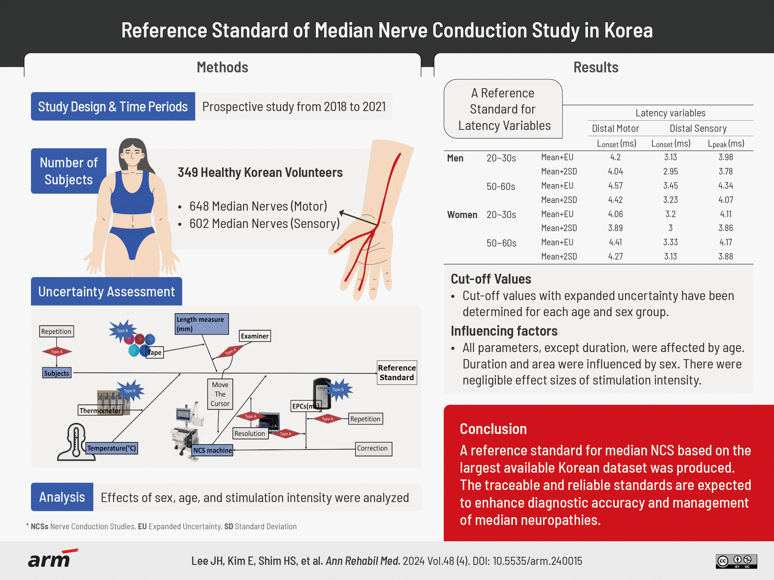
Each measured value of 648 median motor and 602 median sensory nerves were obtained and presented with both mean and expanded uncertainties, as well as mean and standard deviations. The cut-off values with expanded uncertainty were determined for different age and sex groups. After adjusting for anthropometric covariates, all parameters except duration were affected by age, and sex appeared to influence both duration and area. While stimulation intensity significantly affected some parameters including latencies, the effect sizes were negligible.

Conclusion

We propose the median NCS reference standard using the largest Korean dataset ever available. The use of the traceable and reliable reference standard is anticipated to promote more accurate and dependable diagnosis and appropriate management of median neuropathies in Korea.

GRAPHICAL ABSTRACT

INTRODUCTION

The nerve conduction study (NCS) is a crucial component of electrodiagnosis, evaluating the transmission of electrical signals within nerves in the context of various neuromuscular diseases. This diagnostic procedure analyzes waveform properties, including latencies and amplitudes, to gauge the existence and extent of nerve pathology. Ensuring the accurate assessment of alterations in nerve function between healthy and diseased conditions requires a reliable reference standard. Nevertheless, the range of NCS results can vary due to biological variations and factors related to the environment or the procedure itself [1-5].
Efforts have been undertaken to standardize NCS by establishing normative data and clinical guidelines [6-13]. In the United States, the American Association of Neuromuscular & Electrodiagnostic Medicine (AANEM) established the Normative Data Task Force (NDTF), which systematically reviewed articles published between 1990 and 2012. They proposed consensus-based methodological criteria, comprising seven key issues and techniques [14]. Additionally, the AANEM provided normative reference data from the literature that met these criteria in a separate publication [15]. Similarly, in Korea, the Korean Association of Electrodiagnostic Medicine (KAEND) is actively working on standardizing NCS by developing guidelines and administering practitioner qualification tests [9].
Previous efforts to standardize NCSs, however, did not take into account measurement uncertainties, a crucial aspect for securing traceability in laboratory medicine. Traceability refers to the ability to link measurement results to recognized references, such as national or international standards, through an unbroken chain of calibrations [16]. To demonstrate traceability, the uncertainty of measurement must be considered and presented along with the results. This requires calibration of NCS instruments and consideration of all uncertainty components [16]. A reference standard produced with these considerations would improve accuracy in diagnosis and increase reliability among electrodiagnostic laboratories.
Measurement uncertainties are well-documented in laboratory medicine and have been studied in clinical biochemistry, encompassing areas such as serum anion calculation, international normalized ratio monitoring, and creatinine clearance evaluations [17-23]. To date, there have been no previous attempts to measure uncertainty in NCS. Since 2018, we have been working on this and have published reference standards for the ulnar nerve and peripheral nerves in the lower extremities [24,25]. Furthermore, there is a dearth of large-scale studies on median nerve NCS within the Korean population. The aim of this study was to produce a reference standard for median NCS, stratified by age and sex, using the most extensive sample of healthy Korean volunteers ever examined, and to investigate the characteristics of the data.

METHODS

Subjects

Before this study, a survey was conducted among experts within KAEND and the field of rehabilitation medicine to gauge the interest in utilizing standardized reference standard data. The findings indicated a strong preference for age and sex-specific average and cut-off data. Consequently, this study is dedicated to establishing age and sex-specific reference standards for nerve conduction, with a specific focus on individuals in their 50s—the most commonly tested age group in Korea based on the 2016 Health and Medical Bigdata Open portal—alongside healthy representative adults in their 20s for comparative analysis [26]. The number of subjects was determined based on previous reports, ensuring accurate estimation and representation of 3rd (97th) percentiles and 2 standard deviation values [6,27].
During 2018–2021, 349 healthy volunteers participated in this study after responding to recruitment ads posted at four hospitals or on the website of the clinical trial center. The following were the criteria for inclusion: (1) reside in Korea, (2) are in good health, and (3) are at the ages of 20–39 or 50–69 years old. The following were the criteria for exclusion: (1) patients on pacemakers, (2) diagnosed with diabetes, (3) having hypothyroidism or hyperthyroidism, (4) having cancer or on chemotherapy or external beam radiation, (5) pregnant or planning to become, (6) significantly reduced cognitive function making communication difficult, (7) numbness or pain in all or part of the upper or lower limbs for more than a month in the last three months, (8) diagnosed with psychiatric or neurological disease or on medications, (9) having other medical conditions or taking medications that affect the peripheral nerve function as judged by the researchers.
NCS was conducted from the left and right upper extremities of each of the 349 subjects according to the predetermined protocol. Out of a total of 349 subjects, 324 subjects conducted median motor NCS, and 301 subjects conducted median sensory NCS. Among the 648 median motor nerves that were analyzed, 13 were excluded as outliers. As a result, a total of 635 median motor nerves were included in the statistical analysis and reference standard production for distal motor NCS parameters. However, only 629 nerves were analyzed for proximal motor NCS and nerve conduction velocity (NCV) parameters due to the presence of Martin-Gruber anastomosis (MGA) in 6 nerves. Similarly, among the 602 median sensory nerves analyzed, 18 were excluded as outliers, and a total of 584 were included in the analysis. Outliers are determined by more than 1.5 interquartile range from the 3rd quartile or less than 1.5 interquartile range from the 1st quartile according to the distribution characteristics of the parameters from each of the four subgroups by age and sex. All subjects have read and signed an informed consent form and were rewarded after the examination. The present study protocol was reviewed and approved by the Institutional Review Board of Seoul National University Hospital (No. 1804-125-940, 2104-107-1212), Seoul National University Bundang Hospital (No. B-1809-493-301, B-2106-689-402), SMG-SNU Boramae Medical Center (No. 10-2018-82, 10-2021-76), and National Traffic Injury Rehabilitation Hospital (No. NTRH-20017, NTRH-21008).

NCS parameters

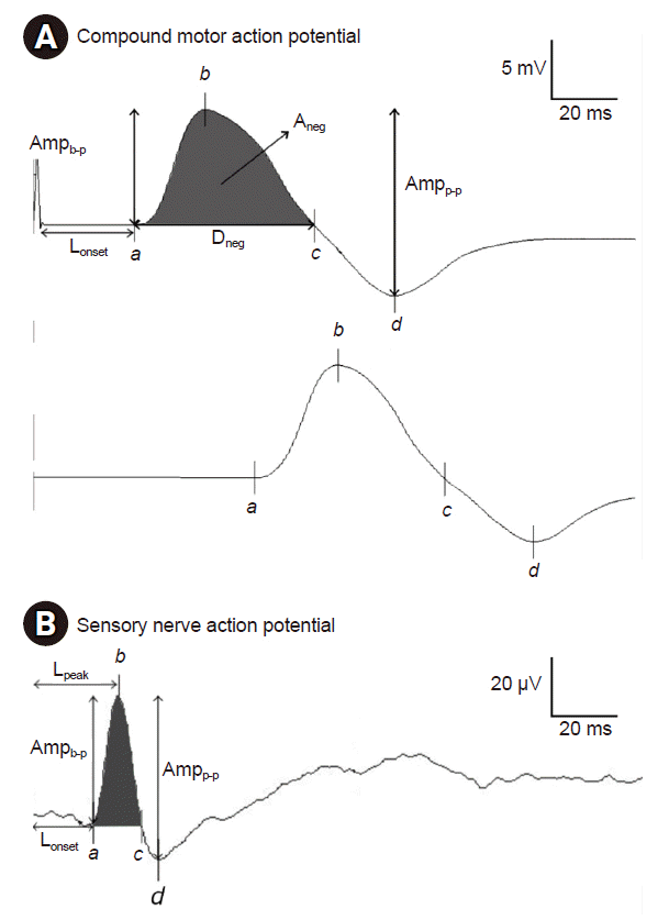
The parameters of compound motor action potential (CMAP) were as follows: onset latency (Lonset); baseline to negative peak amplitude (Ampb-p); negative peak to positive peak amplitude (Ampp-p); negative spike area (Aneg); negative spike duration (Dneg). The Lonset from CMAP elicited by proximal stimulation was only used in calculating the NCV parameter and is not presented as reference standard. The motor NCV was determined by dividing the distance between the proximal and distal stimulation sites by the time difference between the proximal and distal latencies (Fig. 1A). For the sensory nerve action potential (SNAP), Lonset, peak latency (Lpeak), Ampb-p and Ampp-p were presented as parameters. The NCV, Aneg, and Dneg were not presented as they were not included in the current reference standard (Fig. 1B).

NCS instruments and settings

The NCS data were produced using six NCS instruments (Nicolet EDX®; Natus) from four hospitals. Each instrument consisted of a base unit, a hand-held stimulator probe, an amplifier, a control panel, PC and an LCD monitor. The NCS instruments, soft tape measures, thermometers, and indoor thermo-hygrometers had been calibrated annually. The frequency filtration range for motor NCS was 10–10,000 Hz, sweep speed was 2 ms/division, and sensitivity was 2–5 mV/division. The frequency filtration range for sensory nerves was 20–2,000 Hz, sweep speed was 1–2 ms/division, and sensitivity was 5–20 μV/division.
The stimulation intensity was determined by progressively raising the intensity until no further amplitude rise was possible. In order to ensure that the stimulation is supramaximal, 10% to 20% of stimulation intensity was added. If maximal stimulation intensity could not be attained, with stimulation duration of 0.1 ms, even at 100 mA, the process was repeated with the stimulation duration of 0.2 ms.

NCS protocol

First, the surface temperature of both hands was measured to ensure that it is between 33 and 37 degrees Celsius. If necessary, the use of a water bath, a heating lamp, a hot pack, or a hair dryer was permitted prior to beginning NCS. The skin was cleaned with a swab containing 70% isopropyl alcohol to increase electrode contact and decrease impedance. Although the influence of ambient temperature and humidity on NCSs has not yet been precisely established, these variables were likewise kept within tolerable limits of 15 to 25 degrees Celsius and 40 to 60 percent, respectively.
The posture, stimulation locations, and recording sites for median motor NCS were determined according to the predetermined in-house protocol, which is in accordance with widely accepted methodology in Korea and other countries [1,27,28]. In brief, for median motor NCS, the active electrode was attached to the midpoint of the line between the first metacarpophalangeal joint and the median point of the distal wrist crease, on the most prominent belly of the abductor pollicis brevis muscle. It can be moved to obtain maximum negative deflections to get as close to the motor point as possible. The reference electrode was attached to the interphalangeal joint of the thumb. The ground electrode was attached adjacent to the palm or the dorsum of hand. The cathode of the stimulator was placed 8 cm proximal to the active electrode along the following path: (1) from the active electrode to the midpoint of the distal volar wrist crease and (2) upward along the midline between the flexor carpi radialis and palmaris longus tendons. Proximal stimulation was just medial to the brachial artery pulsation on the antecubital fossa (Fig. 2A). The MGA was defined as a nerve where Ampp-p is 0.3 greater than Ampb-p. Considering that MGA is an anatomical variant occurring in the forearm region, the upper limbs in which MGA was confirmed were only excluded from the analysis of proximal motor NCS parameters and NCV.
For median sensory NCS, the active electrode was attached to the skin over the second proximal phalanx. The reference electrode was attached at least 4cm distal to the active electrode. The ground electrode was attached adjacent to the active electrode between the active electrode and the cathode of the stimulator or on the hand dorsum. The cathode of the stimulator was placed 14 cm proximal to the active electrode between the flexor carpi radialis and palmaris longus tendons (Fig. 2B).

Uncertainty assessment

The evaluation of uncertainty begins with the definition of measurand. In this study, the measurands included all NCS parameters which mentioned in the previous section. The process of evaluating uncertainty is carried out according to the Guide to the expression of uncertainty in measurement (GUM), which is the international standard for uncertainty evaluation [29]. The evaluation of uncertainty followed the steps outlined in the GUM, including the expression of model equation; investigation of uncertainty components and standard uncertainty for each component; calculation of combined standard uncertainty and expanded uncertainty. The entire procedure for evaluating uncertainty is publicly available in Korean at the Data Center for Korean Reference Nerve Conductions website (https://emgreference.org/43) and we attached additional supplement materials with an English translation for international readers (Supplementary Material S1). Additionally, this approach is akin to that employed in the paper on reference standard data for the other nerves, conducted parallelly during 2018–2019 [24,25].

Statistical analysis

Anthropometric data were summarized by age and sex, then compared using the student-t test. To assess the impact of age and sex on NCS parameters, both univariable and multivariable linear mixed models (LMMs) were employed to effectively address the data gathered from two separate instances within both extremities. The univariable LMM was adjusted only for the side (left or right), while the multivariable LMM accounted for height, weight, soft lean mass, body fat mass, and skeletal muscle mass. Additionally, the effect of stimulation intensity on NCS parameters was also analyzed using a multivariable LMM that accounted for the aforementioned anthropometric factors, as well as sex and age. Normality was confirmed in each model through residual plots, with no models failing to meet this criterion. Statistical significance was established at p-value<0.05. All statistical analyses were conducted using R statistical software (version 3.5.3; R Foundation for Statistical Computing).

RESULTS

Anthropometric data

The anthropometric data, detailed in Table 1, reveal significant differences in measured parameters based on sex within the same age group or age within the same sex group. Specifically, obesity rates, defined as a body mass index exceeding 25, demonstrate notable disparities. In the 20s–30s age group, 51.0% of male and 6.8% of female are classified as obese. In the 50s–60s age group, these rates shift to 46.2% for male and 27.2% for female. Notably, only one subject required an increased stimulation duration of 0.2 ms for supramaximal stimulation.

Reference standards for the NCS parameters

Tables 2 and 3 comprehensively summarize the NCS parameters, with Table 2 summarizes the upper limits of latency parameters and Table 3 summarizes the lower limits of the rest parameters. All parameters are presented as mean with expanded uncertainty and mean with two standard deviations.
Due to the vast amount of data, this section will present the cut-off values with expanded uncertainty on Lonset and NCV which hold clinical significance in the diagnosis of carpal tunnel syndrome. In case of Lonset of SNAP, the cut-off value was 3.13 ms for male in their 20s–30s and 3.45 ms for those in their 50s–60s. For female, these values were 3.20 and 3.33 ms for the respective age groups. Regarding CMAP, the cut-off values were 4.20 ms for male in their 20s–30s and 4.57 ms for those in their 50s–60s. For female, these values were 4.06 and 4.41 ms for the respective age groups. As for the cut-off values for NCV, the values were 46.34 and 43.71 m/s for male in their 20s–30s and 50s–60s, respectively. For female, these values were 43.97 and 41.85 m/s for the respective age groups.

The effects of age and sex on NCS parameters

In the CMAP parameters, such as Lonset, Ampb-p, Ampp-p, Aneg, and NCV showed significant age effects in both univariable and multivariable analyses, with p-values less than 0.001. However, Dneg did not exhibit a significant age effect in either analysis. In the SNAP parameters, Lonset, Lpeak, Ampb-p, and Ampp-p showed significant age effects in both analyses. In particular, SNAP amplitude parameters exhibited significant effect sizes, presenting -18.49 (p<0.001) for Ampb-p and -27.86 (p<0.001) for Ampp-p.
Regarding the effect of sex on all NCS parameters, there were no parameters that showed the sex effect, except for Aneg and Dneg, which were observed as significant in both types of analyses. In case of Lonset of CMAP and Ampb-p and Ampp-p of SNAP, they displayed statistical significance in the univariable analysis. However, these effects did not persist in the multivariable analysis once anthropometric factors were controlled for. These findings are summarized in Table 4.

The effects of stimulation intensity on NCS parameters

When exploring the effects of stimulation intensity on CMAP parameters, Lonset showed a negative effect size of -0.002. Ampb-p demonstrated a effect in both distal and proximal CMAPs with effect sizes of -0.011 and -0.018, respectively. Dneg also showed significant differences with effect sizes of 0.004 in distal and 0.003 in proximal CMAP. These parameters show p-values indicating statistical significance. However, Ampp-p in the distal CMAP did not reach statistical significance (-0.015, p=0.077), contrasting with its significant effect in the proximal CMAP (-0.028, p<0.001). In the SNAP parameters, Lonset showed a significant effect size of -0.003 (p=0.004), and Lpeak was also significant with an effect size of -0.003 (p=0.032).
The remaining parameters did not show significant differences, and the results are summarized in Table 5.

DISCUSSION

We conducted median motor and sensory NCSs, producing the largest reference standard for the Korean population to date. We comprehensively evaluated the effect of age, sex and stimulation intensity on median NCS in Koreans, while considering as many uncertainty components as possible, encompassing subject characteristics and testing procedures.

The significance of developing a reliable new reference standard for the Korean population

In this study, we produced the NCS dataset comprising 648 median motor nerves and 602 sensory nerves, along with 14 NCS parameters and 6 anthropometric factors, making it the largest normative dataset currently available. A few previous studies in Korea have presented normative data for the median nerve, however they were subject to certain limitations.
For example, in 1982, Hahn and Chang [30] reported normative data on distal latency, Ampb-p, and conduction velocity for both motor and sensory median nerves. However, their study was limited by a small sample size of only 120 subjects, with some subgroups containing as few as 10 subjects. Later, in 1998, Lee et al. [31] reported the terminal latency and amplitudes of median motor and sensory nerves in 242 normal Korean subjects. Yet, their study did not provide normative data segmented by age and sex, nor did it examine the effects of these factors. In 2002, a meta-analysis conducted by Lee and Ra [32] reviewed data from 36 articles, with subject populations ranging from 296 to 500. However, only one of these articles focused on the Korean population, which was the study conducted by Hahn and Chang [30]. Our study addresses these gaps by providing a more extensive and detailed dataset, featuring over 100 nerves in each groups, categorized by sex and age. This extensive dataset could enriches the understanding of median nerve characteristics in the Korean population.
Furthermore, this study not only establishes the largest reference standard for median NCS but also introduces the concept of “expanded uncertainty,” a value commonly used in other medical fields [17-23], yet novel in the context of NCS. Previous attempts by physicians, researchers, and electrodiagnostic societies to create normative data involved skilled practitioners and standardized procedures [6-15]. However, achieving “metrological traceability” remains. Metrological traceability can be achieved by developing measurement standards that include measurement uncertainty and by establishing an unbroken calibration chain to the international unit system through collaboration with the National Metrology Institute in Korea. For instance, we have developed a standard signal generator that mimics neural waveforms, used for calibrating electrodiagnosis machines. This signal generator undergoes yearly calibration by a nationally certified institution and is accompanied by calibration certificates. In our pursuit of robust NCS reference data, we calculated the expanded uncertainty by considering a wide range of comprehensive uncertainty components. These components were meticulously integrated and presented in a formula that encompasses each standard uncertainty. Considering these various potential variabilities could enhance reliability across different laboratories. Electrodiagnostic laboratories not only refer to these standards for clinical tests but also have the flexibility to adapt this reference data to various laboratory environments by adjusting the standard uncertainty with their specific uncertainty components from our openly disclosed protocol. Consequently, reference data presented with expanded uncertainty becomes versatile, making it applicable to diverse situations. To achieve this goal, more hospitals must actively participate, labs must become proficient in evaluating nerves with uncertainties, and international collaboration is necessary to establish international reference standards.

The effect of aging

Our study was conducted on adult populations with two age groups: those in their 20–30’s and those in their 50–60s. The latter group is commonly tested for NCS in Korea, and we sought to examine the impact of age by comparing them with the younger group. Thus, the age effects reported in our study are based on the comparison between these distinct adult groups. Although direct comparisons with studies from other age populations are limited, discovering similar trends could still hold significant implications. Our data could be compared with two previous studies, due to the appropriateness of the study populations and the methodological completeness. The first study, conducted by Hahn and Chang [30], was the only one that attempted to produce the reference of median NCS for a healthy Korean population. The second study, conducted by Buschbacher [27], was the only study to meet the NDTF criteria set by AANEM for the median motor nerve. Hahn and Chang [30] compared NCS parameters of individuals in their 10–40s with those in their 50–60s, while Buschbacher [27] compared individuals in their 20–40s with those in their 50–70s.
In our study, the effect of aging on all NCS parameters, except Dneg, was significant in the univariable analysis. Upon further multivariable analysis, all parameters remained statistically significant, but the effect sizes of CMAP parameters were slightly reduced, and the effect sizes of SNAP parameters were slightly increased.
At first, all latency parameters and NCV had an aging effects that delayed and slowing down. Previously, NORRIS et al. [33] have explained the aging changes of conduction velocity in terms of changes in the endoneurial permeability because of vascular changes and local ischemia, WAGMAN and LESSE [34] have explained it in terms of changes in metabolism and circulation. The previous Korean study showed a similar trend but reported sensory NCV, which was not included in our study and was not statistically significant. It is possibly due to the smaller sample size [30].
All amplitudes parameters and Aneg are larger in younger group. This age effect on Ampb-p are previously reported and appear to be relevant [15]. However, reports regarding Ampp-p and Aneg are scarce. The previous Korean data, which only reported statistically significant aging effects on the Ampb-p of SNAP, contrasts with our study [30]. Nevertheless, our findings, with its sufficient sample size and inclusion of various covariates, demonstrated aging effects on all amplitude parameters, corroborating with Buschbacher's [27] data.
The absence of an age effect in Dneg suggests a relatively consistent aging process in nerve fibers, because inhomogeneous aging in either the fastest or slowest fibers could conceivably result in a decrease or increase in Dneg. Additionally, the observed decrease in amplitudes with aging suggests that it is primarily attributable to a reduction in the source of action potentials, rather than dispersion of conduction duration. Previous findings revealed that aging, including in adults ranging from their 40s to 70s, had no impact on Dneg, further reinforcing the idea of a homogeneous aging process in nerves post-adulthood [27,30]. Conversely, some studies have shown that children have shorter durations compared to adults, which suggests that immature nerves may conduct electrical signals more synchronously [30,35].

The effect of sex

In the previous studies, sex effects have been inconsistent. Notably, when considering anthropometric covariates comprehensively, the apparent effect of sex tends to vanished [3,36]. There have been reports of minor morphological and certain electrophysiological variations based on sex [5,37,38]. A particular study on cadavers focusing on sexual dimorphism in morphometric characteristics found no significant differences in the total count, average cross-sectional area, and circularity ratio of myelinated axons [37]. Gøransson et al. [38] noted a higher density of epidermal nerve fibers in females, while Meh and Denišlič [39] observed a heightened sensitivity to minor temperature shifts in females. Some researchers have hypothesized that these differences might be attributable to environmental factors, such as higher rates of alcohol consumption, occupational neurotoxic exposure, and physical trauma, which are more common in males [5,38]. In our study, all six examined anthropometric parameters showed substantial sex-based differences, necessitating multivariate analysis in assessing the impact of sex on NCS data, to adequately account for physical disparities between the sexes.
Initially, multivariable analysis, accounting for anthropometric factors, revealed no sex differences in all latency parameters of motor and sensory nerves. This suggests that median nerve fibers transmit electrical signals at equivalent speeds in both sexes, given similar physical traits. There was no sex effect in NCV, as it is calculated based on latency. These findings align with previous Korean data, however, contrast with the study by Buschbacher [27], which reported sex differences in Lonset and NCV [30]. This inconsistency could stem from different racial backgrounds of subjects. However, Buschbaher's [27] study only considered height as a covariate, overlooking other sex-specific anthropometric variables like lean muscle mass, body fat mass, and appendicular muscle mass.
Among all amplitude parameters, only SNAP amplitudes were significantly larger in female in univariable analysis. However, this significance vanished after adjusting for anthropometric factors in multivariable analysis. Aneg was significantly small in females. Although Aneg decreases in both females and the aging group, the result in females is due to shorter durations without a difference in amplitude, while in the aging group, it is due to smaller amplitudes without a difference in duration, presenting a contrasting appearance. In case of Dneg, it was the only parameter displaying no aging effect but a significant sex effect, even after adjusting for anthropometric factors. This implies that besides the physique differences, other neuromuscular physiologic factors such as dimorphic neurotransmitter release at the neuromuscular junction [40] might contribute to the observed disparities between sexes.
In summary, latencies and NCV were faster, and amplitudes and Aneg were higher at the subjects in their 20–30’s. The smaller Aneg and shorter Dneg of female subjects show the statistical significance but the difference of other parameters were vanished after adjusting for anthropometric factors.

The effect of stimulation intensity

In our previous study, we firstly describe supramaximal stimulus intensity on Asian populations, and stimulation intensity show correlation with body fat mass and faster Lonset [25]. The present study also found statistical significance between stimulation intensity and NCS parameters such as all latency parameters, all duration parameters, and CMAP amplitude parameters, although the effect sizes were small. Aneg were same on both distal and proximal stimulations, so that whole median nerve fibers are depolarized and supramaximal stimulus are appropriately achieved. Interestingly, at higher stimulation intensities, durations were longer and amplitudes were smaller. This could be due to the requirement for higher intensity to achieve supramaximal stimulation, indicating the presence of more conducting soft tissue between the stimulator and the median nerve, leading to slight but definite unsynchronized excitation. Furthermore, even after achieving supramaximal stimulation, higher intensity shortened all latency parameters. Nevertheless, the effect size is notably small, amounting to less than one percent compare to age effects, suggesting that the clinical significance may be limited. Therefore, while these results are intriguing, further comprehensive research is necessary to fully understand their clinical implications.

Uncertainty components

There are several dominant uncertainty components. The most dominant source of uncertainty came from the “difference between subjects including repeated observation on a subject.” For example, the expanded uncertainties of distal motor Lonset ranged from 0.89 to 1.03 according to groups, and the uncertainty components from “difference between subjects” accounted for 45.04% to 50.03%.
Meanwhile, the second most dominant source of uncertainty, the “selection of points on the waveform,” accounted for 23.93% to 26.32%. Notably, in NCV parameter where these components are calculated twice, the expanded uncertainty becomes enlarged compared to other parameters. This outcome could be an inherent aspect of the current practice, where practitioners manually identify the inflection points of each Lonset. Factors such as using a consistent scale of sensitivity and sweep speed, as well as ensuring similar skill levels among practitioners could help mitigate these uncertainty components.
The uncertainty components of “measurements from skin temperature” accounted for 12.39%–13.63%. To address this aspect, the conversion coefficients were adopted to convert surface temperature and distance into milliseconds, as reference in existing literatures [1,4]. For example, Kouyoumdjian et al.’s [4] study stated that a 1°C difference in skin temperature corresponds to a 0.2-millisecond change in motor latency. To aid in comprehension, the uncertainty components of input quantities for each measurand of the median motor nerve are presented as an example (Supplementary Table S1).

Limitations

This study faces several limitations. Firstly, it examines non-continuous adult age groups, omitting data for individuals in their 40s, those over 70, and those under 20. However, through preliminary expert surveys, the study primarily presented data comparing standards for individuals aged 50 to 60, where median nerve conduction studies are most prevalent, to those in the 20 to 30 age group. Secondly, the actual distance and composition of the conducting soft tissue between the stimulator and the median nerve are not directly measured. However, this component was at least indirectly reflected in the differences between subjects, and while direct measurement using imaging technique such as ultrasonography is feasible, it is time-intensive and thus hinders clinical application. In the future, such detailed measurement standards might be considered for specialized research situations requiring precise data. Lastly, employing a single device type suggests the potential for variability in uncertainty components when using devices from other manufacturers. Nevertheless, the overall resemblance in the setup of NCS machines implies that these disparities may be insignificant. If necessary, the expanded uncertainties using other NCS devices could be determined by substituting their specific uncertainty components and standard uncertainties, given that reference standard have traceability. Future research could expand to include NCS devices from multiple manufacturers. Additionally, it is feasible to develop a model formula that accounts for the uncertainties of each separate element of NCS machines.

Conclusions

In conclusion, this study produced the measurement and reference standards for median NCS in the Korean population, considering appropriate uncertainty components. It also revealed effects of age, sex and stimulation intensity among Korean population beside physique difference from the produced reference standard data. All measurement tools are connected to the calibration chain, the protocol is reproducible, and the data is traceable to the international measurement standard. The resulting data has been approved as the national standard reference data of Korea, and it is expected to improve reliability among laboratories, decrease the need for redundant re-tests, reduce medical costs, and be utilized in various clinical situations.

Notes

CONFLICTS OF INTEREST

Byung-Mo Oh is the Editor-in-Chief, Jae-Young Lim is a Section Editor, and Sang Yoon Lee and Shi-Uk Lee are Editorial Board members of Annals of Rehabilitation Medicine. The authors did not engage in any part of the review and decision-making process for this manuscript. Otherwise, no potential conflict of interest relevant to this article was reported.

FUNDING INFORMATION

This work was supported by the Technology Innovation Program (No. 20002169, No. 20003641, No. 20010587 and No. 20016225, Development and Dissemination on National Standard Reference Data) funded by the Ministry of Trade, Industry, and Energy (MOTIE, Korea).

AUTHOR CONTRIBUTION

Conceptualization: Kim K, Lee SY, Lee SU, Lim JY, Chung SG, Oh BM. Data curation: Lee JH, Kim E, Shim HS. Formal analysis: Lee JH, Kim E, Shim HS. Funding acquisition: Oh BM, Kim K. Methodology: Lee JH, Kim E, Shim HS, Kim K, Lee SY, Lee GJ, Lee SU, Lim JY, Chung SG, Oh BM. Project administration: Oh BM, Kim K. Investigation: Lee JH, Oh BM, Kang MG. Writing – original draft: Lee JH, Shim HS. Writing – review and editing: all authors. Approval of final manuscript: all authors.

ACKNOWLEDGMENTS

The authors would like to acknowledge the support of the Ministry of Trade, Industry, and Energy (MOTIE, Korea) for this research. We also thank Dr. Gwang-pyo Jeong, Dr. Jae-Hyun Yun, Dr. Ji Soo Choi, and Dr. Inpyo Jeon for their valuable contributions in performing NCS and collecting data. This work would not have been possible without their support and cooperation.

SUPPLEMENTARY MATERIALS

Supplementary materials can be found via https://doi.org/10.5535/arm.240015.

Supplementary Material S1.

Uncertainty Evaluation Procedure
arm-240015-Supplementary-Material-1.pdf

Supplementary Table S1.

Uncertainty components of input quantities for each measurand of the median motor nerve
arm-240015-Supplementary-Table-1.pdf

REFERENCES

1. Dumitru D, Amato AA, Zwarts MJ. Nerve conduction studies. In : Dumitru D, Amato AA, Zwarts MJ, editors. Electrodiagnostic medicine. 2nd ed. Hanley & Belfus;2002. p. 159–223.
2. Dorfman LJ, Bosley TM. Age-related changes in peripheral and central nerve conduction in man. Neurology. 1979; 29:38–44.
crossref
3. Robinson LR, Rubner DE, Wahl PW, Fujimoto WY, Stolov WC. Influences of height and gender on normal nerve conduction studies. Arch Phys Med Rehabil. 1993; 74:1134–8.
crossref
4. Kouyoumdjian JA, Ribeiro AT, Grassi LV, Spressão M. Influence of temperature on comparative nerve conduction techniques for carpal tunnel syndrome diagnosis. Arq Neuropsiquiatr. 2005; 63:422–6.
crossref
5. Kommalage M, Gunawardena S. Influence of age, gender, and sidedness on ulnar nerve conduction. J Clin Neurophysiol. 2013; 30:98–101.
crossref
6. Benatar M, Wuu J, Peng L. Reference data for commonly used sensory and motor nerve conduction studies. Muscle Nerve. 2009; 40:772–94.
crossref
7. American Association Of Electrodiagnostic Medicine. Guidelines in electrodiagnostic medicine. Muscle Nerve. 1992; 15:229–53.
8. Bolton CF, Benstead TJ, Grand'Maison F, Tardif GS, Weston LE. Minimum standards for electromyography in Canada: a statement of the Canadian Society of Clinical Neurophysiologists. Can J Neurol Sci. 2000; 27:288–91.
crossref
9. Sohn MK, Choi YC, Seo JH, Shim DS, Kwon HK. An evidence-based electrodignositc guideline for the diagnosis of radiculopathy. J Korean Assoc EMG Electrodiagn Med. 2012; 14:1–9.
crossref
10. Awang MS, Abdullah JM, Abdullah MR, Tahir A, Tharakan J, Prasad A, et al. Nerve conduction study of healthy Asian Malays: the influence of age on median, ulnar, and sural nerves. Med Sci Monit. 2007; 13:CR330–2.
11. Pawar SM, Taksande AB, Singh R. Normative data of upper limb nerve conduction in Central India. Indian J Physiol Pharmacol. 2011; 55:241–5.
12. Owolabi LF, Adebisi SS, Danborno BS, Buraimoh AA. Median nerve conduction in healthy Nigerians: normative data. Ann Med Health Sci Res. 2016; 6:85–9.
crossref
13. Ryan CS, Conlee EM, Sharma R, Sorenson EJ, Boon AJ, Laughlin RS. Nerve conduction normal values for electrodiagnosis in pediatric patients. Muscle Nerve. 2019; 60:155–60.
crossref
14. Dillingham T, Chen S, Andary M, Buschbacher R, Del Toro D, Smith B, et al. Establishing high-quality reference values for nerve conduction studies: a report from the Normative Data Task Force of the American Association Of Neuromuscular & Electrodiagnostic Medicine. Muscle Nerve. 2016; 54:366–70.
crossref
15. Chen S, Andary M, Buschbacher R, Del Toro D, Smith B, So Y, et al. Electrodiagnostic reference values for upper and lower limb nerve conduction studies in adult populations. Muscle Nerve. 2016; 54:371–7.
crossref
16. BIPM, IEC, IFCC, ILAC, ISO, IUPAC, et al. International vocabulary of metrology - Basic and general concepts and associated terms (VIM). 3rd ed. Joint Committee for Guides on Metrology; 2012. p. 1-91.
17. Fraser CG, Hyltoft Petersen P, Libeer JC, Ricos C. Proposals for setting generally applicable quality goals solely based on biology. Ann Clin Biochem. 1997; 34(Pt 1):8–12.
crossref
18. Dorwart WV, Chalmers L. Comparison of methods for calculating serum osmolality form chemical concentrations, and the prognostic value of such calculations. Clin Chem. 1975; 21:190–4.
19. Bhagat CI, Garcia-Webb P, Fletcher E, Beilby JP. Calculated vs measured plasma osmolalities revisited. Clin Chem. 1984; 30:1703–5.
crossref
20. Levey AS, Coresh J, Greene T, Stevens LA, Zhang YL, Hendriksen S, Chronic Kidney Disease Epidemiology Collaboration, et al. Using standardized serum creatinine values in the modification of diet in renal disease study equation for estimating glomerular filtration rate. Ann Intern Med. 2006; 145:247–54.
crossref
21. Levey AS, Bosch JP, Lewis JB, Greene T, Rogers N, Roth D. A more accurate method to estimate glomerular filtration rate from serum creatinine: a new prediction equation. Modification of Diet in Renal Disease Study Group. Ann Intern Med. 1999; 130:461–70.
crossref
22. Florkowski CM, Chew-Harris JS. Methods of estimating GFR - different equations including CKD-EPI. Clin Biochem Rev. 2011; 32:75–9.
23. Jones GR. Estimating renal function for drug dosing decisions. Clin Biochem Rev. 2011; 32:81–8.
24. Chae HJ, Kim E, Lee JH, Lee GJ, Park J, Lim JY, et al. Ulnar nerve conduction studies: reference standards with extended uncertainty in healthy South Korean adults. J Electrodiagn Neuromuscul Dis. 2021; 23:1–10.
crossref
25. Kim JY, Kim E, Shim HS, Lee JH, Lee GJ, Kim K, et al. Reference standards for nerve conduction studies of individual nerves of lower extremity with expanded uncertainty in healthy Korean adults. Ann Rehabil Med. 2022; 46:9–23.
crossref
26. HIRA Bigdata Open portal: medical procedures statistics [Internet]. Health Insurance Review & Assessment Service; 2010 [cited 2017 May 25]. Available from: https://opendata.hira.or.kr/op/opc/olapDiagBhvInfoTab3.do.
27. Buschbacher RM. Median nerve motor conduction to the abductor pollicis brevis. Am J Phys Med Rehabil. 1999; 78(6 Suppl):S1–8.
28. Lee HJ, Kwon HK, Kim DH, Pyun SB. Nerve conduction studies of median motor nerve and median sensory branches according to the severity of carpal tunnel syndrome. Ann Rehabil Med. 2013; 37:254–62.
crossref
29. Working Group 1 of the Joint Committee for Guides in Metrology. Evaluation of measurement data — Guide to the expression of uncertainty in measurement. Joint Committee for Guides on Metrology;2008. p. 1–120.
30. Hahn MS, Chang JK. A study on the conduction veloctiy of the median and ulnar nerves in healthy Korean. J Korean Orthop Assoc. 1982; 17:575–87.
crossref
31. Lee KY, Kim WK, Kwon SH, Cho TY, Lee SH, Cheong KH, et al. The usefulness of standardization of the nerve conduction study in the diagnosis and follow up of the demyelinating polyneuropathy. J Korean Neurol Assoc. 1998; 16:510–8.
32. Lee KM, Ra YJ. Estimation of reference values of median nerve conduction study: a meta-analysis. J Korean Acad Rehabil Med. 2002; 26:717–27.
33. NORRIS AH, SHOCK NW, WAGMAN IH. Age changes in the maximum conduction velocity of motor fibers of human ulnar nerves. J Appl Physiol. 1953; 5:589–93.
34. WAGMAN IH, LESSE H. Maximum conduction velocities of motor fibers of ulnar nerve in human subjects of various ages and sizes. J Neurophysiol. 1952; 15:235–44.
35. Di Benedetto M. Evoked sensory potentials in peripheral neuropathy. Arch Phys Med Rehabil. 1972; 53:126–31 passim.
36. Stetson DS, Albers JW, Silverstein BA, Wolfe RA. Effects of age, sex, and anthropometric factors on nerve conduction measures. Muscle Nerve. 1992; 15:1095–104.
crossref
37. Moriyama H, Hayashi S, Inoue Y, Itoh M, Otsuka N. Sex differences in morphometric aspects of the peripheral nerves and related diseases. NeuroRehabilitation. 2016; 39:413–22.
38. Gøransson LG, Mellgren SI, Lindal S, Omdal R. The effect of age and gender on epidermal nerve fiber density. Neurology. 2004; 62:774–7.
crossref
39. Meh D, Denišlič M. Quantitative assessment of thermal and pain sensitivity. J Neurol Sci. 1994; 127:164–9.
crossref
40. Yan Z, Cheng X, Li Y, Su Z, Zhou Y, Liu J. Sexually dimorphic neurotransmitter release at the neuromuscular junction in adult caenorhabditis elegans. Front Mol Neurosci. 2022; 14:780396.
crossref

Fig. 1.
Demonstration of the waveform of action potentials obtained from median motor (A) and sensory (B) nerves stimulation. a, initial point of negative deflection; b, peak point of negative deflection: c, the point that negative potentials turn into the iso-electrical line; d, peak point of positive deflection; Lonset, onset latency which is time from initial point to marker a; Ampb-p, baseline to negative peak amplitude which is vertical distance from marker a to b; Ampp-p, negative peak to positive peak amplitude which is vertical distance from marker b to d; Aneg, negative spike area which is shaded; Dneg, negative spike duration which is time from marker a to c; Lpeak, peak latency which is time from initial point to marker b.
arm-240015f1.tif
Fig. 2.
Schematic illustration of the motor (A) and sensory (B) median nerve conduction studies. (A) Distal and proximal stimulation sites on the median motor nerve and recording electrodes attached to the abductor pollicis brevis muscle. (B) Stimulation site on the median sensory nerve and recording electrodes attached to the index finger. G1, active electrode; G2, reference electrode; Ground, ground electrode; Cathode, cathode of stimulator.
arm-240015f2.tif
arm-240015f3.tif
Table 1.
Anthropometric data stratified by age and sex
Young adults (20–30s) Middle-aged (50–60s) p-value
Male (N=98) Female (N=103) Male (N=74) Female (N=74) Male vs. Female 20s vs. 50s
20s 50s Male Female
Height (cm) 174.46±5.55 160.26±4.81 169.18±5.67 156.70±4.74 <0.001 <0.001 <0.001 <0.001
Weight (kg) 77.01±10.52 55.30±7.11 70.68±9.79 58.28±7.15 <0.001 <0.001 <0.001 0.007
SLM (kg) 55.02±6.33 35.62±4.16 50.36±6.02 36.18±3.35 <0.001 <0.001 <0.001 0.338
BFM (kg) 18.92±7.53 17.33±4.55 17.23±5.30 19.83±5.41 0.073 0.004 0.085 0.001
SMM (kg) 33.01±4.13 20.37±2.70 29.81±3.80 20.62±2.10 <0.001 <0.001 <0.001 0.454
BMI (kg/m2) 25.27±3.02 21.51±2.38 24.65±2.86 23.64±2.58 <0.001 0.04 0.172 <0.001

Values are presented as mean±standard deviation.

N, number of subjects with at least one nerve examined; SLM, soft lean mass; BFM, body fat mass; SMM, skeletal muscle mass; BMI, body mass index.

Table 2.
Latency variables stratified by sex and age of median nerve conduction studies
Latency variable
Distal motor Distal sensory
Lonset (ms) Lonset (ms) Lpeak (ms)
Male
 20–30s Mean+EU 4.20 3.13 3.98
Mean+2SD 4.04 2.95 3.78
 50–60s Mean+EU 4.57 3.45 4.34
Mean+2SD 4.42 3.23 4.07
Female
 20–30s Mean+EU 4.06 3.20 4.11
Mean+2SD 3.89 3.00 3.86
 50–60s Mean+EU 4.41 3.33 4.17
Mean+2SD 4.27 3.13 3.88

648 for motor nerves, 602 for sensory nerves.

Lonset, onset latency; Lpeak, peak latency; EU, expanded uncertainty; SD, standard deviation.

Table 3.
The variables except latencies, stratified by sex and age of median nerve conduction studies
Parameters except latency
Distal motor Proximal motor Proximal to distal Distal sensory
Ampb-p (mV) Ampp-p (mV) Aneg (mVms) Dneg (ms) Ampb-p (mV) Ampp-p (mV) Aneg (mVms) Dneg (ms) NCV (m/s) Ampb-p (μV) Ampp-p (μV)
Male
 20–30s Mean-EU 6.97 10.49 19.47 4.39 6.63 10.08 18.39 4.52 46.34 27.08 34.40
Mean-2SD 7.01 10.50 19.47 4.39 6.67 10.09 18.39 4.52 53.45 27.43 34.72
 50–60s Mean-EU 6.47 9.78 15.83 4.12 6.19 9.23 15.34 4.33 43.71 16.17 19.75
Mean-2SD 6.52 9.79 15.83 4.12 6.24 9.24 15.34 4.33 50.17 15.94 17.71
Female
 20–30s Mean-EU 7.72 11.61 21.48 4.27 7.22 10.91 19.94 4.37 43.97 34.06 37.30
Mean-2SD 7.76 11.62 21.48 4.27 7.27 10.92 19.94 4.37 51.85 35.23 38.42
 50–60s Mean-EU 6.35 9.31 15.35 3.93 6.12 8.90 15.04 4.13 41.85 23.85 31.42
Mean-2SD 6.40 9.32 15.35 3.93 6.17 8.92 15.04 4.13 49.26 26.42 35.61

648 for motor nerves, 602 for sensory nerves.

Ampb-p, baseline to negative peak amplitude; Ampp-p, negative peak to positive peak amplitude; Aneg, negative spike area; Dneg, negative spike duration; NCV, nerve conduction velocity; EU, expanded uncertainty; SD, standard deviation.

Table 4.
Effects of age and sex for nerve conduction study parameters on univariable and multivariable linear mixed model analysis
Age effect Sex effect
Univariable Multivariable Univariable Multivariable
b p-value b p-value b p-value b p-value
Distal motor
 Lonset (ms) 0.25 <0.001*** 0.23 <0.001*** -0.16 <0.001*** 0.03 0.728
 Ampb-p (mV) -1.60 <0.001*** -1.53 <0.001*** -0.16 0.519 -0.51 0.290
 Ampp-p (mV) -3.16 <0.001*** -2.85 <0.001*** -0.61 0.156 -1.37 0.083
 Aneg (mVms) -6.42 <0.001*** -5.42 <0.001*** -2.76 0.002** -4.03 0.013*
 Dneg (ms) -0.09 0.188 0.01 0.850 -0.32 <0.001*** -0.33 0.007**
Proximal motor
 Ampb-p (mV) -1.57 <0.001*** -1.53 <0.001*** -0.23 0.360 -0.56 0.234
 Ampp-p (mV) -3.08 <0.001*** -2.82 <0.001*** -0.71 0.091 -1.40 0.069
 Aneg (mVms) -5.98 <0.001*** -5.12 <0.001*** -2.63 0.002** -3.94 0.012*
 Dneg (ms) -0.05 0.468 0.06 0.391 -0.30 <0.001*** -0.26 0.040*
Proximal-to-distal
 NCV (m/s) -2.29 <0.001*** -2.09 <0.001*** 0.02 0.952 -0.91 0.175
Distal sensory
 Lonset (ms) 0.08 0.003** 0.10 0.0015** -0.05 0.075 0.03 0.527
 Lpeak (ms) 0.07 0.029* 0.09 0.0052** -0.04 0.211 0.04 0.512
 Ampb-p (μV) -18.20 <0.001*** -18.49 <0.001*** 11.99 <0.001*** 4.51 0.152
 Ampp-p (μV) -27.17 <0.001*** -27.86 <0.001*** 19.32 <0.001*** 9.00 0.102

b, estimate; Lonset, onset latency; Ampb-p, baseline to negative peak amplitude; Ampp-p, negative peak to positive peak amplitude; Aneg, negative spike area; Dneg, negative spike duration; NCV, nerve conduction velocity; Lpeak, peak latency.

*p<0.05, **p<0.01, ***p<0.001.

Table 5.
Effect of stimulation intensity on nerve conduction study parameters
Effect of stimulation intensity
Effect size p-value
Distal motor
 Lonset (ms) -0.002 0.016*
 Ampb-p (mV) -0.011 0.042*
 Ampp-p (mV) -0.015 0.077
 Aneg (mVms) -0.001 0.964
 Dneg (ms) 0.004 0.012*
Proximal motor
 Ampb-p (mV) -0.018 <0.001***
 Ampp-p (mV) -0.028 <0.001***
 Aneg (mVms) -0.027 0.083
 Dneg (ms) 0.003 0.021*
Proximal-to-distal
 NCV (m/s) -0.012 0.145
Distal sensory
 Lonset (ms) -0.003 0.004**
 Lpeak (ms) -0.003 0.032*
 Ampb-p (μV) -0.023 0.675
 Ampp-p (μV) 0.031 0.751

Lonset, onset latency; Ampb-p, baseline to negative peak amplitude; Ampp-p, negative peak to positive peak amplitude; Aneg, negative spike area; Dneg, negative spike duration; NCV, nerve conduction velocity, Lpeak, peak latency.

*p<0.05, **p<0.01, ***p<0.001.

TOOLS
Similar articles