Abstract
Objective
To examine the areas of the maxillary tuberosity (MT) (coronal, apical, width, and height) with respect to the presence or absence of the third molar to establish possible anatomical limitations for molar distalization.
Methods
A total of 277 tuberosities were evaluated through sagittal computed tomography (CT) images, divided for measurement into coronal (free of bone), apical (area of influence of the maxillary sinus), and tuberosity (bony area) zones, and stratified by the presence or absence of the third molar, sex, and two age subgroups. Mann–Whitney U test was used to compare the groups considering the third molar.
Results
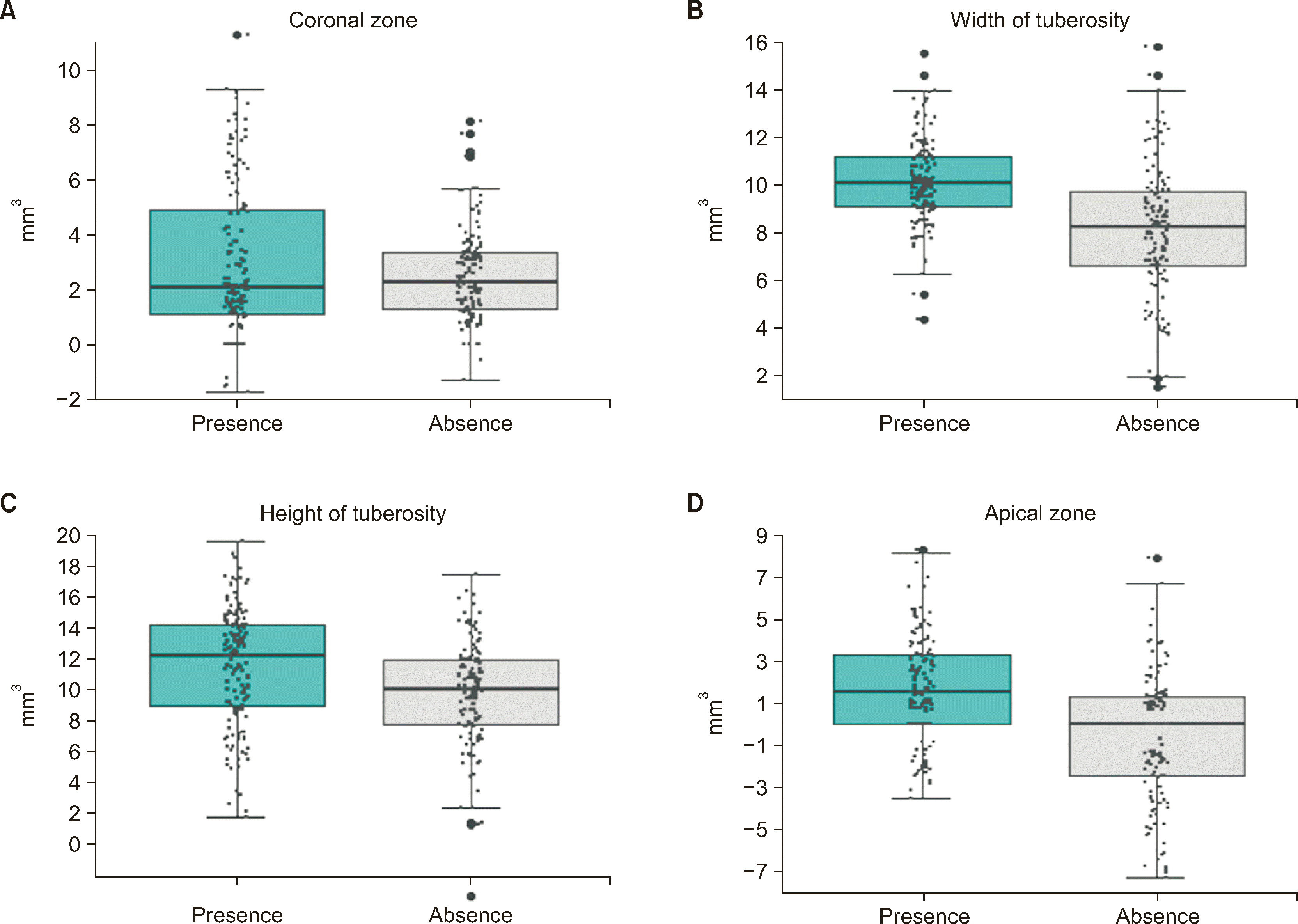
The medians of the width and height of the tuberosity decreased significantly in the absence of the third molar (P < 0.001). The apical area also showed differences, with negative values in the absence of the third molar and positive values in the presence of the third molar (P < 0.001). However, no differences were observed for the coronal area (P > 0.05).
Conclusions
In the absence of the third molar, the size of the MT, represented by its width and height, was smaller and negative values (decrease) were observed for the maxillary sinus. The sagittal CT provides useful information regarding the amount of bone tissue available for distalization and relationship of the second molar with respect to the maxillary sinus, which allows individualizing each case in relation to the amount and type of movement expected.
The maxillary tuberosity (MT) is a bilateral anatomical structure that corresponds to the distal and inferior border of the infratemporal surface of the upper maxilla, where normally the alveoli of the third molars are located, with its posterior and superior limits being the pterygomaxillary fissure and the floor of the maxillary sinus, respectively.1,2 This anatomical configuration facilitates an adequate biomechanical approach for molar distalization because it allows the orthodontic en masse retraction of the upper dentition. It can be applied bilaterally or unilaterally (Class II malocclusion with subdivision) with good results when the appropriate protocols are followed.3 Likewise, it can be used to correct space discrepancies without extractions or when the use of other appliances, such as a pendulum or a headgear, is not desired.4 These distalization movements involve moving the molars towards the MT; therefore, the amount of distalization depends on the available bone volume in the direction where the roots are being displaced. Its success depends on the topography of the MT.5
Current treatment proposals, such as the use of temporary anchorage devices (TADs), including interradicular or infrazygomatic TADs using skeletal anchorage or clear aligners using reinforced dental anchorage promote distalization mechanics in the treatment of Class II malocclusion.6-8 However, it is not only the mechanical element but also the anatomical characteristics of the bony corridor through which the displacement will occur that limits the magnitude of the displacement. Therefore, MT topography is an important factor to consider in planning molar distalization.
Three-dimensional assessment of the molar alveolar bone and MT topography through both computed axial tomography (CAT) and cone beam computed tomography (CBCT) provides an accurate representation of the anatomical configuration and its influence on distalization, assisting the clinician in both the diagnostic and treatment planning process.9
Considering the above, the objective of this study was to examine the MT zones (coronal, apical, width, and height) in relation to the presence or absence of the third molar, age, and sex, as these factors can potentially influence the magnitude and efficiency of molar distalization.
A descriptive cross-sectional study was conducted based on the available records. All CAT scans of patients between 11 and 53 years of age sent for facial morphological assessment by indication of their treating specialists during the period between January 2015 and January 2022 were selected from the Imbanaco Clinic’s imaging processing center. The study was approved by the Institutional Ethics Committee (Imbanaco Clinics approval code: CEI-782) and was conducted according to the latest version of the Declaration of Helsinki. At admission, all patients signed an informed consent for the use and disclosure of information and images for research purposes. Images were anonymized to ensure patient privacy.
Patients with a history of maxillary orthognathic surgery, with absence of the first and/or second molar or with any of these teeth impacted, with primary or mixed dentition, with a history of orthodontic treatment or periodontal disease, with craniofacial syndromes, or with inadequate quality images were excluded.
CAT images were acquired using a Biograph mCT20 PET/CT scanner (Siemens, Erlangen, Germany). The images of the skull were obtained without contrast medium, from the vertex to the sternal fork using the following parameters: slice thickness of 0.75 mm, a pitch of 1.0, and 512 × 512 cubic matrix with an isotropic voxel (size: 0.58 × 0.58 × 0.87 mm) that avoids image distortion in the different planes. This protocol was applied for both adult and growing patients. CAT images were reconstructed using a low-dose homogeneous B26F filter for anatomical localization. All patients were positioned with a head immobilizer to avoid motion artifacts and facilitate image fusion.
The CAT images were stored in DICOM format and transferred to the Horos software version 4.0.0 (Nimble Co., LLC, Annapolis, MD, USA), which is a free and open source, fully functional 64-bit medical image viewer for OS X, and were processed to evaluate the two-dimensional measurements of the MT. A maxillary occlusal plane was traced from the intercuspation of the second premolar to beyond the second molar and used as the reference plane for the MT measurements, as described in Table 1. The images were analyzed in the 3D multiplanar reconstruction module with a slice thickness of 0.4 mm. The sagittal plane was adjusted so that it passed through the vestibular roots of the first and second molars and extended to the posterior part of the tuberosity (Supplementary Figure 1).
The statistical power of this study was calculated using two different sample sizes, a group of 151 records with the presence of the third molar and another group of 126 records with the absence of the third molar. A power greater than 98% was obtained for MT width, height, and apical area (Supplementary Figure 2).
Measurements were performed by an operator skilled in the use of the software and in evaluating the maxillary anatomy. Each set of images and data per patient was reviewed and classified, jointly by an operator and a specialist in craniofacial imaging. Twenty patients were randomly selected for a second measurement four weeks apart. The reproducibility of the measurements of the width and height of the tuberosity, and the coronal and apical zones was evaluated to determine the level of intraobserver agreement in the measurements. A concordance correlation coefficient of higher than 98% was observed for all variables, indicating an almost perfect concordance (Supplementary Table 1).
Quantitative variables are summarized as median and interquartile range (P25–P75) and qualitative variables as relative and absolute frequencies. Mann–Whitney U test was used to compare the medians of the groups considering the presence or absence of the third molar. Statistical significance was set at P < 0.05. All analyses were performed with R software version 4.3.1 (The R Foundation, Vienna, Austria).
We analyzed a total of 277 tuberosities corresponding to the right and left sides of 140 patients (three were excluded), including 85 (61%) female with a median age of 18 (15–27) years and 55 male with a median age of 22 (17–28) years. The prevalence of third molars was higher in patients aged 22 years or younger for both sexes, 83.9% for females and 70.7% for males (Table 2).
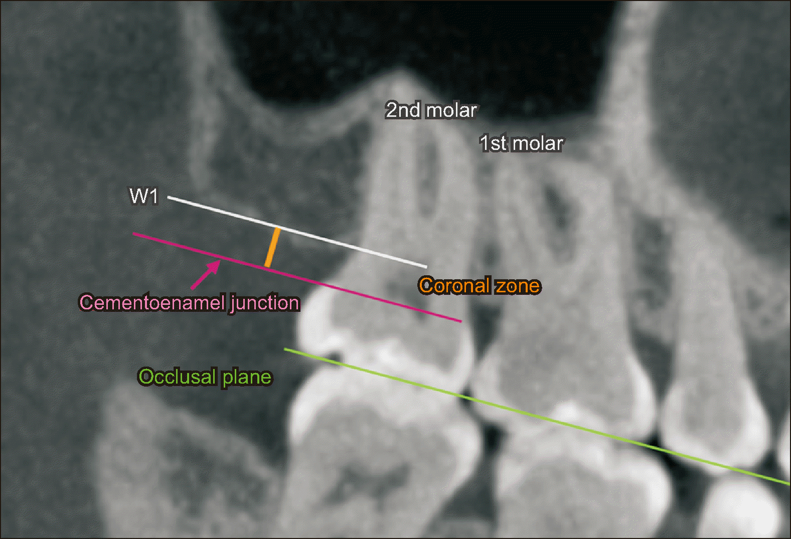
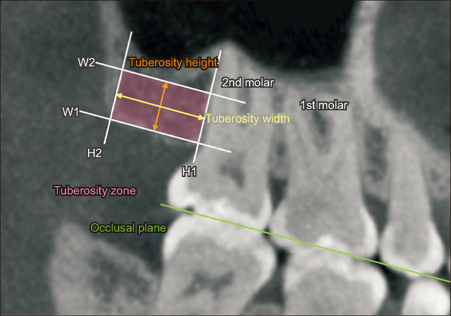
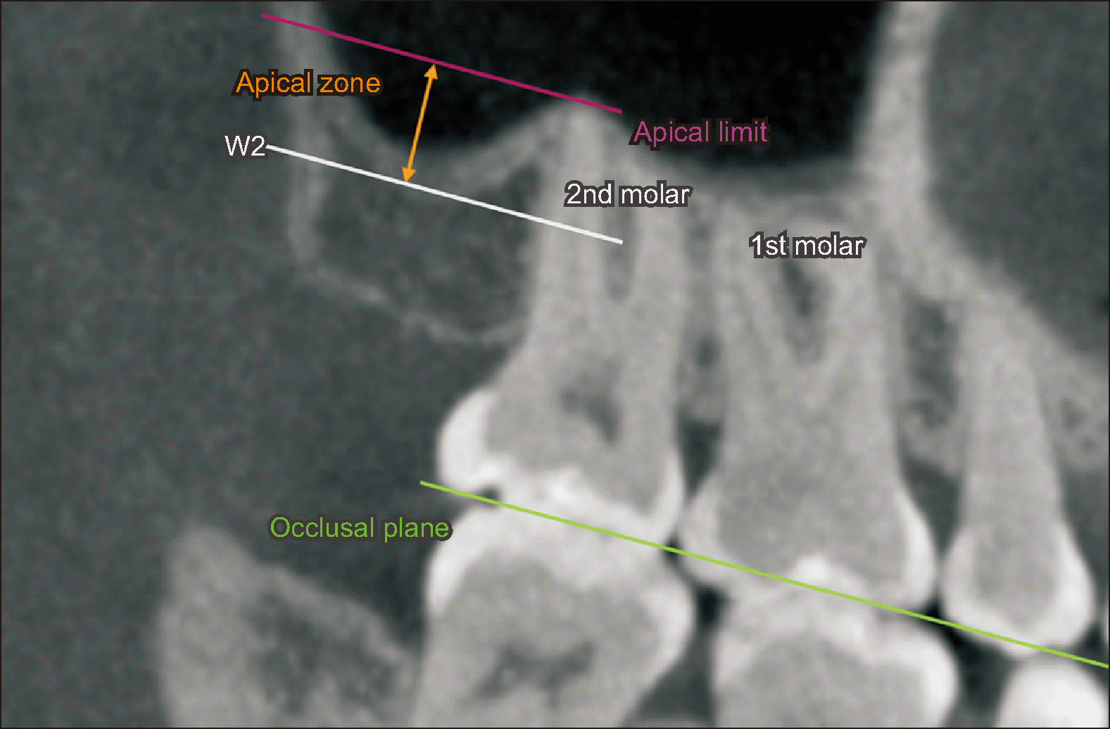
The anatomical characteristics of the MT were described according to the measurement of the coronal zone (bone-free area; Figure 1), tuberosity zone (bony area; Figure 2), and apical zone (area of influence of the maxillary sinus; Figure 3), with statistically large significant differences between the medians of both the height and width in cases of the presence of the third molar (P < 0.001). The apical area also showed significant differences with negative values in the absence of the third molar and positive values in the presence of the third molar (P < 0.001). In contrast, for the coronal area, no differences were found (P > 0.05) (Table 3 and Figure 4).
The MT anatomical characteristics according to sex showed reduced lengths in the MT height and width in the absence of the third molar in female (P < 0.001), as well as reduced values in the apical area (P < 0.001). In male, no significant differences were observed in the MT height (P = 0.116). However, significant differences were observed in the width and apical zone in the absence of the third molar (Table 3).
Concerning age and sex, female aged 22 years or younger presented significant differences (P < 0.001), with decreased measurements of the apical zone and MT width and height in the absence of the third molar. Female older than 22 years presented significant differences for the coronal zone (P = 0.019) and MT width (P < 0.001). In male, significant differences were only observed for the MT width in the age range of 22 years or younger. In the absence of the third molar the MT width measurement was lower (Table 4).
The distance between the distal root of the second molar and the internal cortex of the MT depicts the bony corridor through which the teeth can move distally.10 This area can be affected by the presence or absence of the third molar; therefore, it should be considered since it can influence the amount of bone tissue and facilitate pneumatization of the maxillary sinus, affecting the distal displacement potential of the maxillary molars. Thus, along with determining the amount of movement from the appliance used or the procedure performed, the clinician should individualize each case and be aware of the anatomical limitations of tooth movement around the MT.
Our study results suggest that the coronal, apical, and bony areas of the MT can be evaluated on the sagittal CAT scan. This will provide individual information on the patient who requires maxillary molar distalization. The imaging can demonstrate the limits to which the molars can be distalized before colliding with the bone cortex, as well as the relationship of the roots with the maxillary sinus cortex.
Using CBCT, Hui et al.11 evaluated the MT width at 3, 6, and 9 mm from the cementoenamel line and found differences with the coronal zone closest to the distal root in patients with Class II malocclusion. They also detected a smaller distance in hyperdivergent subjects, showing the individual anatomical variations in MT. They emphasized the need for future studies to evaluate the relationship of the maxillary sinus to the roots of the second molar as it may be another anatomical limitation in molar distalization.
Although the MT bone ridge is the “channel” of molar displacement during distalization, most related studies have focused on the mechanical element, i.e., the magnitude of the forces and the biomechanics applied to determine the amount of distalization. Chiu et al.12 recorded a distalization of 3 mm using tooth-supported appliances (pendulum and distal Jet). Considering contemporary approaches, such as the use of interradicular TADs, Abdelhady et al.13 reported a greater distalization with an average of 4.09 mm. For infrazygomatic implants, a maximum distalization of 4 mm has also been reported with asymmetric mechanics.14 This was supported by the meta-analyses by Bayome et al.3 and Raghis et al.,15 who reported 4 mm of displacement during bilateral mechanics. However, studies performed with esthetic aligners have reported smaller distalization amounts (2.5 mm in the study by Ravera et al.7) and only achieving 68% reliability for programmed movements of up to 2 mm.16
Spena and Turatti17 performed complementary surgical procedures, such as localized corticotomies in the molar area, to trigger osteopenia and facilitate distalization, reporting a total distalization of 4.1 mm. However, their study evaluated the technical aspect; however, the amount of bone tissue that allows movement and proximity of the maxillary sinus to the molar roots to be displaced were overlooked.
Concerning sexual dysmorphism, a smaller overall MT width was evident in female than in male. These findings were similar to those reported by Manzanera et al.18 in their study on the anatomical characterization of the MT and Gapski et al.19 in their histomorphometry study where they observed a lower percentage of bone surface area in female. Additionally, female ≤ 22 years of age and without the third molar presented all decreased measurements (MT height and width and a negative apical zone). Regarding the negative relationship between root apices and maxillary sinus in the young population, Pei et al.20 postulated that the distances between the roots of the second molar and the floor of the maxillary sinus increase with age; therefore, there is a closer relationship in the adolescent and young population as observed in the present study. The allocation by age subgroups was established considering that residual growth only ends at 22 years of age.21
Regarding the maxillary sinus, the study showed that in the absence of the third molar, there was a statistically significant (P < 0.001) descent of the sinus behind the second molar, projecting the apical portion of the distal root against the sinus cortex. In relation to this finding, it is important to note that the bony cortex limits, slows down, and makes tooth movement more complex.22-24 Furthermore, it favors the distal inclination of the crown25 and increases the risk of root resorption.26 Therefore, it should be considered when planning orthodontic distalization of the molar.
A limitation of the study is that it did not evaluate the transverse MT dimension and its relationship to the vestibulo-palatal width of the teeth.18 This is a relevant aspect in the assessment of the bony corridor. Future studies should assess the MT osseous metabolism and the density of the cortical bone, which are important variables in the amount and type of dental movement.
In the absence of the third molar, the size of the MT, represented by its width and height, was smaller. Further, the maxillary sinus showed negative values (decrease) behind the distal roots of the second molar.
Female aged 22 years or younger who lacked the third molar had significantly lower values of the width and height of the tuberosity and a negative apical zone.
Sagittal CT provides useful information to the clinician regarding the amount of alveolar bone available for distalization and the relationship of the second molar roots with respect to the maxillary sinus, allowing the clinician to individualize each case in relation to the amount and type of distal movement.
ACKNOWLEDGEMENTS
The authors appreciate the Research Institute of Imbanaco Clinic for its support during the development of this project.
Notes
AUTHOR CONTRIBUTIONS
Conceptualization: DFL, DAO. Data curation: DAO. Formal analysis: MAM. Investigation: DFL, DAO. Methodology: MAM. Project administration: DFL. Resources: MAM. Software: DAO, MAM. Supervision: DFL. Validation: DFL, MAM. Visualization: DFL. Writing–original draft: DFL, DAO. Writing–review & editing: DFL, MAM.
References
1. Cheung LK, Fung SC, Li T, Samman N. 1998; Posterior maxillary anatomy: implications for Le Fort I osteotomy. Int J Oral Maxillofac Surg. 27:346–51. https://doi.org/10.1016/s0901-5027(98)80062-3. DOI: 10.1016/S0901-5027(98)80062-3. PMID: 9804196.

2. Apinhasmit W, Chompoopong S, Methathrathip D, Sangvichien S, Karuwanarint S. 2005; Clinical anatomy of the posterior maxilla pertaining to Le Fort I osteotomy in Thais. Clin Anat. 18:323–9. https://doi.org/10.1002/ca.20131. DOI: 10.1002/ca.20131. PMID: 15971227.

3. Bayome M, Park JH, Bay C, Kook YA. 2021; Distalization of maxillary molars using temporary skeletal anchorage devices: a systematic review and meta-analysis. Orthod Craniofac Res. 24 Suppl 1:103–12. https://doi.org/10.1111/ocr.12470. DOI: 10.1111/ocr.12470. PMID: 33484608.

4. Chae JM. 2006; A new protocol of Tweed-Merrifield directional force technology with microimplant anchorage. Am J Orthod Dentofacial Orthop. 130:100–9. https://doi.org/10.1016/j.ajodo.2005.10.020. DOI: 10.1016/j.ajodo.2005.10.020. PMID: 16849080.
5. Garib DG, Yatabe MS, Ozawa TO, Silva Filho OG. 2010; Alveolar bone morphology under the perspective of the computed tomography: defining the biological limits of tooth movement. Dental Press J Orthod. 15:192–205. Português. https://pesquisa.bvsalud.org/portal/resource/pt/lil-562911. DOI: 10.1590/S2176-94512010000500023. PMID: d2fe53efb95a45d2b5a8ab6eef782798.
6. Rosa WGN, de Almeida-Pedrin RR, Oltramari PVP, de Castro Conti ACF, Poleti TMFF, Shroff B, et al. 2023; Total arch maxillary distalization using infrazygomatic crest miniscrews in the treatment of Class II malocclusion: a prospective study. Angle Orthod. 93:41–8. https://doi.org/10.2319/050122-326.1. DOI: 10.2319/050122-326.1. PMID: 36126679. PMCID: PMC9797147.

7. Ravera S, Castroflorio T, Garino F, Daher S, Cugliari G, Deregibus A. 2016; Maxillary molar distalization with aligners in adult patients: a multicenter retrospective study. Prog Orthod. 17:12. https://doi.org/10.1186/s40510-016-0126-0. DOI: 10.1186/s40510-016-0126-0. PMID: 27041551. PMCID: PMC4834290. PMID: 970deba192ee417f921a2b9ccfcdac13.

8. Marcelino V, Baptista S, Marcelino S, Paço M, Rocha D, Gonçalves MDP, et al. 2023; Occlusal changes with clear aligners and the case complexity influence: a longitudinal cohort clinical study. J Clin Med. 12:3435. https://doi.org/10.3390/jcm12103435. DOI: 10.3390/jcm12103435. PMID: 37240538. PMCID: PMC10219537. PMID: 55db70ba4aa74e718498c3f48149cd3b.
9. Karatas OH, Toy E. 2014; Three-dimensional imaging techniques: a literature review. Eur J Dent. 8:132–40. https://doi.org/10.4103/1305-7456.126269. DOI: 10.4103/1305-7456.126269. PMID: 24966761. PMCID: PMC4054026.
10. Manojna NL, Sunil G, Ramya K, Ranganayakulu I, Raghu Ram R. 2023; Three-dimensional assessment and comparison of the maxillary tuberosity between skeletal and dental Class I and Class II adults in maxillary third molar agenesis using cone beam computed tomography: a descriptive cross-sectional human study. Cureus. 15:e42232. https://doi.org/10.7759/cureus.42232. DOI: 10.7759/cureus.42232. PMID: 37605685. PMCID: PMC10440149.

11. Hui VLZ, Xie Y, Zhang K, Chen H, Han W, Tian Y, et al. 2022; Anatomical limitations and factors influencing molar distalization. Angle Orthod. 92:598–605. https://doi.org/10.2319/092921-731.1. DOI: 10.2319/092921-731.1. PMID: 35604682. PMCID: PMC9374358.
12. Chiu PP, McNamara JA Jr, Franchi L. 2005; A comparison of two intraoral molar distalization appliances: distal jet versus pendulum. Am J Orthod Dentofacial Orthop. 128:353–65. https://doi.org/10.1016/j.ajodo.2004.04.031. DOI: 10.1016/j.ajodo.2004.04.031. PMID: 16168332.

13. Abdelhady NA, Tawfik MA, Hammad SM. 2020; Maxillary molar distalization in treatment of angle class II malocclusion growing patients: uncontrolled clinical trial. Int Orthod. 18:96–104. https://doi.org/10.1016/j.ortho.2019.11.003. DOI: 10.1016/j.ortho.2019.11.003. PMID: 31974060.

14. Ishida T, Yoon HS, Ono T. 2013; Asymmetrical distalization of maxillary molars with zygomatic anchorage, improved superelastic nickel-titanium alloy wires, and open-coil springs. Am J Orthod Dentofacial Orthop. 144:583–93. https://doi.org/10.1016/j.ajodo.2012.10.028. DOI: 10.1016/j.ajodo.2012.10.028. PMID: 24075667.

15. Raghis TR, Alsulaiman TMA, Mahmoud G, Youssef M. 2022; Efficiency of maxillary total arch distalization using temporary anchorage devices (TADs) for treatment of Class II-malocclusions: a systematic review and meta-analysis. Int Orthod. 20:100666. https://doi.org/10.1016/j.ortho.2022.100666. DOI: 10.1016/j.ortho.2022.100666. PMID: 35871982.

16. Mao B, Tian Y, Xiao Y, Li J, Zhou Y. 2023; The effect of maxillary molar distalization with clear aligner: a 4D finite-element study with staging simulation. Prog Orthod. 24:16. https://doi.org/10.1186/s40510-023-00468-1. DOI: 10.1186/s40510-023-00468-1. PMID: 37183221. PMCID: PMC10183381. PMID: 21ecd418be8b4de78db3c527f063f796.
17. Spena R, Turatti G. 2011; Upper molar distalization and periodontally facilitated orthodontics. Rev Española Ortod. 41:246–54. Spanish. https://www.revistadeortodoncia.com/frame_esp.php?id=1149.
18. Manzanera E, Llorca P, Manzanera D, García-Sanz V, Sada V, Paredes-Gallardo V. 2018; Anatomical study of the maxillary tuberosity using cone beam computed tomography. Oral Radiol. 34:56–65. https://doi.org/10.1007/s11282-017-0284-x. DOI: 10.1007/s11282-017-0284-x. PMID: 30484092.

19. Gapski R, Satheesh K, Cobb CM. 2006; Histomorphometric analysis of bone density in the maxillary tuberosity of cadavers: a pilot study. J Periodontol. 77:1085–90. https://doi.org/10.1902/jop.2006.050118. DOI: 10.1902/jop.2006.050118. PMID: 16734586.

20. Pei J, Liu J, Chen Y, Liu Y, Liao X, Pan J. 2020; Relationship between maxillary posterior molar roots and the maxillary sinus floor: cone-beam computed tomography analysis of a western Chinese population. J Int Med Res. 48:300060520926896. https://doi.org/10.1177/0300060520926896. DOI: 10.1177/0300060520926896. PMID: 32489120. PMCID: PMC7278324. PMID: 8d1d327d98c84a958ab6e6d636b077e0.

21. Aarts BE, Convens J, Bronkhorst EM, Kuijpers-Jagtman AM, Fudalej PS. 2015; Cessation of facial growth in subjects with short, average, and long facial types- implications for the timing of implant placement. J Craniomaxillofac Surg. 43:2106–11. https://doi.org/10.1016/j.jcms.2015.10.013. DOI: 10.1016/j.jcms.2015.10.013. PMID: 26548528.

22. Zachrisson BU. 2005; Bjorn U. Zachrisson, DDS, MSD, PhD, on current trends in adult treatment, part 2. Interview by Robert G. Keim. J Clin Orthod. 39:285–96. quiz 315https://pubmed.ncbi.nlm.nih.gov/15961889/.
23. Park JH, Tai K, Kanao A, Takagi M. 2014; Space closure in the maxillary posterior area through the maxillary sinus. Am J Orthod Dentofacial Orthop. 145:95–102. https://doi.org/10.1016/j.ajodo.2012.07.020. DOI: 10.1016/j.ajodo.2012.07.020. PMID: 24373659.
24. Oh H, Herchold K, Hannon S, Heetland K, Ashraf G, Nguyen V, et al. 2014; Orthodontic tooth movement through the maxillary sinus in an adult with multiple missing teeth. Am J Orthod Dentofacial Orthop. 146:493–505. https://doi.org/10.1016/j.ajodo.2014.03.025. DOI: 10.1016/j.ajodo.2014.03.025. PMID: 25263152.
25. Kim S, Lee NK, Park JH, Ku JH, Kim Y, Kook YA, et al. 2022; Treatment effects after maxillary total arch distalization using a modified C-palatal plate in patients with Class II malocclusion with sinus pneumatization. Am J Orthod Dentofacial Orthop. 162:469–76. https://doi.org/10.1016/j.ajodo.2021.04.033. DOI: 10.1016/j.ajodo.2021.04.033. PMID: 35773112.

26. Sun W, Xia K, Huang X, Cen X, Liu Q, Liu J. 2018; Knowledge of orthodontic tooth movement through the maxillary sinus: a systematic review. BMC Oral Health. 18:91. https://doi.org/10.1186/s12903-018-0551-1. DOI: 10.1186/s12903-018-0551-1. PMID: 29792184. PMCID: PMC5966888. PMID: 932a8c9a278843fea3e84b62bbc486e0.

Figure 1
Coronal zone. Reference lines to measure the length of coronal zone.
W1, top of the tuberosity.

Figure 2
Tuberosity zone. Reference lines to measure the width and height of the tuberosity.
W1, top of the tuberosity; W2, floor of the tuberosity; H1, tangent to the distal root of the second molar; H2, tangent to the inner cortex of the tuberosity.

Figure 3
Apical zone. Reference lines to measure the length of the apical zone. If the floor of the tuberosity is below the apical end of the second molar root, the measurement is negative. If it is above, the measurement is positive.
W2, floor of the tuberosity.

Figure 4
Coronal (A), tuberosity (B, C), and apical (D) measurements in the presence or absence of the third molar.

Table 1
Description of the measurements of the analyzed variables
| Variable | Description |
|---|---|
| Coronal zone | The coronal zone is defined as the bone-free zone and is delimited by drawing a line parallel to the occlusal plane from the amelocemental junction and another from the top of the tuberosity (W1). The distance between the two lines is measured in mm and is the length of the coronal zone (Figure 1) |
| Tuberosity zone |
The tuberosity zone is defined as the bone zone and is delimited horizontally by drawing a parallel to the occlusal plane from the top of the tuberosity (W1) and another parallel from the floor of the tuberosity (W2). Vertically, it is delimited by tracing a tangent to the distal root of the second molar (H1) and a tangent to the inner cortex of the tuberosity (H2). Both extending from the apical zone to the coronal zone The width of the tuberosity is measured (mm) with a horizontal line, parallel and equidistant to the lines (W1 to W2), extending from line H1 to line H2 The height of the tuberosity is measured (mm) with a vertical line, parallel and equidistant to the lines (H1 and H2), extending from line W1 to line W2 (Figure 2) |
| Apical zone |
The apical zone is defined as the zone of influence of the maxillary sinus and is delimited by drawing a line parallel to the occlusal plane from the floor of the tuberosity (W2) and another parallel from the uppermost point of the root apex of the distal root of the second molar The distance between the two lines (mm) is the length of the apical zone. When the floor of the tuberosity is below the root apex of the second molar, the result will be negative (–) and when it is above the distal root, the result will be positive (+) (Figure 3) |
Table 2
Comparison of groups based on age and sex considering the presence or absence of the third molar
Table 3
Description of the anatomical characteristics of the maxillary tuberosity according to the presence or absence of the third molar
| General | Presence of 3rd molar | Absence of 3rd molar | P value | Female (n = 167) | Male (n = 110) | ||||||
|---|---|---|---|---|---|---|---|---|---|---|---|
|
Presence of 3rd molar (n = 93)* |
Absence of 3rd molar (n = 74)* |
P value |
Presence of 3rd molar (n = 58)* |
Absence of 3rd molar (n = 52)* |
P value | ||||||
| Coronal zone | 2.17 (1.24:3.82) | 2.06 (1.14:4.95) | 2.27 (1.32:3.37) | 0.667 | 1.89 (1.06:5.43) | 1.78 (1.10:3.06) | 0.306 | 2.36 (1.34:4.81) | 3.12 (2.09:3.73) | 0.647 | |
|
Width of tuberosity |
9.51 (8.01:9.27) | 10.14 (9.16:11.29) | 8.31 (6.61:9.73) | < 0.001 | 9.91 (9.04:10.51) | 7.79 (6.16:9.14) | < 0.001 | 10.96 (9.67:12.34) | 8.54 (7.21:10.46) | < 0.001 | |
|
Height of tuberosity |
10.89 (8.08:13.26) | 12.10 (8.74:14.10) | 9.89 (7.59:11.84) | < 0.001 | 12.11 (8.47:14.06) | 9.45 (7.47:11.84) | < 0.001 | 11.47 (9.23:14.12) | 10.64 (7.90:11.96) | 0.116 | |
| Apical zone | 0.97 (–1.35:2.57) | 1.53 (0:3.29) | 0 (–2.46:1.36) | < 0.001 | 1.72 (0.72:3.32) | –1.35 (–3.12:1.11) | < 0.001 | 1.03 (0:3.19) | 0.79 (–1.56:1.51) | 0.039 | |
Table 4
Anatomical characteristics of the maxillary tuberosity based on age and sex according to the presence or absence of the third molar
| Female (n = 167) | Male (n = 110) | ||||||||||||||
|---|---|---|---|---|---|---|---|---|---|---|---|---|---|---|---|
| ≤ 22 yr (n = 108) | > 22 yr (n = 59) | ≤ 22 yr (n = 60) | > 22 yr (n = 50) | ||||||||||||
|
Presence (n = 78)* |
Absence (n = 30)* |
P value |
Presence (n = 15)* |
Absence (n = 44)* |
P value |
Presence (n = 41)* |
Absence (n = 19)* |
P value |
Presence (n = 17)* |
Absence (n = 33)* |
P value | ||||
|
Coronal zone |
1.74 (0.98:5.48) |
2.29 (1.67:3.20) |
0.345 |
2.63 (1.75:4.08) |
1.46 (0.87:2.52) |
0.019 |
2.84 (1.22:4.81) |
3.11 (1.99:3.84) |
0.679 |
2.22 (1.77:4.23) |
3.14 (2.11:3.68) |
0.689 | |||
|
Width of tuberosity |
9.80 (8.88:10.36) |
7.16 (5.84:8.76) |
< 0.001 |
10.79 (9.49:11.49) |
8.31 (6.76:9.32) |
< 0.001 |
10.80 (9.64:12.19) |
7.55 (5.46:8.77) |
< 0.001 |
11.24 (9.75:12.39) |
9.68 (8.00:11.98) |
0.063 | |||
|
Height of tuberosity |
12.33 (8.68:14.26) |
9.22 (6.87:11.66) |
< 0.001 |
11.19 (7.68:12.45) |
9.66 (7.58:12.05) |
0.536 |
11.51 (9.63:14.08) |
9.85 (7.09:12.15) |
0.060 |
9.72 (7.79:14.14) |
10.80 (9.30:11.83) |
0.910 | |||
|
Apical zone |
2.53 (0.99:3.88) |
0.83 (–2.13:1.30) |
< 0.001 |
0 (–1.32:0.93) |
–1.57 (–3.63:0.26) |
0.083 |
0.92 (0:2.67) |
0.91 (–1.19:1.84) |
0.272 |
2.01 (–2.19:3.39) |
0 (–1.50:1.47) |
0.210 | |||


 PDF
PDF Citation
 Print
Print


 XML Download