Abstract
Background
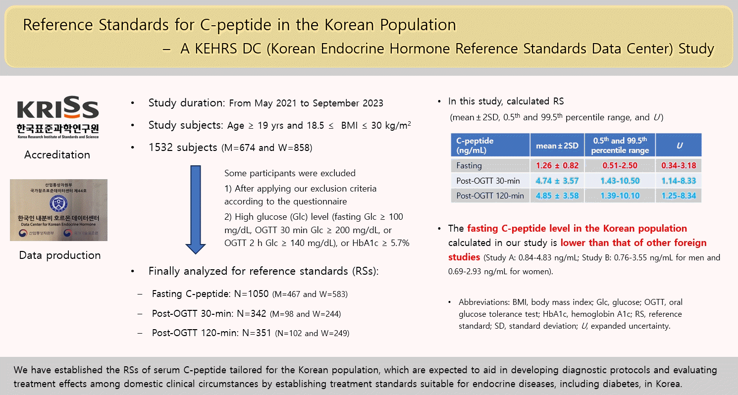
The Korean Endocrine Hormone Reference Standard Data Center (KEHRS DC) has created reference standards (RSs) for endocrine hormones since 2020. This study is the first of its kind, wherein the KEHRS DC established RSs for serum C-peptide levels in a healthy Korean population.
Methods
Healthy Korean adults were recruited from May 2021 to September 2023. After excluding participants according to our criteria, serum samples were collected; each participant could then choose between fasting glucose only or fasting glucose plus an oral glucose tolerance test (OGTT). If their sample showed high glucose (≥100 mg/dL) or hemoglobin A1c (HbA1c) (≥5.70%), their C-peptide levels were excluded from analyzing the RSs.
Results
A total of 1,532 participants were recruited; however, only the data of 1,050 participants were analyzed after excluding those whose samples showed hyperglycemia or high HbA1c. Post-30-minute OGTT data from 342 subjects and post-120-minute OGTT data from 351 subjects were used. The means±2 standard deviations and expanded uncertainties of fasting, post-30-minute and 120-minute OGTT C-peptide levels were 1.26±0.82 and 0.34–3.18, 4.74±3.57 and 1.14–8.33, and 4.85±3.58 and 1.25–8.34 ng/mL, respectively. Serum C-peptide levels correlated with obesity, serum glucose levels, and HbA1c levels.
Reference intervals (RIs) are benchmark points through which clinicians can distinguish healthy individuals from those with disease; an accurate setting of RIs is essential for rapid diagnosis and the establishment of a proper treatment plan [1]. However, RIs that have been established satisfactorily for most hormone items remain scarce [2]. Most are cited and derived from Western papers or literature [3,4] and do not reflect the real situation of the Korean population. There are many restrictions on their direct application to Korean patients’ diagnosis and treatment process. In addition, hormones are highly likely to fluctuate according to the situation, such as diurnal, seasonal, and postural variations [5-7]. Furthermore, the hormone levels varied according to age and gender [1,8]. Therefore, it is necessary to derive reference values from various groups in consideration of the above, which is essential for basic and clinical research as well as the development of the domestic medical industry in Korea.
In 2020, the Korean Endocrine Hormone Reference Standard Data Center (KEHRS DC) was designated by the Korea Institute of Standards and Science. And, we have created reference standards (RSs) for some endocrine hormones through various activities over the past several years, as supported by the Korean Endocrine Society and 10 university hospitals in Korea. The data created from our center will be uploaded to the National Center for Standard Reference Data of Korea.
Type 2 diabetes mellitus (T2DM) is a disease caused by various etiologies; it is thought that there will be differences in the phenotype of diabetes depending on the major etiologic process; there will be differences in the response to therapeutic drugs and the risk of complications [9]. However, T2DM is diagnosed simply by confirming hyperglycemia and excluding other types of diabetes; no subtype classification or appropriate treatment methods have been established [10,11]. Insulin treatment may be required from the beginning depending on the degree of insulin secretion defects, even in T2DM; this is due to phenotypic differences according to race, social environment, and complex etiologies [9]. However, only Western standards are applied to the prevention, management, and treatment of diabetes, and treatment is determined based on blood glucose levels. For the proper management of diabetes, prevention and treatment guidelines tailored to the characteristics of diabetes in Koreans are needed; for this, it is necessary to evaluate insulin secretion defects. For these reasons, the KEHRS DC developed Korean RSs for serum C-peptide as the first project for the Korean population. Serum C-peptide is a major marker for insulin secretion in patients with T2DM and is particularly useful in distinguishing between insulinoma from exogenous hyperinsulinemia [12].
This study is the first result of our activities in the data center, which can be used for diabetes-related research by preparing diagnostic criteria for prediabetes and prediabetes using C-peptide hormone reference data tailored to Korean characteristics; our results can be expected to develop new research fields through a review of the literature. The establishment of normal RSs for endocrine hormones suitable for Koreans can increase the accuracy of diagnostic results as well as reduce medical and social expenses, such as additional examination costs incurred by applying existing foreign diagnostic standards. In this study, we established RSs for C-peptide in the Korean population. Our results may aid in developing diagnostic protocols and in evaluating treatment effects among domestic clinical circumstances, by establishing treatment standards suitable for the Korean characteristics of endocrine-related diseases such as T2DM.
This study was conducted using the KEHRS DC from May 2021 to September 2023. Study subjects aged 19 and over as well as with a body mass index (BMI) ≥18.5 and ≤30 kg/m2 were recruited from four hospitals in Korea; Wonju Severance Christian Hospital (Wonju), Keimyung University Dongsan Hospital (Daegu), Hallym University Chuncheon Sacred Heart Hospital (Chuncheon), and Nowon Eulji Medical Center (Seoul). Only healthy, non-pregnant Koreans without diagnosed diseases who met the exclusion criteria were included in this study (Table 1). However, even if patients had a history of diseases C to G based on the exclusion criteria, those who were cured for more than a year were included in this study. Information about medical and family histories was obtained from direct interviews with the study subjects. Each participant could choose between fasting glucose alone or fasting glucose plus oral glucose tolerance test (OGTT). This study was approved by the Institutional Review Board of Wonju Severance Christian Hospital (IRB No. CR321010). All the participants voluntarily participated in the study and provided written informed consent.
Blood samples were collected after an overnight fast. To minimize the pre-analytical factors possibly interfering with the hormone levels, each participant was asked to do the following: (1) fasting for at least 10 hours, but with allowed anti-hypertensive drug intake; (2) refraining from sweaty exercises other than daily activities (e.g., gym, jogging, weight training, zumba dance, running, etc.) for at least 1 day; (3) limiting blood collection time between 8:00 and 12:00 AM to minimize circadian variations; (4) smoking cessation for at least 1 day, applicable to cigarettes and electronic cigarettes, chewing cigarette, and smoking cessation plants; and (5) collecting blood in a sitting position on a chair and at least 30 minutes after waking up. Blood samples were collected in serum separation tubes (SSTs) and BD Vacutainers SST II Advance Plus blood collection tubes (Becton-Dickinson Biosciences, Franklin Lakes, NJ, USA). For OGTT testing, Diasol-S solutions (Taejoon Pharm., Seoul, Korea) were used; samples were collected 30 minutes and 2 hours after administration.
Three tubes (two SSTs and one K2-ethylenediaminetetraacetic acid [EDTA] tube) were used for each collection. One EDTA and one SST tubes were sent to Seoul Clinical Laboratories and analyzed for hemoglobin A1c (HbA1c) levels and routine chemistry tests, including serum glucose levels. Even if patients were included in this study because they did not have diseases according to the exclusion criteria, their data were excluded from the calculation of RSs in the following cases: (1) serum glucose concentrations were high of if any of the following were met (fasting glucose ≥100 mg/dL, OGTT 30 minutes glucose ≥200 mg/dL, OGTT 2 hours glucose ≥140 mg); or (2) the baseline (fasting) HbA1c is ≥5.7%. HbA1c and serum glucose levels were measured on the day of collection and analyzed using Cobas c513 for HbA1c and Cobas c702 (Roche Diagnostics, Basel, Switzerland) analyzers, respectively.
Within an hour of sample collection, the remaining SST samples were centrifuged at 1,200 g (3,000 rpm) for 10 minutes. After centrifugation, serum was aliquoted into some 1 mL microtubes and stored in a deep freezer at –70℃. Each aliquot was thawed at room temperature and subsequently analyzed. Serum C-peptide levels were measured by a chemiluminescent assay using the Atellica IM C-peptide assay on the Atellica IM platform (Siemens Healthcare Diagnostics, Tarrytown, NY, USA) in Wonju Severance Christian Hospital. The analytical measuring interval of this assay ranged from 0.05 to 30 ng/mL; the reference range was initially from 0.81 to 3.85 ng/mL.
The study participants were divided based on sex into groups at 10-year intervals: 20s (19–29), 30s (30–39), 40s (40–49), 50s (50–59), 60s (60–69), and over 70 (70 and above). In addition, statistics were analyzed by grouping adjacent age groups, the total number of males and females, and the total number of subjects. The uncertainty was evaluated according to the GUM (International Organization for Standardization Guide to the Expression of Uncertainty in Measurement) [13]. Type A uncertainty was estimated based on the statistical analysis of the results and was estimated as the standard deviation (SD) in each subgroup. Type B uncertainty was estimated by means other than statistical analysis; the uncertainties of the internal quality control (IQC) material and calibrator (cal) of this study were used (). The combined uncertainty (uc) was calculated by the formula of ; the expanded uncertainty (U) was obtained by multiplying the combined uncertainty with a coverage factor k (approximately k=2 at the 95% level of confidence).
For each subgroup, extreme outliers exceeding three times the interquartile range (IQR) in Q1 and Q3 were excluded. Values lesser than Q1–3×IQR or greater than Q3+3×IQR were also excluded. The Shapiro-Wilk test was then used to confirm the normality of C-peptide values according to subgroups; the distribution of data was determined as parametric when the P value was >0.05. The results were presented as mean±2SD, U, 2.5th and 97.5th, and 0.5th and 99.5th percentile values, and minimum and maximum values. U was obtained as the formula of mean±k ·uc. The results between the two groups were compared using Student’s t test for parametric data and the Mann-Whitney U test for non-parametric data. The results of more than two groups were compared using the analysis of variance (ANOVA) test for parametric data and the Kruskal-Wallis test for non-parametric data. All statistical analyses were performed using the Analyse-it version 6.15 (Analyse-it Software Ltd., Leeds, UK) add-in in Microsoft Excel 2019 (Microsoft Corp., Redmond, WA, USA).
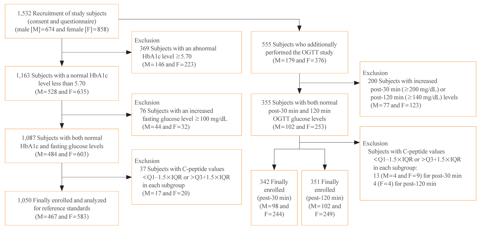
A total of 1,532 healthy, non-pregnant Korean subjects were recruited after excluding participants with diseases according to the criteria: 1,282 subjects from Wonju Severance Christian Hospital, 239 subjects from Keimyung University Dongsan Hospital, six subjects from Hallym University Chuncheon Sacred Heart Hospital, and five subjects from Nowon Eulji Medical Center. Blood samples were collected after enrollment in this study. However, only 1,087 subjects were included in the analysis after excluding patients according to their serum glucose levels and HbA1c. Furthermore, 37 subjects were excluded after applying the Tukey’s test for outlier evaluation per age subgroup. Finally, C-peptide data from 1,050 subjects were used to calculate the RSs. In the same way, 555 participants initially performed the OGTT test; however, only 355 participants remained after excluding according to serum glucose levels and HbA1c. After excluding outliers, the post-30 minutes data of 342 subjects and post-120 minutes data of 351 subjects were used. The enrollment process for the study participants is illustrated in Fig. 1.
The baseline characteristics of the 1,050 study subjects are provided in Table 2. In the fasting C-peptide analysis, 467 (44.5%) patients were male and 583 (55.5%) were female. For the post-OGTT C-peptide analysis of 355 study subjects, 102 (28.7%) were male and 253 (71.3%) were female. BMI, fasting glucose, and HbA1c were significantly higher in males than in females.
All data showed a parametric distribution per each subgroup. For fasting C-peptide levels, RSs (mean±2SD, U, 2.5th–97.5th and 0.5th–99.5th percentile values) for overall participants were 1.26±0.82, 0.34–3.18, 0.63–2.17, and 0.51–2.50 ng/mL, respectively. Male had higher fasting C-peptide levels than Female (P<0.001). The RSs for fasting C-peptide levels according to sex and 10-year intervals are summarized in Table 3(A). There were no statistical significances in fasting C-peptide levels between 20s and 30s, 40s and 50s, and 60s and over 70 (P=0.144, P=0.568, and P=0.673, respectively). However, the fasting C-peptide levels between 30s and 40s, and 50s and 60s were statistically significant (P=0.001 and P=0.0316, respectively). For these reasons, the data were readjusted at 20-year intervals by grouping adjacent age groups: 20–30s (19–39), 40–50s (40–59), and over 60s (60 and above). The results are summarized in Table 3(B). For post-OGTT C-peptide levels, the post 30-minute levels and post 120-minute levels are summarized in Table 4. For post-30 minutes OGTT C-peptide levels, RSs were 4.74±3.57, 1.44–8.33, 2.12–9.35, and 1.43–10.50 ng/mL. For post-120 minutes OGTT C-peptide levels, RSs were 4.85±3.58, 1.25–8.34, 2.14–9.09, and 1.39–10.10 ng/mL. For post-OGTT C-peptide levels, there were no statistically significant differences between male and female or among age groups.
The impact of obesity, serum glucose level, and HbA1c were determined by analyzing 1,497 subjects for fasting C-peptide (1,050 subjects analyzed for calculating RS plus 439 subjects with impaired fasting glucose (IFG) (100 to 126 mg/dL) or prediabetic (HbA1c level between 5.70 and 6.49) and 526 for postOGTT C-peptide levels. In the comparison according to obesity, subjects were divided into normal (BMI <23 kg/m2), overweight (23≤ BMI <25 kg/m2), and obesity (BMI ≥25 kg/m2). For the comparison according to fasting glucose levels, subjects were divided into normal fasting glucose (<100 mg/dL) and IFG groups. For the comparison of HbA1c levels, subjects were divided into normal HbA1c and prediabetic group. The results are summarized in Table 5. The effects of obesity, serum glucose level, and HbA1c were statistically significant in all cases in terms of both fasting and post-OGTT 120 minutes C-peptide. Additionally, the correlations and scatter plots between fasting or post-OGTT 120 minutes C-peptide and these factors are illustrated in Fig. 2. The delta values post-30 minutes (=C-peptide 30 min, C-peptide 0 min) and 120 minutes (=C-peptide 120 min– C-peptide 0 min) OGTT were calculated. The C-peptidogenic indices (=[C-peptide 30 min–C-peptide 0 min]/[glucose 30 min–glucose 0 min]) [14] were also calculated. However, this difference was not statistically significant (Supplemental Table S1).
C-peptide is a protein that connects the A and B chains of insulin from the insulin precursor, proinsulin, and consists of 31 amino acids. Because proinsulin is separated into insulin and C-peptide in pancreatic beta cell granules, C-peptide and insulin are always secreted in the same molar amounts [15]. Approximately 50% of insulin is removed from the liver after being secreted from the pancreas. Even in the physiological environment, there are many changes in peripheral removal rates; if antibodies to insulin are present, they cannot be accurately evaluated during insulin treatment [16]. In contrast, C-peptide has little first-pass effect on the liver [17]. Its average removal rate is constant; its half-life is longer than that of insulin [15,18]. Thus, C-peptide can act as the main indicator for the storage function of pancreatic beta cells [19]. Previously, C-peptide was thought to only promote the folding of proinsulin in beta cells as a feedback mechanism; however, many studies have shown that C-peptide is also a biologically active protein; its level is related to disease progression and complications in patients with diabetes [20].
The RI of C-peptide is known to be 0.5 to 2.0 ng/mL (or 0.17 to 0.67 nmol/L) [19]. For more than 20 years, many studies have been conducted to evaluate the insulin secretion ability of patients with diabetes using C-peptide; this was used to classify the type of disease or to predict the treatment effect and prognosis. Although there are a number of research results, there are no standard figures yet applicable to treatment guidelines, of which most are Western studies. Further studies on normal values are also needed. Considering the characteristics of diabetes in Koreans, it is necessary to accurately evaluate their insulin secretion ability and establish normal C-peptide levels. It has been shown that a decrease in insulin secretion ability is a more important cause of diabetes in Korea than a decrease in insulin sensitivity. These characteristics suggest that insulin secretion ability can act as an important variable in serum glucose control as compared with that in Westerners [21]. In previous studies, attempts have been made to evaluate the clinical performance of C-peptide for T2DM diagnosis and to establish a diagnostic cut-off. A previous report has shown that a cut-off of 0.28 nmol/L (0.84 ng/mL) [22] or 0.42 nmol/L (1.26 ng/mL) [23] for fasting C-peptide has excellent analytical performance for predicting T2DM diagnosis. In T2DM diagnosis, a previous report also suggested a cut-off of 0.3 nmol/L (0.9 ng/mL) if the patient has autoantibodies and 1.0 nmol/L (3 ng/mL) if the patient has none [24].
In this study, based on healthy, non-pregnant Korean subjects, calculated RSs (mean±SD, U, 2.5th–97.5th, and 0.5th–99.5th percentile values) for fasting serum C-peptide were 1.26±0.82, 0.34–3.18, 0.63–2.17, and 0.51–2.50 ng/mL, respectively. However, a direct comparison might not be applicable. This was not a study that calculated the cut-off for diabetes by enrolling diabetic patients and healthy controls. Thus, it is highly likely to be abnormal (IFG or diabetes) at concentrations above 2.50 ng/mL when considering the 99th percentile value. As compared with the RIs calculated in other previous studies, a Danish study conducted in 623 subjects calculated a RI (2.5th–97.5th percentile) of fasting serum C-peptide at 279 to 1,631 pmol/L (0.84 to 4.83 ng/mL) [25]. In another foreign study, RIs were suggested at 252 to 1,176 pmol/L (0.76 to 3.55 ng/mL) for male and 227 to 971 pmol/L (0.69 to 2.93 ng/mL) for female [26]. In a study conducted in 279 Koreans, RI was 0.17 to 0.85 nmol/L (0.51 to 2.55 ng/mL) [27]. The RSs calculated in this study were lower than those from Western studies, but similar to the results of a previous Korean study. This emphasizes the importance of using the C-peptide RSs in the Korean population, which is lower than that of the Western population.
In this study, not only were the RSs of fasting C-peptide based on all Koreans obtained, but these were also calculated separately by dividing the subjects by age and sex. Unlike other hormones, the change in C-peptide levels according to age is not known to be prominent; however, in this study, the differences in fasting C-peptide levels according to age group were significant between some age groups. The differences between the 20s and 30s, 40s and 50s, and 60s and 70s groups were not significant; however, the differences between the 30s and 40s, and 50s and 60s groups were significant. The correlation coefficient between age and C-peptide levels was 0.160 in a previous study [26], which does not clearly explain the results of our study; thus, further studies are required. The difference in C-peptide levels between male and female is that male have a higher BMI and fasting blood sugar than female; however, other foreign studies have shown that male have higher C-peptide levels than female, even in the same BMI group [26]. However, in the present study, no significant differences were observed between male and female in the same BMI group (data not shown).
In addition, the RSs for the post-OGTTs at 30 and 120 minutes were calculated. It is known that the postprandial increase in C-peptide at 2 hours is further increased by 1 to 3 nmol/L (3 to 9 ng/mL) [28]. In one domestic study, the median value of postprandial C-peptide increase at 2 hours is 2.49 ng/mL in patients within 5 years of diabetes diagnosis, 1.94 ng/mL in those after 5 to 10 years of diagnosis, and 1.44 in those more than 10 years of diagnosis [18]. However, there are no known previous studies on the RI of C-peptide after OGTT in the healthy population. In this study, the RSs did not show any significant difference in post-OGTT 30 and 120 minutes; this might be because the function of pancreatic beta cells after meals does not differ significantly in normal people, which may explain the reason for excluding cases where the serum glucose levels of post-OGTT 30 or 120 minutes are high.
On the other hand, we have investigated the impact of obesity, serum glucose level, and HbA1c on serum C-peptide levels. Obesity is known to cause an increase in C-peptide levels because it often requires high insulin levels; increased serum glucose and HbA1c levels are known to be associated with a high level of C-peptide [17]. To further determine the impact of serum glucose or HbA1c on C-peptide levels, data from participants who had high glucose levels or HbA1c and were excluded from RS calculations were also included. However, it was not compared to cases where serum glucose exceeded the cut-off for IFG (serum glucose ≥126 mg/dL) or diabetes mellitus (HbA1c ≥6.5%), which is small in their number. However, if beta cells exceed a certain limit, their secretion ability was known to be reduced [29]. In this study, obesity and serum glucose levels correlated well with C-peptide levels in both fasting C-peptide and post-OGTT 120 minutes, but HbA1c levels did not correlate well with C-peptide levels.
In addition, we analyzed the degree of C-peptide change and C-peptidogenic index after OGTT. A previous study showed that the C-peptidogenic index associated with fasting C-peptide to fasting glucose level was useful for judging the progression of diabetes [14]; however, this study did not show any significant difference because it was a study based on healthy people.
In this study, we present various values and intervals for presenting the RSs. In general, when calculating the RI, the mean plus two SDs are presented if the distribution of the data shows a parametric distribution; the 2.5th–97.5th percentile range is presented if the distribution of the data shows a non-parametric distribution [30]. However, these RI calculations reflect only the middle 95% of healthy people, and values outside this interval are usually considered abnormal. However, because the level of analyte can vary greatly depending on age group or gender, a corresponding reference value may be needed [8]. However, if a value deviates from this interval, it is often not considered normal, even if it is obtained from a clinically normal subject. Therefore, we provided an additional 0.5th–99.5th percentile range because the RI could not be uniformized. In this study, we attempted to minimize the uncertainty by applying strict criteria; an average plus expanded uncertainty was obtained by effectively providing various ranges. Therefore, we presented the RSs as such: mean±2SD, U, 2.5th–97.5th percentile range, 0.5th–99.5th percentile range, and minimum and maximum values, thereby allowing clinicians to utilize them according to various clinical situations.
In this study, we tried to allocate and recruit an almost equal number of participants because the minimum number of samples required to ensure a 95% confidence level is 120 according to the guideline [30]. The purpose of this study was to give statistical meaning by presenting the RSs, and therefore the numbers of each group may not fit the actual age-specific demographic structure in Korea, and this is a limitation of this study.
In conclusion, we have established the RSs of serum C-peptide, which are expected to aid in developing diagnostic protocols and evaluating treatment effects among domestic clinical circumstances by establishing treatment standards suitable for endocrine-related diseases, including diabetes, in Korea.
ACKNOWLEDGMENTS
This study was funded by the Korea Research Institute of Standards and Science (grant no. 20016225).
REFERENCES
1. Kelly J, Raizman JE, Bevilacqua V, Chan MK, Chen Y, Quinn F, et al. Complex reference value distributions and partitioned reference intervals across the pediatric age range for 14 specialized biochemical markers in the CALIPER cohort of healthy community children and adolescents. Clin Chim Acta. 2015; 450:196–202.
2. Farrell CL, Nguyen L, Carter AC. Parathyroid hormone: data mining for age-related reference intervals in adults. Clin Endocrinol (Oxf). 2018; 88:311–7.
3. Chung JH. Update on thyroid hormone levels and thyroid dysfunction in the Korean population based on data from the Korea National Health and Nutrition Examination Survey VI (2013 to 2015). Endocrinol Metab (Seoul). 2020; 35:7–13.
4. Choi R, Lee SG, Lee EH. Reference intervals of anti-Müllerian hormone in Korean women. J Clin Lab Anal. 2022; 36:e24525.
5. Brambilla DJ, Matsumoto AM, Araujo AB, McKinlay JB. The effect of diurnal variation on clinical measurement of serum testosterone and other sex hormone levels in men. J Clin Endocrinol Metab. 2009; 94:907–13.
6. Kuzmenko NV, Tsyrlin VA, Pliss MG, Galagudza MM. Seasonal variations in levels of human thyroid-stimulating hormone and thyroid hormones: a meta-analysis. Chronobiol Int. 2021; 38:301–17.
7. Kaaja RJ, Leinonen A, Moore P, Yandle T, Frampton CM, Nicholls MG. Effect of changes in body posture on vasoactive hormones in pre-eclamptic women. J Hum Hypertens. 2004; 18:789–94.
8. Higgins V, Chan MK, Nieuwesteeg M, Hoffman BR, Bromberg IL, Gornall D, et al. Transference of CALIPER pediatric reference intervals to biochemical assays on the Roche cobas 6000 and the Roche Modular P. Clin Biochem. 2016; 49:139–49.
9. Ohn JH, Kwak SH, Cho YM, Lim S, Jang HC, Park KS, et al. 10-Year trajectory of β-cell function and insulin sensitivity in the development of type 2 diabetes: a community-based prospective cohort study. Lancet Diabetes Endocrinol. 2016; 4:27–34.
10. Ahlqvist E, Prasad RB, Groop L. Subtypes of type 2 diabetes determined from clinical parameters. Diabetes. 2020; 69:2086–93.
11. American Diabetes Association. Diagnosis and classification of diabetes mellitus. Diabetes Care. 2011; 34(Suppl 1):S62–9.
12. Little RR, Rohlfing CL, Tennill AL, Madsen RW, Polonsky KS, Myers GL, et al. Standardization of C-peptide measurements. Clin Chem. 2008; 54:1023–6.
13. Clinical Laboratory Standards Institute. CLSI document EP29-A. Expression of measurement uncertainty in laboratory medicine: approved guideline. Wayne: CLSI;2012.
14. Kim JD, Kang SJ, Lee MK, Park SE, Rhee EJ, Park CY, et al. C-peptide-based index is more related to incident type 2 diabetes in non-diabetic subjects than insulin-based index. Endocrinol Metab (Seoul). 2016; 31:320–7.
15. World Health Organization; WHO Expert Committee on Biological Standardization. WHO International Collaborative Study of the proposed 1st IS for human C-peptide: Expert Committee on Biological Standardization. Geneva: WHO;2015.
16. Jones AG, Hattersley AT. The clinical utility of C-peptide measurement in the care of patients with diabetes. Diabet Med. 2013; 30:803–17.
17. Maddaloni E, Bolli GB, Frier BM, Little RR, Leslie RD, Pozzilli P, et al. C-peptide determination in the diagnosis of type of diabetes and its management: a clinical perspective. Diabetes Obes Metab. 2022; 24:1912–26.
18. Shim WS, Kim SK, Kim HJ, Park SE, Kang ES, Rhee YM, et al. Clinical meaning of postprandial insulin secretory function in Korean type 2 diabetes mellitus. J Korean Diabetes Assoc. 2005; 29:367–77.
19. Chen J, Huang Y, Liu C, Chi J, Wang Y, Xu L. The role of C-peptide in diabetes and its complications: an updated review. Front Endocrinol (Lausanne). 2023; 14:1256093.
20. Luppi P, Geng X, Cifarelli V, Drain P, Trucco M. C-peptide is internalized in human endothelial and vascular smooth muscle cells via early endosomes. Diabetologia. 2009; 52:2218–28.
21. Kim JD, Lee WY. Insulin secretory capacity and insulin resistance in Korean type 2 diabetes mellitus patients. Endocrinol Metab (Seoul). 2016; 31:354–60.
22. Katz LE, Jawad AF, Ganesh J, Abraham M, Murphy K, Lipman TH. Fasting C-peptide and insulin-like growth factor-binding protein-1 levels help to distinguish childhood type 1 and type 2 diabetes at diagnosis. Pediatr Diabetes. 2007; 8:53–9.
23. Berger B, Stenstrom G, Sundkvist G. Random C-peptide in the classification of diabetes. Scand J Clin Lab Invest. 2000; 60:687–93.
24. Torn C, Landin‐Olsson M, Schersten B. Predictability of C‐peptide for autoimmune diabetes in young adult diabetic patients. Pract Diabetes Int. 2001; 18:83–8.
25. Larsen PB, Linneberg A, Hansen T, Friis-Hansen L. Reference intervals for C-peptide and insulin derived from a general adult Danish population. Clin Biochem. 2017; 50:408–13.
26. Borai A, Ichihara K, Masaud A, Tamimi W, Bahijri S, Armbuster D, et al. Establishment of reference intervals for immunoassay analytes of adult population in Saudi Arabia. Clin Chem Lab Med. 2020; 58:1302–13.
27. Oh J, Kim JH, Park HD. Clinical utility and cross-reactivity of insulin and C-peptide assays by the Lumipulse G1200 system. Ann Lab Med. 2018; 38:530–7.
28. Leighton E, Sainsbury CA, Jones GC. A practical review of C-peptide testing in diabetes. Diabetes Ther. 2017; 8:475–87.
29. Chon S, Rhee SY, Oh S, Kim SW, Kim JW, Kim YS, et al. Pancreas beta-cell function impaired in obese normal young adults. Korean J Obes. 2006; 15:146–58.
30. Clinical Laboratory Standards Institute. CLSI document EP28-A3c. Defining, establishing, and verifying reference intervals in the clinical laboratory: approved guideline. 3rd ed. Wayne: CLSI;2010.
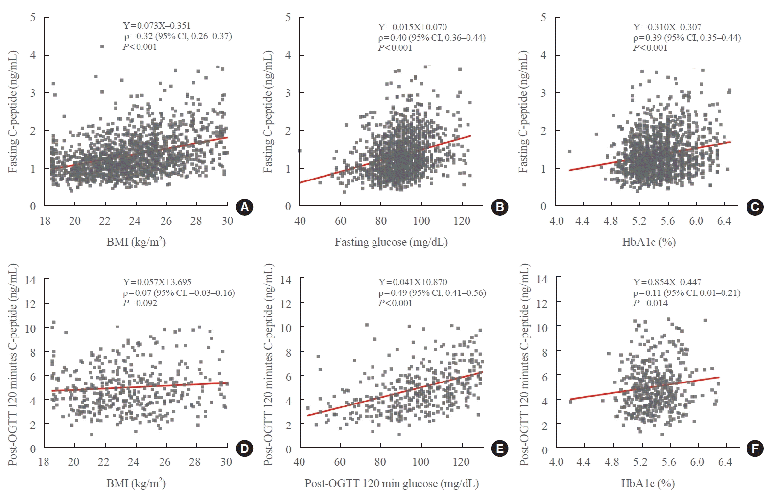
Table 1.
Table 2.
Age, yr |
Number (fasting) |
Number (OGTT) |
BMI, kg/m2 |
Fasting glucose, mg/dL |
HbA1c, % |
||||||||||
---|---|---|---|---|---|---|---|---|---|---|---|---|---|---|---|
Male | Female | Total | Male | Female | Total | Male | Female | P value | Male | Female | P value | Male | Female | P value | |
20s (19–29) | 124 | 166 | 290 | 35 | 75 | 110 | 24.3 (22.3–25.9) | 21.2 (19.9–23.2) | <0.001a | 87 (82–91) | 83 (78–89) | <0.001a | 5.28 (5.15–5.44) | 5.20 (5.09–5.35) | 0.029a |
30s (30–39) | 89 | 93 | 182 | 18 | 41 | 59 | 25.2 (23.5–26.6) | 21.7 (20.1–23.9) | <0.001a | 91 (84–95) | 84 (79–89) | <0.001a | 5.34 (5.20–5.51) | 5.27 (5.10–5.34) | 0.001a |
40s (40–49) | 77 | 96 | 173 | 17 | 44 | 61 | 23.6 (22.4–25.3) | 22.3 (20.6–24.0) | 0.001a | 91 (86–96) | 86 (81–91) | <0.001a | 5.40 (5.25–5.51) | 5.35 (5.19–5.49) | 0.023a |
50s (50–59) | 75 | 102 | 177 | 15 | 47 | 62 | 24.3 (22.7–26.0) | 22.6 (21.1–24.3) | <0.001a | 91 (87–95) | 86 (80–91) | <0.001a | 5.45 (5.30–5.53) | 5.42 (5.28–5.52) | 0.213 |
60s (60–69) | 62 | 97 | 159 | 10 | 36 | 46 | 24.0 (22.8–25.5) | 23.4 (21.9–24.9) | 0.059 | 90 (84–96) | 89 (84–83) | 0.233 | 5.45 (5.28–5.58) | 5.49 (5.32–5.60) | 0.101 |
Over 70 (70 and above) | 40 | 29 | 69 | 7 | 10 | 17 | 23.1 (20.9–25.0) | 23.4 (20.4–25.8) | 0.734 | 91 (86–96) | 87 (82–81) | 0.025a | 5.50 (5.30–5.59) | 5.50 (5.37–5.60) | 0.603 |
Total | 467 | 583 | 1,050 | 102 | 253 | 355 | 24.2 (22.7–26.0) | 22.2 (20.5–24.1) | <0.001a | 90 (84–95) | 85 (80–91) | <0.001a | 5.39 (5.21–5.50) | 5.33 (5.17–5.50) | 0.009a |
Table 3.
Male |
Female |
P value |
||||||||||
---|---|---|---|---|---|---|---|---|---|---|---|---|
Mean±2SD | U |
Percentile rangee |
Min–Max | Mean±2SD | U |
Percentile rangee |
Min–Max | Male vs. female | Subgroup | |||
2.5th–97.5th | 0.5th–99.5th | 2.5th–97.5th | 0.5th–99.5th | |||||||||
(A) Each age, yr | ||||||||||||
20s (19–29) | 1.41±0.91 | 0.42–2.40 | 0.68–2.46 | 0.51–2.61 | 0.51–2.61 | 1.30±0.77 | 0.43–2.16 | 0.70–2.16 | 0.60–2.22 | 0.59–2.23 | 0.016a | 0.144 (20s vs. 30s) |
30s (30–39) | 1.40±0.87 | 0.44–2.36 | 0.60–2.30 | 0.48–2.61 | 0.48–2.61 | 1.37±0.84 | 0.43–2.30 | 0.80–2.43 | 0.42–2.51 | 0.42–2.51 | 0.240 | <0.001 (30s vs. 40s) |
40s (40–49) | 1.33±0.81 | 0.42–2.23 | 0.77–2.32 | 0.54–2.45 | 0.54–2.45 | 1.10±0.67 | 0.32–1.88 | 0.59–1.93 | 0.51–2.13 | 0.51–2.13 | 0.007a | 0.568 (40s vs. 50s) |
50s (50–59) | 1.32±1.00 | 0.24–2.40 | 0.54–2.41 | 0.48–2.60 | 0.48–2.60 | 1.12±0.64 | 0.36–1.88 | 0.65–1.89 | 0.54–2.13 | 0.54–2.13 | 0.002a | 0.032 (50s vs. 60s) |
60s (60–69) | 1.18±0.74 | 0.34–2.02 | 0.64–2.11 | 0.60–2.15 | 0.60–2.15 | 1.10±0.63 | 0.35–1.85 | 0.64–2.11 | 0.50–1.95 | 0.50–1.95 | 0.362 | 0.673 (60s vs. over 70) |
Over 70 (70 and above) | 1.15±0.77 | 0.28–2.01 | 0.54–2.07 | 0.51–2.09 | 0.51–2.09 | 1.11±0.82 | 0.19–2.03 | 0.54–2.07 | 0.59–2.21 | 0.59–2.21 | 0.748 | |
(B) Adjacent age, yr | ||||||||||||
20–30s (19–39) | 1.41±0.89 | 0.43–2.38 | 0.63–2.44 | 0.49–2.61 | 0.48–2.61 | 1.32±0.79 | 0.43–2.21 | 0.71–2.17 | 0.52–2.50 | 0.42–2.51 | 0.007a | 0.007 (20–30s vs. 40–50s) |
40–50s (40–59) | 1.32±0.91 | 0.33–2.32 | 0.60–2.36 | 0.48–2.59 | 0.48–2.60 | 1.11±0.65 | 0.34–1.88 | 0.60–1.89 | 0.52–2.13 | 0.51–2.13 | <0.001a | <0.001 (40–50s vs. over 60) |
Over 60 (60 and above) | 1.17±0.74 | 0.32–2.01 | 0.60–2.09 | 0.51–2.15 | 0.51–2.15 | 1.10±0.68 | 0.31–1.89 | 0.59–1.94 | 0.50–2.21 | 0.50–2.21 | 0.396 | |
Total | 1.33±0.88 | 0.36–2.30 | 0.61–2.28 | 0.50–2.60 | 0.48–2.61 | 1.20±0.75 | 0.35–2.06 | 0.64–2.09 | 0.51–2.36 | 0.42–2.51 | <0.001a | |
Overall | 1.26±0.82 | 0.34–3.18 | 0.63–2.17 | 0.51–2.50 | 0.42–2.61 |
Table 4.
Age, yr |
Male |
Female |
P value (male vs. female) | ||||||||
---|---|---|---|---|---|---|---|---|---|---|---|
Mean±2SD | U |
Percentile rangee |
Min–Max | Mean±2SD | U |
Percentile rangee |
Min–Max | ||||
2.5th–97.5th | 0.5th–99.5th | 2.5th–97.5th | 0.5th–99.5th | ||||||||
Post-30 minutes OGTT | |||||||||||
20–30s (19–39) | 5.08±4.20 | 0.86–9.30 | 2.28–10.68 | 2.10–10.94 | 2.10–10.94 | 5.03±3.57 | 1.44–8.63 | 2.25–9.25 | 2.12–10.36 | 2.12–10.36 | 0.755 |
40–50s (40–59) | 4.58±4.10 | 0.46–8.70 | 1.49–9.71 | 1.42–9.75 | 1.42–9.75 | 4.11±2.54 | 1.53–6.68 | 1.79–6.79 | 1.29–7.67 | 1.29–7.67 | 0.395 |
Over 60 (60 and above) | 4.97±4.04 | 0.91–9.04 | 2.35–8.75 | 2.31–8.95 | 2.31–8.95 | 4.82±3.49 | 1.30–8.33 | 2.21–9.18 | 2.04–9.34 | 2.04–9.34 | 0.771 |
Total | 4.90±4.12 | 0.76–9.05 | 2.09–10.24 | 1.42–10.94 | 1.42–10.94 | 4.67±3.33 | 1.32–8.02 | 2.12–8.94 | 1.54–10.33 | 1.29–10.36 | 0.162 |
Overall | 4.74±3.57 | 1.14–8.33 | 2.12–9.35 | 1.43–10.50 | 1.29–10.94 | ||||||
Post-120 minutes OGTT | |||||||||||
20–30s (19–39) | 4.41±3.27 | 1.12–7.71 | 1.87–7.59 | 1.37–7.75 | 1.37–7.75 | 5.26±3.68 | 1.55–8.96 | 2.65–9.67 | 2.28–10.05 | 2.28–10.05 | 0.002a |
40–50s (40–59) | 5.07±4.72 | 0.34–9.80 | 1.68–10.23 | 1.64–10.30 | 1.64–10.30 | 4.41±2.71 | 1.67–7.16 | 2.17–7.27 | 1.13–8.30 | 1.13–8.30 | 0.196 |
Over 60 (60 and above) | 4.64±3.51 | 1.11–8.17 | 1.92–8.60 | 1.89–8.69 | 1.89–8.69 | 5.15±3.80 | 1.33–8.98 | 2.44–9.69 | 2.30–10.11 | 2.30–10.11 | 0.177 |
Total | 4.66±3.82 | 0.81–8.50 | 1.86–9.17 | 1.37–10.30 | 1.37–10.30 | 4.93±3.46 | 1.45–8.42 | 2.39–9.14 | 1.59–10.07 | 1.13–10.11 | 0.062 |
Overall | 4.85±3.58 | 1.25–8.34 | 2.14–9.09 | 1.39–10.10 | 1.13–10.30 |
Table 5.
Fasting |
Post-OGTT 120 minutes |
|||||||
---|---|---|---|---|---|---|---|---|
Mean±2SD | 2.5th–97.5th percentile range | Min–Max | P value | Mean±2SD | 2.5th–97.5th percentile range | Min–Max | P value | |
BMI, kg/m2 | ||||||||
Normal (<23) | 1.17±0.81 | 0.59–2.09 | 0.42–3.45 | <0.001a | 5.05±3.77 | 2.25–9.72 | 0.87–13.18 | <0.001a |
Overweight (23–25) | 1.32±0.96 | 0.61–2.58 | 0.51–3.35 | 5.55±5.08 | 2.21–11.67 | 1.37–16.11 | ||
Obesity (≥25) | 1.63±1.18 | 0.82–3.18 | 0.58–3.98 | 5.96±4.87 | 2.26–11.38 | 1.64–13.25 | ||
Glucose level, mg/dL | ||||||||
Normal (<100) | 1.33±1.02 | 0.64–2.57 | 0.42–3.98 | <0.001a | 5.33±4.32 | 2.21–10.38 | 0.87–13.44 | <0.001a |
IFG (100–125) | 1.66±1.19 | 0.84–3.24 | 0.61–3.81 | 6.55±4.95 | 2.60–10.64 | 2.53–13.24 | ||
HbA1c, % | ||||||||
Normal (<5.70) | 1.32±0.98 | 0.64–2.46 | 0.42–3.98 | <0.001a | 5.26±4.30 | 2.20–10.39 | 0.87–13.97 | <0.001a |
Prediabetic (5.70–6.49) | 1.54±1.28 | 0.68–3.17 | 0.56–3.85 | 6.62±5.49 | 2.48–13.25 | 2.21–13.44 |