Gastric subepithelial tumors grow within the gastric wall; therefore, endoscopic removal requires advanced techniques and experienced hands. Gastrointestinal stromal tumors (GISTs) are the most common gastric subepithelial tumors with malignant potential. Endoscopic ultrasonography is the most accurate noninvasive method for evaluating layers and echo patterns.1 GIST originates from the muscularis propria layer, which is divided into submucosal, intramuscular, or subserosal types according to endoscopic ultrasonography findings. Conventional endoscopic submucosal dissection (ESD) is a useful technique for removing the submucosal type and is associated with a low risk of adverse events. Endoscopic full-thickness resection (EFTR) may sometimes be required for submucosal-growing GIST.2 The intramuscular and subserosal patterns indicate that the tumor has an extensive muscularis involvement or penetration beyond the gastric wall. Endoscopic resection is associated with a high possibility of full-thickness resection, which involves peritoneal tumor cell seeding and gastric juice leakage.3 In addition, the complete resection (R0 resection) rate is relatively low for tumors originating from the muscularis propria compared to those reported in studies on early gastric cancers. GISTs have pseudocapsules to prevent the tumor cell spillage, and microscopic incomplete resection (R1 resection) is not significant for local recurrence in many studies.4,5 Recently, the European Society of Gastrointestinal Endoscopy guidelines recommended EFTR as an alternative to laparoscopic wedge resection for gastric GIST <3.5 cm.6
An ideal EFTR must have the least peritoneal tumor cell exposure, minimal gastric juice leakage, and a high complete resection rate. Conventional EFTR is based on an ESD technique combined with clip closure; however, it has limitations in viewing the dissecting area and achieving good closure with clipping. Submucosal tunneling endoscopic resection results in good closure after full-thickness resection and is associated with a low risk of gastric juice leakage. Submucosal tunneling endoscopic resection is only possible in the cardia and esophagogastric junction, which is a serious limitation.3,7 The gastric full-thickness resection device is a modified version of the Over-The-Scope Clip, which has the advantages of good closure and low risk of peritonitis. However, it may be useful for tumors <1 cm. Although various full-thickness resection techniques have been developed, they are challenging.3 Precise muscular dissection must be achieved for en bloc resection without injuring the pseudocapsule. Complete closure is important to avoid tumor cell seeding and peritonitis after full-thickness resection. Most techniques using clips, snares, and threads have been reported to provide a good dissection view and a low risk of peritoneal tumor cell exposure.7,8
In a study by Kim et al.,9 EFTR using a modified technique, termed clip-and-cut EFTR (cc-EFTR), was performed in 32 patients. Most lesions (86.3%) were in the upper third of the stomach, and 71.9% of the tumors had subserosal growth patterns. The R0 resection rate was 84.4%, and two cases of localized peritonitis were treated conservatively. No recurrence was observed 25 months after the treatment. In cc-EFTR, a clip connected to dental floss is attached to the dissected flap edge for tumor mass traction. The clip is fixed at both ends of the dissected area for a good approximation of the penetrated muscularis propria, followed by stepwise clipping immediately after the transmural cut.
EFTR has been used to treat GISTs but has not yet been standardized. Submucosal and muscular dissections were performed using a technique like that used in ESD. Full-thickness resection and closure are the most important steps that must be addressed. The cc-EFTR technique is relatively simple and easy to perform compared to previously reported methods. Complete closure was made in most cases with a high R0 resection rate. I believe that cc-EFTR will be a useful technique for full-thickness resection of fundus and upper body tumors.
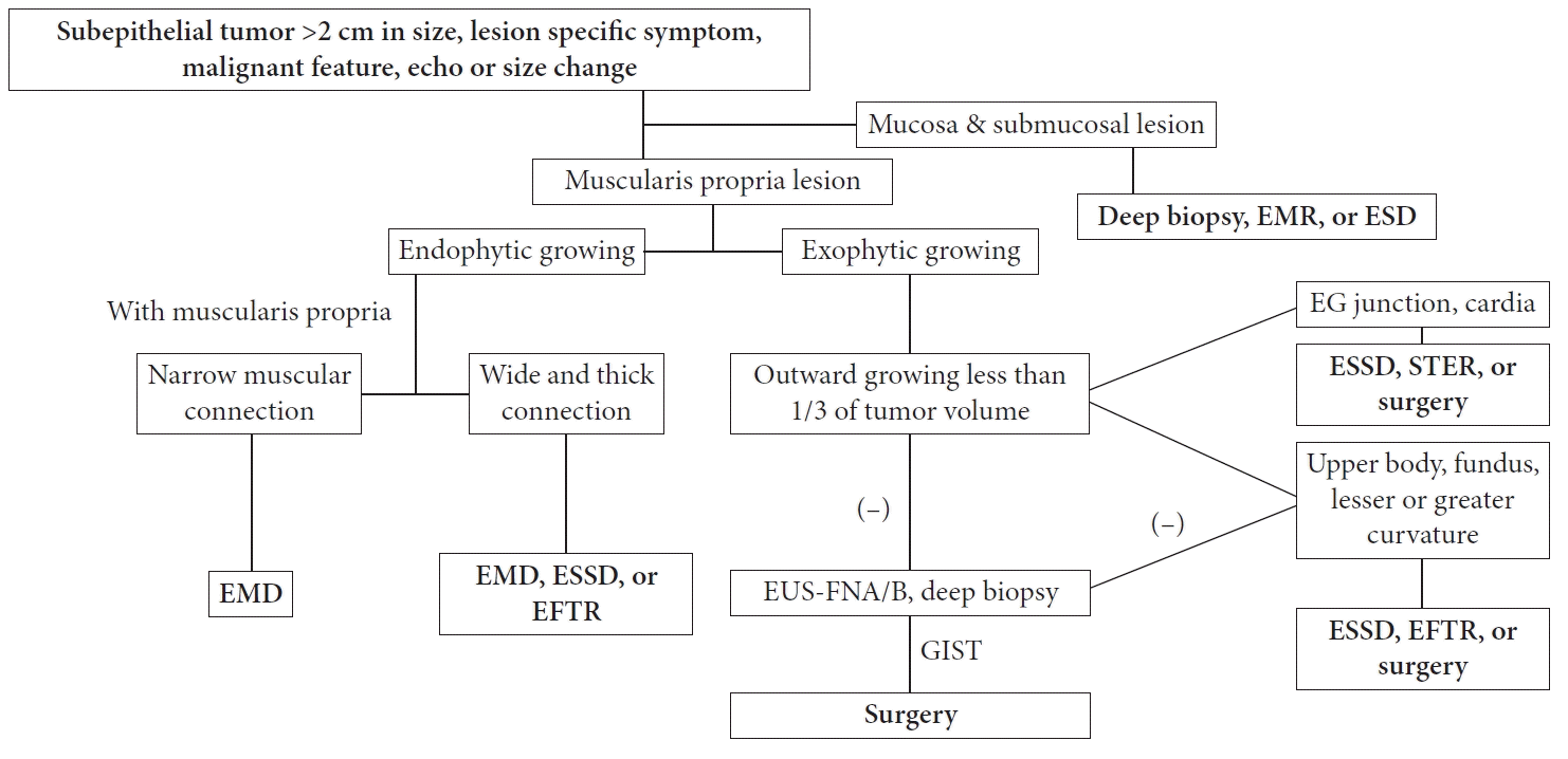
Endoscopy is increasingly used to treat gastric GISTs. Small gastric (<3.5 cm) subepithelial tumors with endophytic growth are good indications for endoscopic resection, even those with significant muscularis propria involvement. The treatment of choice for exophytic growing tumors is laparoscopic wedge resection, which has significant normal tissue loss and some limitations for tumors in special locations, such as the esophagogastric junction, cardia, or lesser curvature.5 Endoscopic resection minimizes gastric tissue loss and prevents gastroparesis and gastric deformities. cc-EFTR can be a standard technique for fundus or upper body tumors with exophytic growth of less than one-third of the tumor volume. Recently, endoscopic subserosal dissection was introduced to treat tumors with exophytic growth. Endoscopic subserosal dissection is a technique that penetrates the muscularis propria layer and dissects the subserosal layer, which is useful in the esophagogastric junction, cardia, fundus and upper body parts, lesser curvature, and greater curvature.10
Small gastric subepithelial tumors can be safely treated with endoscopic resection (Fig. 1). Studies on EFTR for gastric GISTs have been increasing, and technical advancements have been achieved. Endoscopic resection is expected to become a standard treatment for GIST soon.


 PDF
PDF Citation
 Print
Print


 XML Download