Journal List > Korean J Anesthesiol > v.76(4) > 1516083624

Kang, Park, Yoon, Ji, Jang, Kim, Lee, Lee, Kim, and Kim: Association of the perfusion index with postoperative acute kidney injury: a retrospective study

Abstract

Background

Many studies have examined the risk factors for postoperative acute kidney injury (AKI), but few have focused on intraoperative peripheral perfusion index (PPI) that has recently been shown to be associated with postoperative morbidity and mortality. Therefore, this study aimed to evaluate the relationship between intraoperative PPI and postoperative AKI under the hypothesis that lower intraoperative PPI is associated with AKI occurrence.

Methods

We retrospectively searched electronic medical records to identify patients who underwent surgery at the general surgery department from May 2021 to November 2021. Patient baseline characteristics, pre- and post-operative laboratory test results, comorbidities, intraoperative vital signs, and discharge profiles were obtained from the Institutional Clinical Data Warehouse and VitalDB. Intraoperative PPI was the primary exposure variable, and the primary outcome was postoperative AKI.

Results

Overall, 2,554 patients were identified and 1,586 patients were included in our analysis. According to Kidney Disease Improving Global Outcomes (KDIGO) criteria, postoperative AKI occurred in 123 (7.8%) patients. We found that risks of postoperative AKI increased (odds ratio: 2.00, 95% CI [1.16, 3.44], P = 0.012) when PPI was less than 0.5 for more than 10% of surgery time. Other risk factors for AKI occurrence were male sex, older age, higher American Society of Anesthesiologists physical status, obesity, underlying renal disease, prolonged operation time, transfusion, and emergent operation.

Conclusions

Low intraoperative PPI was independently associated with postoperative AKI.

Introduction

The incidence of postoperative acute kidney injury (AKI) in adult non-cardiac surgical patients reportedly ranges from 3% to 35% [1,2]. Much attention has been paid to postoperative AKI as it can independently result in poor patient outcomes. In particular, some studies have reported that AKI affects both short- and long-term outcomes even in patients with mild severity who recovered after a short period of time following cardiac or non-cardiac surgery [1,3,4]. Therefore, it is important to reduce the incidence of postoperative AKI by identifying high-risk patients through preoperative assessment, intraoperative monitoring, and through the provision of appropriate management during and after surgery [5,6]. There has been extensive research on the risk factors of perioperative AKI and an increasing number of studies are using machine learning and big data to predict AKI [5,7,8]. Known risk factors (some of which are not modifiable) reported by these studies include advanced age; pre-existing kidney disease or elevated preoperative serum creatinine levels; pre-existing comorbidities such as hypertension (HTN), congestive heart failure, and insulin-dependent diabetes mellitus (DM); obesity; sex; intraoperative blood transfusion; hemodynamic instability; and use of vasopressors or diuretics [1,913]. Interestingly, a recent study reported that peripheral perfusion index (PPI) during surgery may also be associated with postoperative morbidity and mortality, indicating that PPI may be a risk factor worthy of further investigation [14,15]. To date, though there have been many studies that seek to predict high-risk groups for AKI or provide recommendations for controlling risk factors, there have been no studies that assessed the relationship between intraoperative PPI and postoperative AKI.
Therefore, in this study, we hypothesized that low PPI is related to the occurrence of postoperative AKI and aimed to determine whether this was an independent risk factor.

Materials and Methods

Patients and data collection

After obtaining approval from the Institutional Review Board of Seoul National University Hospital for retrospective record review and a waiver for informed consent for this study (H-2112-044-1280), we retrospectively reviewed medical records in the Seoul National University Hospital Patients Research Environment (SUPREME) website, searching for patients who underwent elective or emergent operations under general anesthesia at the general surgery department of Seoul National University Hospital and also underwent postoperative laboratory tests between May and November in 2021. Patients who underwent more than two surgeries under any type of anesthesia during a single hospital admission and those who had inadequate anesthetic records were excluded. Through a SUPREME search and electronic medical record review, patient baseline characteristics, results of preoperative and postoperative laboratory tests, anesthetic and perioperative variables, and discharge profiles were collected. Vital signs including mean blood pressure (MBP), heart rate, peripheral oxygen saturation, body temperature, and PPI value during surgery were continuously monitored by patient monitors (Philips IntelliVue MX800, Koninklijke Philips, Netherlands) and compiled in the VitalDB database (http://vitaldb.net/) [16]. Patients with incomplete data (where data recording was stopped before the end of the surgery), no data, or no PPI values were excluded. All vital signs were recorded at 1 s intervals and averaged every 15 min in accordance with the methodology provided by a previous study [14]. If MBP was less than 65 mmHg in more than 10% of the averaged sections, the patient was defined as having intraoperative hypotension. Patients who had a low PPI value (< 1.5 or 0.5) [14] during surgery for more than 10% of the sections were defined as having a low mean PPI.

Variables and statistical analysis

The primary outcome variable was postoperative AKI, and the primary exposure variable was intraoperative PPI. Chi-squared tests were conducted on categorical variables. To obtain odds ratios (ORs), a chi-square test was performed by dividing American Society of Anesthesiologists (ASA) physical status into two categories: lower physical status (I, II) and higher physical status (III, IV, V); the fluid infusion rate was divided into two categories: “liberal or restrictive infusion rate” and “non-liberal and non-restrictive infusion rate.”
AKI occurrence was determined according to the Kidney Disease Improving Global Outcomes (KDIGO) definitions [17]. To identify predictive factors for postoperative AKI, patients were classified into two groups based on whether postoperative AKI occurred. The risk factors for postoperative AKI were examined by univariate and multivariate logistic regression analyses that included the following variables: old age (> 65 years); sex (male); higher ASA status (III, IV, V) [14]; obesity (body mass index > 25 kg/m2) [9,10]; medical history of HTN, DM, heart disease, liver disease, neurovascular disease, or renal disease; moderate or severe anemia (hemoglobin < 11 g/dl) [18]; low serum albumin (< 3.5 g/dl); anesthetic type (total intravenous anesthesia); duration of surgery (> 3 h); transfusion (positive); liberal (> 12 ml/kg/h) or restrictive (< 4 ml/kg/h) fluid administration [19,20]; emergent operation; low PPI; and hypotension [21]. Multivariate logistic regression was used to calculate ORs and 95% CIs after simultaneously controlling for potential confounders. To ascertain the multicollinearity of the last remaining variables, we checked variance inflation factors and confirmed that none were greater than five. Furthermore, Pearson’s correlation analysis was conducted to find correlation between PPI and MBP. In case the low PPI is associated with the occurrence of AKI, we decided to conduct receiver operating characteristic curve analysis to find the cut-off value.
All data are expressed as mean ± standard deviation (SD) or median (Q1, Q3) unless otherwise specified; normality was tested using the Shapiro–Wilk test. Continuous variables of the study population were evaluated using the independent t test or the Mann–Whitney U test. All statistical analyses were performed using SPSS® (SPSS® 26.0; IBM Inc., USA) software, and a two-sided P value < 0.05 was considered statistically significant.

Results

Patient data

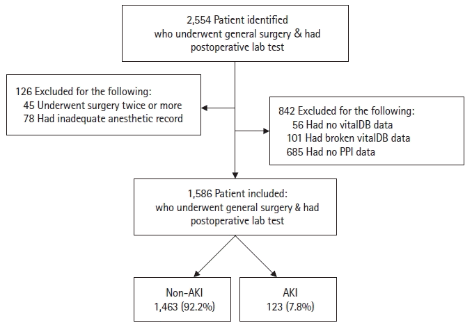
A total of 1,586 patients who underwent general surgery were analyzed, 123 (7.8%) of whom developed postoperative AKI (Fig. 1). Departmental data is shown in Supplementary Table 1. Among them, 101 (6.4%) developed Grade 1 AKI, 17 (1.1%) developed Grade 2 AKI, and five (0.3%) developed Grade 3 AKI. Demographic and baseline variables are shown in Table 1. AKI was more common in men (P < 0.001). Patients in the AKI group were taller (P < 0.001) and heavier than those in the non-AKI group (P < 0.001). Furthermore, more patients in the AKI group had underlying renal disease (P < 0.001) and higher ASA status (P < 0.001). In terms of preoperative blood test results, the AKI group had significantly lower albumin and hemoglobin levels, and higher creatinine and blood urea nitrogen levels (P < 0.001 for all variables).

Operative and postoperative data

Total duration of surgery was longer in the AKI group (mean difference: 81, 95% CI [59, 104], P < 0.001), and there were more emergent operations in the AKI group as well (OR: 2.97, 95% CI, [1.58, 5.6], P < 0.001). There was no difference in MBP during operations between the two groups, but heart rates were faster in the AKI group. Mean PPI during operations were lower in the AKI group. The total amount of infused fluid and blood was higher in the AKI group, and liberal or restrictive infusion was more common in the AKI group (X2 [2, n = 1586] = 20.04, P < 0.001). The postoperative outcomes are shown in Table 2. Planned or unplanned intensive care unit (ICU) admission was more common in the AKI group. The continuous renal replacement therapy was newly initiated in five patients within seven days postoperatively. Among them, two patients in the non-AKI group received hemodialysis due to severe metabolic acidosis, and others in the AKI group received hemodialysis due to decreased renal function. The length of hospital stay was longer in the AKI group. The all-cause in-hospital mortality was higher in the AKI group (OR: 10.98; 95% CI [3.91, 30.8], P < 0.001).

Risk factors of postoperative AKI

The results of the univariate and multivariate logistic regression analyses are shown in Table 3. We found that the risk of postoperative AKI increased if intraoperative mean PPI was less than 0.5 for more than 10% of the sections (OR: 2.00, 95% CI [1.16, 3.44], P = 0.012). Hypotension was not a risk factor for AKI occurrence. Pearson’s correlation analysis found no correlation between mean PPI and MBP (r = 0.029, P = 0.251). Other risk factors were male sex (OR: 3.17, 95% CI [1.90, 0.29], P < 0.001), older age (OR: 1.65, 95% CI [1.08, 2.51], P = 0.020), higher ASA status (OR: 1.72, 95% CI [1.10, 2.49], P = 0.018), obesity (OR: 1.66, 95% CI [1.11, 2.49], P = 0.014), underlying renal disease (OR: 5.82, 95% CI [3.37, 10.05, P < 0.001), prolonged operation time (OR: 2.21, 95% CI [1.28, 3.83], P = 0.004), transfusions (OR: 3.64, 95% CI [2.27, 5.83], P < 0.001), and emergent operations (OR: 2.35, 95% CI [1.12, 4.93], P = 0.024). The risk factors for higher grade AKI (Grades 2 and 3) shown in Table 4 included higher ASA (OR: 3.24, 95% CI [1.35, 7.79], P = 0.009), previous history of HTN (OR: 2.76, 95% CI [1.03, 7.39], P = 0.001), transfusion (OR: 2.90, 95% CI [1.15, 7.36], P = 0.024), and PPI < 0.5 (OR: 5.11, 95% CI [2.03, 12.83], P < 0.001). Moreover, when the total duration of surgery was divided by 20% intervals to determine the proportion where PPI < 0.5, the OR was found to be 8.63 at 20%–40% compared with 0%–20% (95% CI [2.17, 34.26], P = 0.002), and OR was 10.24 for 40%–60% (95% CI [2.68, 39.16], P = 0.001). When another logistic regression analysis was conducted with PPI < 1.5 instead of 0.5, the results were not statistically significant (Supplementary Table 2).

PPI cut-off evaluation

The receiver operating characteristic curve analysis of the association between intraoperative mean PPI and AKI occurrence yielded an area under the receiver operating characteristic curve of 0.581. The optimal cut-off value of intraoperative mean PPI when maximizing sensitivity and specificity using Youden’s index was 3.13, with a sensitivity of 68.29% and specificity of 46.14% (P = 0.002). The proportion of patients with a mean PPI < 3.13 was higher in the AKI group (68.3% vs. 53.9%; OR: 1.85, 95% CI [1.24, 2.72], P = 0.002). However, this factor was not included as a predictor of postoperative AKI based on the results of multivariate logistic regression analysis.

Discussion

This study revealed that low intraoperative PPI was associated with postoperative AKI. The PPI value is known to reflect two main determinants: the cardiac output and the balance between the sympathetic and parasympathetic nervous systems [22,23]. Its usefulness as a vital sign for risk stratification or for determining early intervention has been studied in various scenarios such as emergency departments, ICUs, and operating rooms. PPI can be used to evaluate disease severity or fluid responsiveness in critically ill patients, and can also be used to predict hypotension or poor outcomes [2427]. Moreover, fluid responsiveness can be evaluated in patients undergoing surgery as PPI reflects cardiac output [23,28]. From the perspective of oxygen delivery and tissue perfusion, it is important to maintain adequate cardiac output rather than the more commonly monitored blood pressure, because blood pressure is affected not only by cardiac output, but also by systemic vascular resistance [29]. Similarly, in critically ill patients with a low flow state, organ dysfunctions such as AKI are likely to occur due to a decrease in tissue perfusion even if blood pressure is maintained at normal with increased vascular resistance [30,31]. While hypotension was a risk factor for postoperative AKI in some previous studies [14,32], hypotension did not significantly increase AKI occurrence in our study. It may be because the cut-off value for hypotension is not a fixed MBP level but a value that differs for each patient. Another reason could be because blood pressure is not a good indicator of cardiac output that is more critical for organ perfusion. Since PPI could reflect cardiac output [23,28], monitoring of PPI during surgery could provide additional information on tissue perfusion and oxygenation. As cardiac output was not measured for the patients in our study, we were unable to evaluate the correlation between cardiac output and PPI. However, low PPI may reflect a decrease in cardiac output that may increase AKI occurrence by altering kidney perfusion; furthermore, it may cause poor outcomes by altering perfusion of other major organs; further research on this will be needed for a better understanding of this mechanism [31].
Other risk factors showed similar results to those reported in previous studies [7,9,12]. In terms of ORs, pre-existing renal disease had the highest OR at 5.82, followed by intraoperative transfusion at 3.64, followed in turn by male sex, emergent operation, prolonged duration of surgery, higher ASA physical status, obesity, and older age. Unfortunately, anesthesiologists are unable to modify most of these factors. Additionally, the effects of sex on AKI occurrence are controversial. A study on patients who underwent cardiac surgery suggested that male sex had a protective effect [12], but another study on patients who underwent general surgery showed the opposite result [1]. In this study, we found that men were 3.17 times more likely to develop AKI. This is consistent with the results of the previous studies that showed a higher likelihood of AKI development in male hospitalized patients during follow-up [33,34]. The reasons behind this should be investigated in the future.
Patients who developed AKI had a higher likelihood of ICU admission, new initiation of renal replacement therapy, longer hospital stays, and higher all-cause mortality. Considering that the incidence of in-hospital mortality or postoperative complications was not high in our study, additional studies with larger number of patients are needed to determine whether low intraoperative PPI or AKI itself are causes or predictors of these outcomes.
The present study had several limitations. First, our study was based on a review of recorded data, and the SUPREME system was unable to filter erroneous data or incorrectly entered information. In the case of vital signs collected automatically during surgery, errors may occur due to mechanical or electrical stimulation. To reduce such errors, the SUPREME system information was reconfirmed individually through electronic medical records if the input information was judged to be an outlier. Through this process, it was possible to identify cases where patient height and weight were entered in the reverse order, and where laboratory results from body fluid and not blood were entered incorrectly. Additionally, we checked whether blood pressure and plethysmography waveforms collected in vitalDB had a normal shape to ensure that there were no errors in vital sign data.
Second, the participants of this study were limited to patients who underwent general surgery to reduce the number of confounding variables such as intraoperative cardiopulmonary bypass, one-lung ventilation, or different targets of surgery. Additionally, a postoperative laboratory test was routinely performed by the surgeon that may have biased patient selection. Further studies are required to confirm these results in other patients.
Lastly, although it is known that PPI is associated with poor prognosis, it is not known how PPI can be improved since many of the risk factors for AKI identified so far, including the results of this study, cannot be modified. If a method to improve upon these risk factors can be found and applied to patients with intraoperative and preoperative PPI, this can help reduce postoperative AKI, and in turn morbidity and mortality.
In conclusion, we found that low PPI is a risk factor for postoperative AKI. Future studies based on this finding may be able to construct a more comprehensive model for AKI prediction, or aid patient prognosis through improvement of PPI.

Notes

Funding

This work was supported by the Korea Medical Device Development Fund grant funded by the government of Republic of Korea (in particular, the Ministry of Science and ICT; the Ministry of Trade, Industry and Energy; the Ministry of Health and Welfare; and the Ministry of Food and Drug Safety) (Project Number: 202011823).

Conflicts of Interest

Ji-Hyun Lee has been an editor for the Korean Journal of Anesthesiology since 2021. However, she was not involved in any process of review for this article, including peer reviewer selection, evaluation, or decision-making. There were no other potential conflicts of interest relevant to this article.

Data Availability

Data will be shared with other investigators through academically established means, an necessary and appropriate. On reasonable request, the dataset from this study can be provided by the principal investigator.

Author Contributions

Pyoyoon Kang (Conceptualization; Data curation; Formal analysis; Investigation; Methodology; Software; Writing – original draft; Writing – review & editing)

Jung-bin Park (Data curation; Formal analysis; Software; Writing – original draft)

Hyun-Kyu Yoon (Data curation; Formal analysis; Investigation; Methodology; Software)

Sang-Hwan Ji (Formal analysis; Investigation; Software; Supervision; Validation)

Young-Eun Jang (Data curation; Software; Writing – review & editing)

Eun-Hee Kim (Conceptualization; Methodology; Validation; Writing – review & editing)

Ji-Hyun Lee (Conceptualization; Methodology; Writing – review & editing)

Hyung Chul Lee (Data curation; Formal analysis; Investigation; Writing – review & editing)

Jin-Tae Kim (Conceptualization; Writing – review & editing)

Hee-Soo Kim (Conceptualization; Funding acquisition; Software; Supervision; Writing – review & editing)

Supplementary Materials

Supplementary Table 1.
Departments of general surgery.
kja-22620-Supplementary-Table-1.pdf
Supplementary Table 2.
Univariate and multivariate analysis between potential risk factors including PPI < 1.5 with postoperative AKI.
kja-22620-Supplementary-Table-2.pdf

References

1. Gameiro J, Fonseca JA, Neves M, Jorge S, Lopes JA. Acute kidney injury in major abdominal surgery: incidence, risk factors, pathogenesis and outcomes. Ann Intensive Care. 2018; 8:22.
2. Quan S, Pannu N, Wilson T, Ball C, Tan Z, Tonelli M, et al. Prognostic implications of adding urine output to serum creatinine measurements for staging of acute kidney injury after major surgery: a cohort study. Nephrol Dial Transplant. 2016; 31:2049–56.
3. Hansen MK, Gammelager H, Mikkelsen MM, Hjortdal VE, Layton JB, Johnsen SP, et al. Post-operative acute kidney injury and five-year risk of death, myocardial infarction, and stroke among elective cardiac surgical patients: a cohort study. Crit Care. 2013; 17:R292.
4. Linder A, Fjell C, Levin A, Walley KR, Russell JA, Boyd JH. Small acute increases in serum creatinine are associated with decreased long-term survival in the critically ill. Am J Respir Crit Care Med. 2014; 189:1075–81.
5. Woo SH, Zavodnick J, Ackermann L, Maarouf OH, Zhang J, Cowan SW. Development and validation of a web-based prediction model for AKI after surgery. Kidney360. 2020; 2:215–23.
6. Meersch M, Schmidt C, Zarbock A. Perioperative acute kidney injury: an under-recognized problem. Anesth Analg. 2017; 125:1223–32.
7. Nishimoto M, Murashima M, Kokubu M, Matsui M, Eriguchi M, Samejima KI, et al. External validation of a prediction model for acute kidney injury following noncardiac surgery. JAMA Netw Open. 2021; 4:e2127362.
8. Rank N, Pfahringer B, Kempfert J, Stamm C, Kühne T, Schoenrath F, et al. Deep-learning-based real-time prediction of acute kidney injury outperforms human predictive performance. NPJ Digit Med. 2020; 3:139.
9. Grams ME, Sang Y, Coresh J, Ballew S, Matsushita K, Molnar MZ, et al. Acute kidney injury after major surgery: a retrospective analysis of veterans health administration data. Am J Kidney Dis. 2016; 67:872–80.
10. Suneja M, Kumar AB. Obesity and perioperative acute kidney injury: a focused review. J Crit Care. 2014; 29:694.e1-6.
11. Weingarten TN, Gurrieri C, McCaffrey JM, Ricter SJ, Hilgeman ML, Schroeder DR, et al. Acute kidney injury following bariatric surgery. Obes Surg. 2013; 23:64–70.
12. Kheterpal S, Tremper KK, Heung M, Rosenberg AL, Englesbe M, Shanks AM, et al. Development and validation of an acute kidney injury risk index for patients undergoing general surgery: results from a national data set. Anesthesiology. 2009; 110:505–15.
13. Kim CS, Oak CY, Kim HY, Kang YU, Choi JS, Bae EH, et al. Incidence, predictive factors, and clinical outcomes of acute kidney injury after gastric surgery for gastric cancer. PLoS ONE. 2013; 8:e82289.
14. Agerskov M, Thusholdt AN, Holm-Sørensen H, Wiberg S, Meyhoff CS, Højlund J, et al. Association of the intraoperative peripheral perfusion index with postoperative morbidity and mortality in acute surgical patients: a retrospective observational multicentre cohort study. Br J Anaesth. 2021; 127:396–404.
15. Okada H, Tanaka M, Yasuda T, Okada Y, Norikae H, Fujita T, et al. Decreased peripheral perfusion measured by perfusion index is a novel indicator for cardiovascular death in patients with type 2 diabetes and established cardiovascular disease. Sci Rep. 2021; 11:2135.
16. Lee HC, Jung CW. Vital Recorder-a free research tool for automatic recording of high-resolution time-synchronised physiological data from multiple anaesthesia devices. Sci Rep. 2018; 8:1527.
17. Section 2: AKI definition. Kidney Int Suppl (2011). 2012; 2:19–36.
18. Kassebaum NJ; GBD 2013 Anemia Collaborators. The global burden of anemia. Hematol Oncol Clin North Am. 2016; 30:247–308.
19. Lobo SM, Ronchi LS, Oliveira NE, Brandão PG, Froes A, Cunrath GS, et al. Restrictive strategy of intraoperative fluid maintenance during optimization of oxygen delivery decreases major complications after high-risk surgery. Crit Care. 2011; 15:R226.
20. Nisanevich V, Felsenstein I, Almogy G, Weissman C, Einav S, Matot I. Effect of intraoperative fluid management on outcome after intraabdominal surgery. Anesthesiology. 2005; 103:25–32.
21. Mikkelsen TB, Schack A, Oreskov JO, Gögenur I, Burcharth J, Ekeloef S. Acute kidney injury following major emergency abdominal surgery - a retrospective cohort study based on medical records data. BMC Nephrol. 2022; 23:94.
22. Elshal MM, Hasanin AM, Mostafa M, Gamal RM. Plethysmographic peripheral perfusion index: could it be a new vital sign? Front Med (Lausanne). 2021; 8:651909.
23. Hasanin A, Karam N, Mukhtar AM, Habib SF. The ability of pulse oximetry-derived peripheral perfusion index to detect fluid responsiveness in patients with septic shock. J Anesth. 2021; 35:254–61.
24. He H, Long Y, Liu D, Wang X, Zhou X. Clinical classification of tissue perfusion based on the central venous oxygen saturation and the peripheral perfusion index. Crit Care. 2015; 19:330.
25. He HW, Liu DW, Long Y, Wang XT. The peripheral perfusion index and transcutaneous oxygen challenge test are predictive of mortality in septic patients after resuscitation. Crit Care. 2013; 17:R116.
26. Mostafa H, Shaban M, Hasanin A, Mohamed H, Fathy S, Abdelreheem HM, et al. Evaluation of peripheral perfusion index and heart rate variability as early predictors for intradialytic hypotension in critically ill patients. BMC Anesthesiol. 2019; 19:242.
27. Rasmy I, Mohamed H, Nabil N, Abdalah S, Hasanin A, Eladawy A, et al. Evaluation of perfusion index as a predictor of vasopressor requirement in patients with severe sepsis. Shock. 2015; 44:554–9.
28. de Courson H, Michard F, Chavignier C, Verchère E, Nouette-Gaulain K, Biais M. Do changes in perfusion index reflect changes in stroke volume during preload-modifying manoeuvres? J Clin Monit Comput. 2020; 34:1193–8.
29. Sun J, Yuan J, Li B. SBP is superior to MAP to reflect tissue perfusion and hemodynamic abnormality perioperatively. Front Physiol. 2021; 12:705558.
30. Vincent JL, Pelosi P, Pearse R, Payen D, Perel A, Hoeft A, et al. Perioperative cardiovascular monitoring of high-risk patients: a consensus of 12. Crit Care. 2015; 19:224.
31. Hasanin A, Mukhtar A, Nassar H. Perfusion indices revisited. J Intensive Care. 2017; 5:24.
32. Maheshwari K, Turan A, Mao G, Yang D, Niazi AK, Agarwal D, et al. The association of hypotension during non-cardiac surgery, before and after skin incision, with postoperative acute kidney injury: a retrospective cohort analysis. Anaesthesia. 2018; 73:1223–8.
33. Loutradis C, Pickup L, Law JP, Dasgupta I, Townend JN, Cockwell P, et al. Acute kidney injury is more common in men than women after accounting for socioeconomic status, ethnicity, alcohol intake and smoking history. Biol Sex Differ. 2021; 12:30.
34. Neugarten J, Golestaneh L, Kolhe NV. Sex differences in acute kidney injury requiring dialysis. BMC Nephrol. 2018; 19:131.

Fig. 1.
Flow diagram describing the retrospective study design.
kja-22620f1.tif
Table 1.
Baseline Characteristics, Comorbidities, Preoperative Laboratory Variables, and Operative Information
Variable Total (n = 1,586) Non-AKI group (n = 1,463) AKI group (n = 123) Effect size* (95% CI) P value
Patient’s characteristics
 Age (yr) 61.1 ± 14.0 60.9 ± 14.1 63.2 ± 12.8 2.00 (0.00, 4.00) 0.094
 Sex (M/F) 953 (60) / 633 (40) 851 (58) / 612 (42) 102 (83) / 21 (17) 0.29 (0.18, 0.46) < 0.001
 Height (cm) 163.48 ± 8.97 163.0 (156.0, 170.0) 167.0 (162.0, 173.0) 3.80 (2.20, 5.4) < 0.001
 Weight (kg) 65.05 ± 13.02 63.6 (55.8, 71.9) 69.0 (60.5, 76.3) 4.95 (2.70, 7.2) < 0.001
 BMI 24.23 ± 3.83 23.9 (21.7, 26.2) 24.6 (22.1, 26.9) 0.69 (0.03, 1.33) 0.041
Comorbidities
 ASA (I/II/III/IV/V) 114/1124/321/27/0 110/1054/253/25/0 4/59/58/2/0 3.89 (2.67, 5.7) < 0.001
 HTN 197 (12) 175 (12) 22 (18) 1.60 (0.98, 2.61) 0.056
 DM 138 (9) 130 (9) 8 (7) 0.71 (0.34, 1.49) 0.368
 Heart disease 65 (4) 59 (4) 6 (5) 1.22 (0.52, 2.89) 0.650
 Liver disease 106 (7) 96 (7) 10 (8) 1.26 (0.64, 2.48) 0.504
 Neurovascular disease 35 (2) 31 (2) 4 (3) 1.55 (0.54, 4.47) 0.412
 Renal disease 120 (8) 84 (6) 36 (29) 6.79 (4.35, 10.6) < 0.001
Preoperative lab test
 Hemoglobin 12.60 ± 2.05 12.9 (11.4, 14.1) 11.6 (10.4, 13.5) 0.80 (0.40, 1.30) < 0.001
 Albumin 4.15 ± 0.51 4.2 (3.9, 4.5) 4.0 (3.7, 4.3) 0.20 (0.10, 0.30) < 0.001
 GOT 33.52 ± 173.06 21.0 (17.0, 27.0) 23.0 (17.0, 31.5) 1.00 (0.00, 3.00) 0.087
 GPT 28.55 ± 89.50 18.0 (13.0, 27.0) 20.0 (14.0, 31.0) 2.00 (0.00, 4.00) 0.025
 BUN 19.33 ± 13.13 16.0 (13.0, 20.0) 20.0 (15.0, 34.0) 5.00 (3.00, 7.0) < 0.001
 Creatinine 1.37 ± 2.18 0.81 (0.71, 0.97) 0.97 (0.82, 2.53) 0.19 (0.13, 0.26) < 0.001
Operative variables
 Operation time, min 245.57 ± 120.40 226.0 (153.0, 302.0) 305.0 (226.0, 415.5) 81 (59, 104) < 0.001
 Emergency operation 69 (4) 56 (4) 13 (11) 2.97 (1.58, 5.6) < 0.001
 Volatile/TIVA 1536 (97) / 50 (3) 1418 (97) / 45 (3) 118 (96) / 5 (4) 1.34 (0.52, 3.43) 0.547
 MBP (mmHg) 84.25 ± 8.13 83.6 (78.9, 88.8) 82.0 (77.7, 87.8) 1.10 (-0.33, 2.53) 0.131
 HR (beats/min) 71.36 ± 12.69 69.1 (62.1, 77.8) 72.5 (64.7, 82.0) 3.18 (0.87, 5.5) 0.007
 PPI 3.18 ± 1.65 3.0 (2.0, 4.2) 2.6 (1.6, 3.6) 0.41 (0.14, 0.69) 0.003
 Infused fluid
  Total amount of fluid (ml) 1574.97 ± 1577.45 1100.0 (650.0, 1760.0) 2100.0 (1225.0, 3575.0) 900 (650, 1150) < 0.001
  Averaged rate (ml/kg/h) 5.61 ± 3.41 4.7 (3.4, 6.6) 6.4 (4.3, 8.9) 1.39 (0.84, 1.94) < 0.001
 Fluid infusion rate 1.86 (1.21, 2.86) 0.005
  < 4 (restrictive) 953 (60) 873 (60) 80 (65)
  4–12 562 (35) 533 (36) 29 (24)
  > 12 (liberal) 71 (5) 57 (4) 14 (11)
 Transfused blood (ml) 149.00 ± 849.01 0.0 (0.0, 0.0) 0.0 (0.0, 400.0) 0.00 (0.00, 0.00) < 0.001

Values are presented as mean ± SD, number (%), or median (Q1, Q3). AKI: acute kidney injury, BMI: body mass index, ASA: American Society of Anesthesiology physical status classification system, HTN: hypertension, DM: diabetes mellitus, GOT: glutamic oxaloacetic transaminase, GPT: glutamic pyruvic transaminase, BUN: blood urea nitrogen, TIVA: total intravenous anesthesia, MBP: mean blood pressure, HR: heart rate, PPI: peripheral perfusion index. *Effect measures are odds ratio in categorical data and mean difference in continuous data. Heart disease include ischemic heart disease, valvular heart disease, and congestive heart failure. Renal disease include grade 3 or higher chronic kidney disease with or without renal replacement therapy.

Table 2.
Outcome Variables
Variable Total (n = 1,586) Non-AKI group (n = 1,463) AKI group (n = 123) Effect size* (95% CI) P value
ICU admission 225 (14) 182 (12) 43 (35) 3.78 (2.53, 5.7) < 0.001
Postoperative RRT 5 (0.3) 2 (0.1) 3 (2) 18.26 (3.02, 110.36) 0.002
Hospital length of stay (days) 13.03 ± 1.65 12.43 ± 10.95 20.13 ± 18.19 4.00 (3.00, 5.0) < 0.001
In-hospital death 15 (1) 8 (0.6) 7 (6) 10.98 (3.91, 30.8) < 0.001

Values are presented as number (%), median (Q1, Q3) or mean ± SD. AKI: acute kidney injury, ICU: intensive care unit, RRT: renal replacement therapy. *Effect measures are odds ratio in categorical data and mean difference in continuous data.

Table 3.
Univariate and Multivariate Analysis between Potential Risk Factors including PPI < 0.5 with Postoperative AKI
Variable Univariate analysis Multivariate analysis
OR (95% CI) P value OR (95% CI) P value
Sex M 2.00 (0.00, 4.00) 0.094 3.17 (1.90, 5.29) < 0.001
Age (yr) > 65 1.26 (0.87, 1.83) 0.217 1.65 (1.08, 2.51) 0.020
ASA PS > 2 3.89 (2.67, 5.66) < 0.001 1.72 (1.10, 2.49) 0.018
Obesity BMI > 25 1.45 (0.99, 2.12) 0.013 1.66 (1.11, 2.49) 0.014
HTN Positive 1.60 (0.98, 2.61) 0.056
DM Positive 0.71 (0.34, 1.49) 0.368
Heart disease* Positive 1.22 (0.52, 2.89) 0.650
Liver disease Positive 1.26 (0.64, 2.48) 0.504
Neurovascular disease Positive 1.55 (0.54, 4.47) 0.412
Renal disease Positive 6.79 (4.35, 10.62) < 0.001 5.82 (3.37, 10.05) < 0.001
Anemia Hb < 11 1.96 (1.32, 2.90) 0.001
Hypoalbuminemia < 3.5 2.31 (1.41, 3.80) 0.001
Anesthetic type TIVA 1.34 (0.52, 3.43) 0.547
Duration of surgery > 3 h 2.77 (1.70, 4.53) < 0.001 2.21 (1.28, 3.83) 0.004
Transfusion Positive 4.78 (3.18, 7.18) < 0.001 3.64 (2.27, 5.83) < 0.001
Fluid infusion rate (ml/kg/h) Restrictive (< 4) 1.68 (1.09, 2.61) 0.020
Liberal (> 12) 4.51 (2.26, 9.04) < 0.001
Emergency Positive 4.00 (3.00, 5.00) < 0.001 2.35 (1.12, 4.93) 0.024
PPI < 0.5 > 10% 2.64 (1.64, 4.24) < 0.001 2.00 (1.16, 3.44) 0.012
Hypotension (MBP < 65) > 10% 2.51 (1.50, 4.22) < 0.001

PPI: peripheral perfusion index, AKI: acute kidney injury, OR: odds ratio, ASA PS: American Society of Anesthesiology physical status classification, BMI: body mass index, HTN: hypertension, DM: diabetes mellitus, TIVA: total intravenous anesthesia, MBP: mean blood pressure. *Heart diseases include ischemic heart disease, valvular heart disease, and congestive heart failure. Renal disease includes grade 3 or higher chronic kidney disease with or without renal replacement therapy.

Table 4.
Univariate and Multivariate Analysis between Potential Risk Factors including PPI < 0.5 with High Grade AKI
Variable Univariate analysis Multivariate analysis
OR (95% CI) P value OR (95% CI) P value
Sex M 3.03 (1.02, 8.99) 0.046
Age (yr) > 65 1.40 (0.60, 3.24) 0.438
ASA PS > 2 4.39 (1.88, 10.24) < 0.001 3.24 (1.35, 7.79) 0.009
Obesity BMI > 25 1.17 (0.50, 2.76) 0.717
HTN Positive 2.70 (1.04, 6.97) 0.041 2.76 (1.03, 7.39) 0.001
DM Positive 1.05 (0.24, 4.54) 0.948
Heart disease* Positive 3.83 (1.10, 13.27) 0.035
Liver disease Positive 0.66 (0.09, 4.97) 0.689
Neurovascular disease Positive 0.96 (0.06, 16.10) 0.976
Renal disease Positive 1.23 (0.28, 5.31) 0.786
Anemia Hb < 11 2.59 (1.10, 6.11) 0.030
Hypoalbuminemia < 3.5 4.71 (1.89, 11.74) < 0.001
Anesthetic type TIVA 1.47 (0.19, 11.17) 0.708
Duration of surgery > 3 h 1.73 (0.63, 4.71) 0.285
Transfusion Positive 5.26 (2.22, 12.47) < 0.001 2.90 (1.15, 7.36) 0.020
Fluid infusion rate (ml/kg/h) < 4 0.60 (0.22, 1.69) 0.337
> 12 2.67 (0.75, 9.51) 0.129
Emergency Positive 2.23 (0.51, 9.76) 0.285
PPI < 0.5 > 10% 6.78 (2.85, 16.12) < 0.001 5.11 (2.03, 12.83) < 0.001
Hypotension (MBP < 65) > 10% 1.17 (0.27, 5.07) 0.832

PPI: peripheral perfusion index, AKI: acute kidney injury, OR: odds ratio, ASA PS: American Society of Anesthesiology physical status classification, BMI: body mass index, HTN: hypertension, DM: diabetes mellitus, TIVA: total intravenous anesthesia, MBP: mean blood pressure. *Heart disease include ischemic heart disease, valvular heart disease, and congestive heart failure. Renal disease include grade 3 or higher chronic kidney disease with or without renal replacement therapy.

TOOLS
Similar articles