Journal List > Ann Lab Med > v.43(5) > 1516082628

Wei, Guo, Ou, Lin, Su, Li, Wu, and Cai: Clinical Significance of Serum IgG4 in the Diagnosis and Treatment Response of IgG4-Related Disease in Adults of Southwest China: A Retrospective Study

Abstract

Background

There is no standard cut-off value of serum IgG4 concentration and serum IgG4/total IgG ratio for the diagnosis of IgG4-related disease (IgG4-RD) or as a marker of treatment responses. We aimed to explore this issue through a retrospective cohort analysis of adults in southwest China.

Methods

The diagnostic performance of serum IgG4 concentration and IgG4/IgG ratio for IgG4-RD was evaluated in a retrospective analysis of 177 adults newly diagnosed as having IgG4-RD and 877 adults without IgG4-RD. Dynamic analysis was performed to evaluate the significance of serum IgG4 concentration on IgG4-RD treatment responses.

Results

The serum IgG4 concentration differed according to sex. The optimal cut-off values of serum IgG4 concentration and IgG4/IgG ratio for IgG4-RD diagnosis were 1.92 g/L and 0.12 in males and 1.83 g/L and 0.11 in females, respectively. For patients with serum IgG4 concentration >2.01 g/L, the cut-off values in the total population were >3.00 g/L and 0.19, respectively. The median serum IgG4 concentration decreased over time, and the decrease rate increased over time. The serum IgG4 concentration significantly decreased at >1 week post-treatment (P=0.004), and the median decrease rate was close to 50% at >4 weeks post-treatment.

Conclusions

Serum IgG4 can be a good indicator for IgG4-RD diagnosis; however, different diagnostic cut-off values should be determined according to sex. The decreasing rate is more conducive than the serum IgG4 concentration to monitor treatment efficacy. The IgG4/IgG ratio did not improve the diagnostic efficacy for IgG4-RD.

INTRODUCTION

IgG4-related disease (IgG4-RD) is an immune-mediated chronic inflammatory fibrosis disease characterized by tissue infiltration of IgG4+ cells in the lesion and an elevated serum IgG4 concentration, resulting in masses, destructive tissue damage, and organ failure [1, 2]. IgG4-RD may present in nearly any anatomical site, but it most commonly occurs in the pancreas, bile duct, retroperitoneum, kidneys, lungs, salivary and lacrimal glands, orbit, and lymph nodes [3-5]. Due to the non-specific symptoms of this disease and its weak recognition by clinicians, IgG4-RD is often easily misdiagnosed as a tumor or other autoimmune disease, resulting in unnecessary surgery and inappropriate medication.
The guideline for IgG4-RD diagnosis was first established in 2011 in Japan, including a serum IgG4 concentration cut-off value of >1.35 g/L (conversion factor: 1 g/L=6.85E-06 mol/L for IgG4) for a definite IgG4-RD diagnosis [6]. Recently, the 2019 American College of Rheumatology (ACR)/European League Against Rheumatism (EULAR) published new and more comprehensive rheumatism classification criteria for IgG4-RD [7]. However, these new criteria do not offer a precise cut-off value of serum IgG4 concentration but rather use multiples of the upper limit to acquire the numeric weight. Thus, the diagnostic performance of serum IgG4 concentration requires further investigation.
High concentrations of serum IgG4 occur in other diseases [8-10], and wide variability in the diagnostic performance of serum IgG4 for IgG4-RD has been reported owing to different sample sizes, test methods, and ethnic dissimilarities among studies [11-14]. Given the lack of in-depth understanding of this disease, there are limited studies on the diagnostic efficacy of serum IgG4 in patients with IgG4-RD, especially in China, with the majority of published studies being case reports [15, 16]. The majority of clinical descriptions of IgG4-RD have been concentrated on the Japanese and Caucasian populations and are confined to studies of a single organ [17-20]. Increased ratios of serum IgG4 to total IgG (>10%) can improve diagnostic specificity, especially when IgG4 concentrations are only slightly elevated [21]. Nevertheless, according to a survey performed by Carruthers, et al. [22], the serum IgG4/IgG ratio did not improve the specificity or result in a better positive predictive value (PPV) for the diagnosis of IgG4-RD.
In this study, we investigated the clinical characteristics of 177 adult IgG4-RD patients in southwest China to explore the diagnostic performance of serum IgG4 concentration and the serum IgG4/total IgG ratio, along with their effect on treatment responses.

MATERIALS AND METHODS

Patients

For this retrospective analysis, we reviewed the medical records of all patients who underwent serum IgG4 and total IgG measurements at the same time at West China Hospital of Sichuan University, Chengdu, China, from April 2014 to November 2021. We identified 177 adult (≥18 years old) patients newly diagnosed as having IgG4-RD and 877 untreated non-IgG4-RD patients. The comprehensive diagnostic criteria and organ-specific criteria established by Japan in 2011 and by the ACR/EULAR in 2019 were used to diagnose IgG4-RD [6, 7].
This study followed a retrospective design that had no potential to interfere with the diagnosis or treatment process of patients and was approved by the Institutional Ethics Committee of West China Hospital of Sichuan University (reference No. 759, 2020). The study was performed in accordance with the 2013 guidelines of the Helsinki Declaration.

Laboratory testing

Data on laboratory examinations, clinical manifestations, and pathological biopsy results were collected through the hospital information system and laboratory information system of West China Hospital, Sichuan University. The first test of serum IgG4 and other laboratory tests were all performed within 3 days after a diagnosis and before the start of treatment. The test results during follow-up were obtained within 3 days of the outpatient follow-up date, at which point the doctor adjusted the medication according to the results of serum IgG4 measurements as necessary. The serum IgG4 concentration was measured on a Siemens BN II nephelometer (Siemens Healthcare Diagnostics, Malberg, Germany), and the total serum IgG concentration was measured on a Beckman IMMAGE 800 analytical system (Beckman Coulter, Brea, CA, USA). Relevant biochemistry, routine blood parameters, and immunological indicators were measured in compliance with the automated procedures of the clinical laboratories. Tissue biopsy and immunohistochemical staining results were available for all IgG4-RD patients.

Statistical analysis

Data were analyzed using SPSS version 19.0 for Windows (SPSS Inc., Chicago, IL, USA). Categorical variables are expressed as percentages, and continuous variables are expressed as the median and interquartile range. The comparison between two groups was analyzed using the Mann–Whitney U-test. Spearman’s correlation coefficient was calculated to evaluate the correlation of the serum IgG4 concentration with the number of involved organs. ROC analysis was performed to evaluate the diagnostic performance of the serum IgG4 concentration and IgG4/IgG ratio for IgG4-RD. P<0.05 was considered statistically significant. The ROC curve comparison was analyzed using GraphPad Prism 8.4 software (GraphPad Inc., San Diego, CA, USA), which was also used to construct charts and graphs.

RESULTS

Characteristics of patients with and without IgG4-RD

The median age of patients in the IgG4-RD group was 56 years (range: 18-83 years) and that of the non-IgG4-RD group was 49 years (range: 18-88 years). The percentage of males in the IgG4-RD group (68.9%) was significantly higher than that in the non-IgG4-RD group (46.8%) (P<0.001).
The majority (66.1%, 117/177) of the IgG4-RD patients were initially treated with glucocorticoids (GCs), including 32 patients with combination therapy of immunosuppressive agents (ISAs). At the first visit to the hospital, 23.7% of patients received surgical treatment and 7.3% received symptomatic treatment. Two patients initially received anti-infective therapy, one patient received anti-fungal treatment, one patient received anti-tuberculosis treatment, and one patient received anti-parasitic treatment.
Among patients in the non-IgG4-RD group, 200 (22.8%) had kidney disease, 138 (15.7%) had pancreato-biliary diseases, 121 (13.8%) had viral hepatitis, 91 (10.4%) had autoimmune liver disease, 62 (7.1%) had systemic lupus erythematosus, 48 (5.5%) had neurological disease, 43 (4.9%) had inflammatory bowel disease, 29 (3.3%) had Sjogren syndrome, 26 (3.0%) had rheumatoid arthritis, 22 (2.5%) had mixed connective tissue disease, 20 (2.3%) had myasthenia gravis, 17 (1.9%) had antineutrophilic cytoplasmic antibody (ANCA)-associated systemic vasculitis, and 12 (1.4%) had allergic purpura. See Supplemental Data Table S1 for the complete clinical characteristics of the non-IgG4-RD group.
The baseline characteristics of the 177 IgG4-RD adult patients are summarized in Table 1. The median serum IgG4 concentration, serum IgE concentration, C-reactive protein (CRP) concentration, erythrocyte sedimentation rate (ESR), and serum IgG concentration in the IgG4-RD group were all elevated compared with the corresponding reference ranges. The serum IgG4 concentration and IgG4/IgG ratio in the non-IgG4-RD group were 0.48 (0.24-0.93) g/L and 0.04 (0.02-0.07), respectively, which were both significantly lower than those in the IgG4-RD group (P<0.001 and <0.001, respectively; Table 1).
Local mass (31.6%) was the most frequent initial symptom in the 177 IgG4-RD patients, followed by cough (23.2%), jaundice (20.3%), abdominal pain (19.8%), fever (18.1%), expectoration (16.4%), fatigue (11.9%), dyspnea (11.3%), bloating (9.6%), cutaneous pruritus (8.5%), edema of the lower extremity (8.5%), lymphadenectasis (8.5%), and poor appetite (7.9%).
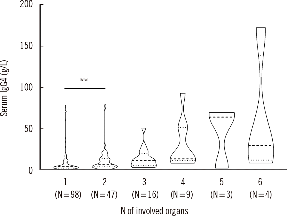
The clinical manifestations of the 177 IgG4-RD patients were systemic and considerably heterogeneous, since the disease may affect nearly every organ system. The number and type of involved organs in IgG4-RD patients varied. The proportion of IgG4-RD patients with involvement of one, two, three, four, five, and six organs was 55.4%, 26.6%, 9.0%, 5.1%, 1.7%, and 2.3%, respectively, with corresponding serum IgG4 concentrations of 4.25 (2.65-8.88) g/L, 6.54 (4.19-13.90) g/L, 11.60 (5.46-16.58) g/L, 13.80 (12.30-51.70) g/L, 64.40 (33.53-67.25) g/L, and 30.10 (19.07-71.53) g/L, respectively. The serum IgG4 concentration in the IgG4-RD group was significantly correlated with the number of organs involved (Spearman’s correlation coefficient=0.40, P<0.001). The top seven involved organs were the lymph nodes (39.0%), pancreas (26.6%), orbit (19.2%), submandibular gland (18.1%), bile duct (14.1%), lung (14.1%), and kidney (10.7%) (Supplemental Data Fig. S1) (Fig. 1).

Diagnostic performance of serum IgG4 concentration and the IgG4/IgG ratio for IgG4-RD

There was a significant difference of age and sex ratio between the IgG4-RD and non-IgG4-RD groups, and multiple regression analysis showed that sex was an independent predictor of the serum IgG4 concentration. This suggested that sex influences the serum IgG4 concentration, with males showing a higher serum IgG4 concentration than females (Supplemental Data Table S2). Therefore, ROC curve analysis was performed separately for each sex to evaluate the diagnostic accuracy of the serum IgG4 concentration and IgG4/IgG ratio in IgG4-RD patients (Supplemental Data Fig. S2). The individual distributions are shown in Supplemental Data Table S3. The area under the ROC curve (AUC) values of serum IgG4 concentration were similar in males and females, whereas the AUC value of the serum IgG4/IgG ratio for males was slightly higher than that for females. The optimal cut-off values of serum IgG4 concentration and the IgG4/IgG ratio for IgG4-RD diagnosis were 1.92 g/L and 0.12, respectively, in males and 1.83 g/L and 0.11, respectively, in females. The negative predictive values (NPVs) of serum IgG4 concentration and the IgG4/IgG ratio were all over than 97%, which were higher than the PPVs (Table 2).
We further performed ROC analysis for IgG4-RD diagnosis in patients with a serum IgG4 concentration >2.01 g/L (reference value) to evaluate whether the serum IgG4/IgG ratio could have better diagnostic performance for IgG4-RD diagnosis in these patients (Supplemental Data Fig. S3). The individual distributions are shown in Supplemental Data Table S3. As shown in Table 3, diagnostic efficiency analysis demonstrated that the AUC values of serum IgG4 concentration or the IgG4/IgG ratio for patients with serum IgG4 concentration >2.01 g/L decreased consistently for males, females, and the total population, with a maximum of 0.88. The cut-off values of serum IgG4 concentration and the IgG4/IgG ratio were >3.00 g/L and 0.19, respectively. In patients with serum IgG4 concentration >2.01 g/L, the PPVs of serum IgG4 concentration or the IgG4/IgG ratio were all over 0.88, which were higher than the NPVs (Table 3).

Association of IgG4 serum concentration with the treatment response in IgG4-RD patients

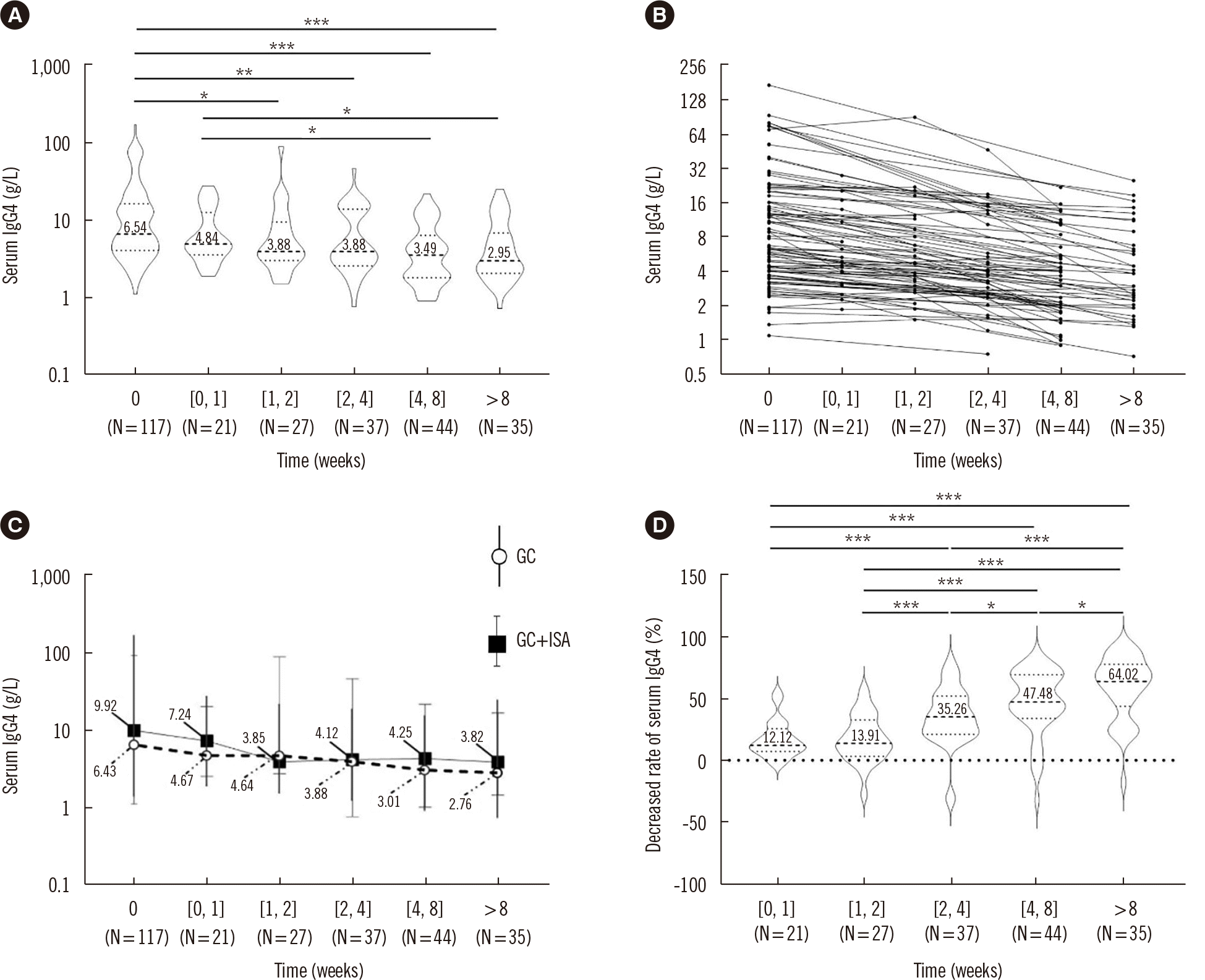
Dynamic analysis of the serum IgG4 concentrations measured at different time points during follow-up was performed for the 117 IgG4-RD patients initially treated with GCs. Patients were divided into a pre-treatment group (N=117), group 1 (0 week<post-treatment ≤1 week; N=21), group 2 (1 week<post-treatment ≤2 weeks; N=27), group 3 (2 weeks<post-treatment ≤4 weeks; N=37), group 4 (4 weeks<post-treatment ≤8 weeks; N=44), and group 5 (post-treatment >8 weeks; N=35). With increased time post-treatment, the median serum IgG4 concentration decreased and the decrease rate increased. The serum IgG4 concentration significantly decreased at >1 week post-treatment (P=0.004), and the change in the serum IgG4 concentration was similar between patients treated with a single GC and those treated with GCs plus ISAs, which indicated that the serum IgG4 concentration significantly decreased with a good response to treatment. Further analysis showed that the median decrease rate was close to 50% at >4 weeks post-treatment (Fig. 2).
We stratified the 117 IgG4-RD patients into four groups according to different pre-treatment serum IgG4 concentrations (upper limit of the reference value, and 2 or 5 times the upper limit of the reference value: IgG4 ≤2.01 g/L, 2.01-4.02 g/L, 4.02-10.05 g/L, >10.05 g/L) to observe the associations between pre-treatment serum IgG4 concentration and the decrease rate of serum IgG4 concentration over time. The decrease rate increased for a longer treatment time. However, the serum IgG4 concentration did not readily recover to the normal range after treatment for patients with a pre-treatment serum IgG4 concentration that was much higher than the reference range, even after 8 weeks of treatment (Supplemental Data Fig. S4).

DISCUSSION

It has been gradually recognized that the traditional criteria for IgG4-RD diagnosis are no longer applicable. The cut-off value of 1.35 g/L is based on a reagent used in radial immunodiffusion (Binding Site, Birmingham, UK) or ELISA (Yoshitomi Pharmaceutical Industries, Osaka, Japan) [6]. Exploration of an appropriate cut-off value and its clinical significance for different regions, nationalities, and reagents has gradually become a focus of attention in several recent studies [23-25].
In our study, the median age of the 177 IgG4-RD patients was 56 years and the male:female ratio was 2.22:1 (males accounted for 68.9% of all patients), which was similar to the results of Lin, et al. [26] and Xia, et al. [27]. Serum IgE and CRP concentrations and ESR were elevated and the complement C3 concentration was decreased in IgG4-RD patients, compared with the reference ranges (Table 1), which was in accordance with the results of Lin, et al. [26], Yamada, et al. [28], and Wallace, et al. [29]. Therefore, IgG4-RD predominantly occurs in older men and is characterized by humoral immune activation.
We retrospectively analyzed the clinical significance of serum IgG4 concentration in assisting in diagnosing IgG4-RD and the clinical characteristics of IgG4-RD patients. In our study, the most common symptom and manifestations were similar to those reported previously [26, 29], in which local mass was the most frequent symptom and the lymph node was the main organ involved. We found that 44.6% of IgG4-RD patients had more than two involved organs, which was similar to the study of Culver, et al. [14] (43.1% IgG4-RD patients had multiple-organ involvement). The serum IgG4 concentration increased with the numbers of involved organs (ρ=0.40, P<0.001), which was similar to the result of Culver, et al. [14]. However, Lin, et al. [26] found that up to 78.8% of IgG4-RD patients had multiple-organ involvement and the mean IgG4 concentration was 15.22 g/L. Together, these results suggest that the involvement of more organs could result in a higher serum IgG4 concentration, which can explain the difference in the results between our study and the study of Lin, et al. [26].
Diagnostic performance analysis indicated that the serum IgG4 concentration and the IgG4/IgG ratio have good diagnostic efficiency in assisting in the diagnosis of IgG4-RD in the adult population of southwest China. The AUC values of serum IgG4 concentration and the IgG4/IgG ratio were both over 0.95. Multi-factor analysis showed that sex was an independent factor influencing the serum IgG4 concentration, which was in line with the results of Harkness, et al. [30] and Puissant-Lubrano, et al. [31]. Harkness, et al. [30] used multivariate linear regression to determine that IgG and IgG subclass concentrations differ according to sex and race and found that male patients had higher IgG4 concentrations than female patients. Puissant-Lubrano, et al. [31] observed lower serum IgG4 concentrations in females than in males. Therefore, diagnostic performance analysis was conducted based on sex stratification. The AUC values of serum IgG4 concentration and the IgG4/IgG ratio were over 0.95 for both males and females. The optimal cut-off values of serum IgG4 concentration and the IgG4/IgG ratio for IgG4-RD diagnosis in males were 1.92 g/L and 0.12, respectively, and those in females were 1.83 g/L and 0.11, respectively. The cut-off value of serum IgG4 was slightly higher than the classic cut-off (1.35 g/L). The higher NPVs for the serum IgG4 concentration and IgG4/IgG ratio indicated that these indicators could be good exclusive biomarkers for IgG4-RD. Due to increased serum IgG4 concentrations in some patients of the non-IgG4-RD group, the diagnostic performance of the IgG4/IgG ratio for IgG4-RD diagnosis was further evaluated in patients with a serum IgG4 concentration >2.01 g/L. The AUC values of the IgG4/IgG ratio in males and females were 0.85 and 0.79, respectively, which did not improve the diagnostic efficacy of the IgG4/IgG ratio for IgG4-RD. This suggests that the IgG4/IgG ratio cannot improve the diagnostic efficiency of an IgG4-RD diagnosis, similar to the result of Carruthers, et al. [22].
Xia, et al. [27] reviewed the serum IgG4 concentrations and IgG4/IgG ratios in 133 IgG4-RD patients and 1,248 patients without IgG4-RD at Peking University People’s Hospital and found that the cut-off values of IgG4 concentrations and the IgG4/IgG ratio were 2.1 g/L and 0.11, with AUC values of 0.96 and 0.97, respectively. Carruthers, et al. [22] reviewed the medical records of 72 patients who had either probable or definite IgG4-RD and 308 patients without IgG4-RD at Massachusetts General Hospital and found that the sensitivity and specificity were 90% and 60% with serum IgG4 >1.35 g/L, which changed to 35% and 91%, respectively, when serum IgG4 concentrations were higher than 2.7 g/L; the specificity and sensitivity were 86% and 59%, respectively, when the IgG4/total IgG ratio was >0.08. They finally concluded that neither doubling the cut-off for serum IgG4 nor examining the serum IgG4/IgG ratio improves the overall test characteristics for the diagnosis of IgG4-RD. Li, et al. [32] defined the optimal cut-off value of 1.58 g/L for IgG4 concentrations by the ROC curve in the Chinese population when including the IgG4-RD group, other diseases groups, and healthy controls. We found that the optimal cut-off values of the serum IgG4 concentration and IgG4/IgG ratio for IgG4-RD diagnosis were 1.92 g/L and 0.12, respectively, in males and 1.83 g/L and 0.11, respectively, in females. For patients with serum IgG4 concentrations >2.01 g/L, the optimal cut-off values of serum IgG4 concentration and the IgG4/IgG ratio for IgG4-RD diagnosis were 3.07 g/L and 0.21, respectively, in males and 3.48 g/L and 0.20, respectively, in females. Our results were slightly different from those of Xia, et al. [27], Carruthers, et al. [22], and Li, et al. [32], which may be due to regional and ethnic differences, different sample sizes, different detection methods, as well as the stratification by sex that was only used in our study.
IgG4-RD patients have good responses to GCs [5]. According to the 2019 ACR/EULAR classification criteria for IgG4-RD [7], a lack of response to GCs serves as an exclusion criterion for the diagnosis of IgG4-RD. We found that serum IgG4 concentrations were significantly decreased in the IgG4-RD patients who showed good responses after initial treatment with GCs. Our results indicated that the serum IgG4 concentration can strikingly decrease at 2 weeks post-treatment and can decrease by nearly 50% at 4 weeks post-treatment. However, serum IgG4 concentrations were less likely to decrease to the reference range when the pre-treatment serum IgG4 concentration was higher than 10.05 g/L, even after 8 weeks of treatment (Supplemental Data Fig. S4). This may result from the long-lived plasma cells that are not affected by current treatments, which have migrated back to the bone marrow and continue to produce IgG4 [5]. More prospective studies are needed to explore the efficiency of serum IgG4 in the therapeutic monitoring of IgG4-RD. Since it is difficult for an elevated serum IgG4 concentration to recover to a normal level, the decrease rate of serum IgG4 may be more useful for monitoring efficacy.
Because this was a retrospective study, there are several limitations. First, it is possible that we may have easily missed some IgG4-RD patients. Second, our dynamic monitoring data were group-based data due to the retrospective design, and individual dynamic monitoring would be more helpful to evaluate the significance of serum IgG4 detection in therapy assessment. Finally, prospective studies with more patients are needed to validate our results.
In conclusion, we evaluated the diagnostic performance of serum IgG4 concentration and the IgG4/IgG ratio for IgG4-RD in adults of southwest China and found that an elevated serum IgG4 concentration was of great value for IgG4-RD diagnosis, whereas the IgG4/IgG ratio cannot improve the diagnostic efficiency for IgG4-RD. Different diagnostic cut-off values of serum IgG4 should be determined according to sex. The decrease rate of serum IgG4 concentration appears to be more conducive to monitoring the therapeutic efficacy than the serum IgG4 concentration alone.

ACKNOWLEDGEMENTS

We are grateful to Limei Luo, Huan Xu, and Yunfei An for their contributions to the detection of serum IgG4 and other laboratory indicators.

Notes

AUTHOR CONTRIBUTIONS

Wei B and Guo Y performed the data analysis and wrote the manuscript. Ou XQ and Lin LY helped perform the analysis with constructive discussions. Su ZZ, Li LX, and Wu XJ helped to review the relevant literature and checked the data and related calculations during the revision process. Cai B contributed to the conception of the study and revised the manuscript. All authors read and approved the final manuscript.

CONFLICTS OF INTEREST

The authors declare no conflicts of interest.

REFERENCES

1. Chen LYC, Mattman A, Seidman MA, Carruthers MN. 2019; IgG4-related disease: what a hematologist needs to know. Haematologica. 104:444–55. DOI: 10.3324/haematol.2018.205526. PMID: 30705099. PMCID: PMC6395313.
crossref
2. Kamisawa T, Zen Y, Pillai S, Stone JH. 2015; IgG4-related disease. Lancet. 385:1460–71. DOI: 10.1016/S0140-6736(14)60720-0. PMID: 25481618.
crossref
3. Miyabe K, Zen Y, Cornell LD, Rajagopalan G, Chowdhary VR, Roberts LR, et al. 2018; Gastrointestinal and extra-intestinal manifestations of IgG4-related disease. Gastroenterology. 155:990–1003.e1. DOI: 10.1053/j.gastro.2018.06.082. PMID: 30012334.
crossref
4. Perugino CA, Stone JH. 2020; IgG4-related disease: an update on pathophysiology and implications for clinical care. Nat Rev Rheumatol. 16:702–14. DOI: 10.1038/s41584-020-0500-7. PMID: 32939060.
crossref
5. Katz G, Stone JH. 2022; Clinical perspectives on IgG4-related disease and its classification. Annu Rev Med. 73:545–62. DOI: 10.1146/annurev-med-050219-034449. PMID: 34669430.
crossref
6. Umehara H, Okazaki K, Masaki Y, Kawano M, Yamamoto M, Saeki T, et al. 2012; Comprehensive diagnostic criteria for IgG4-related disease (IgG4-RD), 2011. Mod Rheumatol. 22:21–30. DOI: 10.3109/s10165-011-0571-z. PMID: 22218969.
crossref
7. Wallace ZS, Naden RP, Chari S, Choi HK, Della-Torre E, Dicaire JF, et al. 2020; The 2019 American College of Rheumatology/European League Against Rheumatism classification criteria for IgG4-related disease. Ann Rheum Dis. 79:77–87. DOI: 10.1136/annrheumdis-2019-216561. PMID: 31796497.
crossref
8. Ngwa T, Law R, Hart P, Smyrk TC, Chari ST. 2015; Serum IgG4 elevation in pancreatic cancer: diagnostic and prognostic significance and association with autoimmune pancreatitis. Pancreas. 44:557–60. DOI: 10.1097/MPA.0000000000000297. PMID: 25785724.
9. Pan Q, Guo L, Wu J, Cai J, Liao H, Lan Q, et al. 2016; Association between IgG4 autoantibody and complement abnormalities in systemic lupus erythematosus. Mediators Inflamm. 2016:2196986. DOI: 10.1155/2016/2196986. PMID: 27597802. PMCID: PMC4997081.
crossref
10. Takeoka S, Kamata M, Hau CS, Tateishi M, Fukaya S, Hayashi K, et al. 2018; Evaluation of IgG4+ plasma cell infiltration in patients with systemic plasmacytosis and other plasma cell-infiltrating skin diseases. Acta Derm Venereol. 98:506–11. DOI: 10.2340/00015555-2909. PMID: 29437186.
crossref
11. Yu KH, Chan TM, Tsai PH, Chen CH, Chang PY. 2015; Diagnostic performance of serum IgG4 levels in patients with IgG4-related disease. Medicine (Baltimore). 94:e1707. DOI: 10.1097/MD.0000000000001707. PMID: 26469909. PMCID: PMC4616795.
crossref
12. Xu WL, Ling YC, Wang ZK, Deng F. 2016; Diagnostic performance of serum IgG4 level for IgG4-related disease: a meta-analysis. Sci Rep. 6:32035. DOI: 10.1038/srep32035. PMID: 27558881. PMCID: PMC4997323.
crossref
13. Usami Y, Ichihara K, Uehara T, Sugano M, Ishimine N, Kawasaki K, et al. 2020; Evaluation of a novel serum IgG4 assay and determination of reference interval for the Japanese population. Clin Chim Acta. 501:136–41. DOI: 10.1016/j.cca.2019.10.032. PMID: 31730813.
crossref
14. Culver EL, Sadler R, Simpson D, Cargill T, Makuch M, Bateman AC, et al. 2016; Elevated serum IgG4 levels in diagnosis, treatment response, organ involvement, and relapse in a prospective IgG4-related disease UK cohort. Am J Gastroenterol. 111:733–43. DOI: 10.1038/ajg.2016.40. PMID: 27091321.
crossref
15. Chibbar R, Wright GR, Dokouhaki P, Dumanski S, Prasad B, Mengel M, et al. 2018; Recurrent IgG4-related tubulointerstitial nephritis concurrent with chronic active antibody mediated rejection: A case report. Am J Transplant. 18:1799–803. DOI: 10.1111/ajt.14758. PMID: 29607610.
crossref
16. Saeger W, Lohe B, Engels CL, Werner U. 2018; IgG4-associated adrenalitis-a case report. Endocr Pathol. 29:294–8. DOI: 10.1007/s12022-018-9531-x. PMID: 29766416.
crossref
17. Nagpal SJS, Sharma A, Chari ST. 2018; Autoimmune pancreatitis. Am J Gastroenterol. 113:1301. DOI: 10.1038/s41395-018-0146-0. PMID: 29910463.
crossref
18. Moon SH, Kim MH, Lee JK, Baek S, Woo YS, Cho DH, et al. 2017; Development of a scoring system for differentiating IgG4-related sclerosing cholangitis from primary sclerosing cholangitis. J Gastroenterol. 52:483–93. DOI: 10.1007/s00535-016-1246-5. PMID: 27470434.
crossref
19. Kamisawa T, Zen Y, Nakazawa T, Okazaki K. 2018; Advances in IgG4-related pancreatobiliary diseases. Lancet Gastroenterol Hepatol. 3:575–85. DOI: 10.1016/S2468-1253(18)30121-3. PMID: 30047448.
crossref
20. Lian M, Li B, Xiao X, Yang Y, Jiang P, Yan L, et al. 2017; Comparative clinical characteristics and natural history of three variants of sclerosing cholangitis: IgG4-related SC, PSC/AIH and PSC alone. Autoimmun Rev. 16:875–82. DOI: 10.1016/j.autrev.2017.05.018. PMID: 28564616.
crossref
21. Lanzillotta M, Mancuso G, Della-Torre E. 2020; Advances in the diagnosis and management of IgG4 related disease. BMJ. 369:m1067. DOI: 10.1136/bmj.m1067. PMID: 32546500.
crossref
22. Carruthers MN, Khosroshahi A, Augustin T, Deshpande V, Stone JH. 2015; The diagnostic utility of serum IgG4 concentrations in IgG4-related disease. Ann Rheum Dis. 74:14–8. DOI: 10.1136/annrheumdis-2013-204907. PMID: 24651618.
crossref
23. Wang H, Wang C, Wan Q, Li L. 2023; Roles of IgG4 and IgG4/IgG ratio to IgG4-related disease in patients with elevated serum IgG4 level. Clin Rheumatol. 42:793–800. DOI: 10.1007/s10067-022-06413-7. PMID: 36305979.
crossref
24. Usami Y, Sugano M, Uehara T, Koinuma M, Ishimine N, Kawasaki K, et al. 2021; Cut-off values of serum IgG4 among three reagents, including a novel IgG4 reagent: a multicenter study. Sci Rep. 11:7280. DOI: 10.1038/s41598-021-86024-5. PMID: 33790306. PMCID: PMC8012344.
crossref
25. Kwon OC, Park MC, Kim YG. 2022; Correlation between serologic parameters and disease activity of IgG4-related disease: differences between patients with normal and elevated serum IgG4 concentrations. Front Immunol. 13:1020459. DOI: 10.3389/fimmu.2022.1020459. PMID: 36311699. PMCID: PMC9608652.
crossref
26. Lin W, Lu S, Chen H, Wu Q, Fei Y, Li M, et al. 2015; Clinical characteristics of immunoglobulin G4-related disease: a prospective study of 118 Chinese patients. Rheumatology (Oxford). 54:1982–90. DOI: 10.1093/rheumatology/kev203. PMID: 26106212.
crossref
27. Xia CS, Fan CH, Liu YY. 2017; Diagnostic performances of serum IgG4 concentration and IgG4/IgG ratio in IgG4-related disease. Clin Rheumatol. 36:2769–74. DOI: 10.1007/s10067-017-3685-7. PMID: 28540606.
crossref
28. Yamada K, Yamamoto M, Saeki T, Mizushima I, Matsui S, Fujisawa Y, et al. 2017; New clues to the nature of immunoglobulin G4-related disease: a retrospective Japanese multicenter study of baseline clinical features of 334 cases. Arthritis Res Ther. 19:262. DOI: 10.1186/s13075-017-1467-x. PMID: 29191210. PMCID: PMC5709928.
crossref
29. Wallace ZS, Deshpande V, Mattoo H, Mahajan VS, Kulikova M, Pillai S, et al. 2015; IgG4-related disease: clinical and laboratory features in one hundred twenty-five patients. Arthritis Rheumatol. 67:2466–75. DOI: 10.1002/art.39205. PMID: 25988916. PMCID: PMC4621270.
crossref
30. Harkness T, Fu X, Zhang Y, Choi HK, Stone JH, Blumenthal KG, et al. 2020; Immunoglobulin G and immunoglobulin G subclass concentrations differ according to sex and race. Ann Allergy Asthma Immunol. 125:190–5.e2. DOI: 10.1016/j.anai.2020.03.018. PMID: 32224206. PMCID: PMC7387162.
crossref
31. Puissant-Lubrano B, Peres M, Apoil PA, Congy-Jolivet N, Roubinet F, Blancher A. 2015; Immunoglobulin IgA, IgD, IgG, IgM and IgG subclass reference values in adults. Clin Chem Lab Med. 53:e359–61. DOI: 10.1515/cclm-2014-1186. PMID: 25720098.
crossref
32. Li P, Liu Z, Wu Z, Wen X, Li L, Zhang S, et al. 2017; Adult reference intervals for IgG subclasses with Siemens immunonephelometric assays in Chinese population. Allergy Asthma Clin Immunol. 13:44. DOI: 10.1186/s13223-017-0216-7. PMID: 29042863. PMCID: PMC5629794.
crossref

Fig. 1
Violin plot showing the serum IgG4 concentrations of IgG4-related disease patients according to the number of involved organs. The tops and bottoms of the violin plots represent the minimum and maximum, respectively. The bold dotted line in the middle represents the median, and the dotted lines on either side represent the interquartile range covering 50% of the values. **P<0.01.
Abbreviation: IgG4-RD, IgG4-related disease.
alm-43-5-461-f1.tif
Fig. 2
Dynamic analysis of serum IgG4 concentration (A-C) and the decrease rate of serum IgG4 concentration post-treatment (D) in IgG4-RD patients treated with glucocorticoids. *P<0.05, **P<0.01, and ***P<0.001. 0: pre-treatment; [0, 1]: 0 weeks <post-treatment ≤1 week; [1, 2]: 1 week <post-treatment ≤2 weeks; [2, 4]: 2 weeks <post-treatment ≤4 weeks; [4, 8]: 4 weeks <post-treatment ≤8 weeks. For A and D, the tops and bottoms of the violin plots represent the minimum and maximum, respectively, the bold dotted line in the middle represents the median, and the dotted lines on either side represent the interquartile range covering 50% of the values.
Abbreviations: GC, glucocorticoid; ISA, immunosuppressive agent.
alm-43-5-461-f2.tif
Table 1
Laboratory findings of IgG4-RD and non-IgG4-RD patients
Reference range IgG4-RD (N=177) Non-IgG4-RD (N=877) P Reference range


N Median Interquartile range Total range N Median Interquartile range Total range
Male, N (%) 122 (68.9) NA NA NA 410 (46.8) NA NA NA < 0.001 NA
Age (yr) 177 56 47-65 18-83 877 49 38-60 18-88 < 0.001 NA
Serum IgG4 (g/L) 177 5.78 3.27-13.50 0.69-173.00 877 0.48 0.24-0.93 0.00-6.28 < 0.001 0.03-2.01
IgG4/IgG 158 0.27 0.17-0.75 0.05-2.69 877 0.04 0.02-0.07 0.00-0.31 < 0.001 NA
Serum IgG (g/L) 158 18.80 15.33-28.88 9.12-88.70 877 12.80 9.92-16.70 2.84-104.00 < 0.001 8.00-15.50
Serum IgE (IU/mL) 135 279 150.00-551.42 1.79-7,120.00 NA NA NA NA NA 5.00-150.00
CRP (mg/L) 116 7.73 2.45-40.58 1.00-321.00 NA NA NA NA NA < 5.00
ESR (mm/hr) 97 52 31-83 2-120 NA NA NA NA NA < 21
Serum C3 (g/L) 121 0.76 0.59-1.03 0.19-2.59 NA NA NA NA NA 0.79-1.52
Serum C4 (g/L) 121 0.16 0.12-0.22 0.02-0.46 NA NA NA NA NA 0.15-0.36
Serum IgA (mg/L) 146 2,015 1,320-3,057.5 436-8,360 NA NA NA NA NA 836-2,900
Serum IgM (mg/L) 146 1,065 718.50-1,557.5 254-5,950 NA NA NA NA NA 700-2,200
Eosinophils (%) 174 3.0 1.0-5.9 0.0-39.6 NA NA NA NA NA 0.4-8.0
Absolute eosinophils (109/L) 174 0.19 0.06-0.37 0.00-4.11 NA NA NA NA NA 0.02-0.52
CD3+ T cells (%) 128 71.6 63.8-77.2 24.6-90.8 NA NA NA NA NA 66.9-83.1
CD3+ CD4+ T cells (%) 128 42.7 34.8-48.8 11.6-68.3 NA NA NA NA NA 33.2-47.9
CD3+ CD8+ T cells (%) 128 22.5 16.9-27.3 6.2-56.0 NA NA NA NA NA 20.4-34.7
Absolute count of CD3+ T cells (cells/µL) 36 1,016 852-1,370 313-1,991 NA NA NA NA NA 941-2,226
Absolute count of CD3+ CD4+ T cells (cells/µL) 36 596 494-744 201-1,460 NA NA NA NA NA 471-1,220
Absolute count of CD3+ CD8+ T cells (cells/µL) 36 300 239-403 82-966 NA NA NA NA NA 303-1,003

Abbreviations: NA, not applicable; CRP, C-reactive protein; ESR, erythrocyte sedimentation rate; IgG4-RD, IgG4-related disease.

Table 2
Diagnostic performance of serum IgG4 concentration and IgG4/IgG ratio in patients with and without IgG4-RD, stratified by sex
Groups Serum IgG4 IgG4/IgG


AUC Cut-off value (g/L) Sensitivity (%) Specificity (%) PPV (%) NPV (%) AUC Cut-off value Sensitivity (%) Specificity (%) PPV (%) NPV (%)
All patients 0.98 1.73 94.9 90.3 66.4 98.9 0.96 0.11 89.2 88.8 59.0 97.9
Males 0.98 1.92 93.4 91.5 76.5 97.9 0.97 0.12 90.6 89.5 69.1 97.4
Females 0.97 1.83 94.6 91.7 57.1 99.3 0.95 0.11 84.6 91.0 51.2 98.2

Abbreviations: AUC, area under the curve; IgG4-RD, IgG4-related disease; PPV, positive predictive value; NPV, negative predictive value.

Table 3
Diagnostic performance of serum IgG4 concentration and IgG4/IgG ratio in patients with and without IgG4-RD, stratified by serum IgG4 concentration >2.01 g/L and sex
Groups Serum IgG4 IgG4/IgG


AUC Cut-off value (g/L) Sensitivity (%) Specificity (%) PPV (%) NPV (%) AUC Cut-off value Sensitivity (%) Specificity (%) PPV (%) NPV (%)
Total (IgG4 > 2.01 g/L) 0.88 3.62 78.9 85.5 92.7 63.4 0.83 0.20 77.10 84.1 91.0 63.7
Males (IgG4 > 2.01 g/L) 0.88 3.07 88.4 76.5 92.5 66.7 0.85 0.21 78.6 82.4 92.8 57.1
Females (IgG4 > 2.01 g/L) 0.88 3.48 79.6 85.7 88.6 75.0 0.79 0.20 69.6 91.4 91.4 69.6

Abbreviations: AUC, area under the curve; IgG4-RD, IgG4-related disease; PPV, positive predictive value; NPV, negative predictive value.

TOOLS
Similar articles