

 PDF
PDF Citation
 Print
Print


INTRODUCTION
Alzheimer’s disease (AD) is one kind of dementia with different clinical manifestations of cognitive impairment [1]. It is recognized as a series of age-associated abnormalities, such as heterogeneous interactions of neurodegeneration and vessel-related pathologies. Increased intracranial artery injury and capillary damage, such as atherosclerosis, arteriosclerosis, atrophy of the arterial walls, and reduction in the density of microvessels, have been found in AD patients [2]. This cerebrovascular dysfunction might be the initiative or synergistic factors of chronic hypoxia/ischemia resulting in neurodegeneration and cognitive impairment [3]. Therefore, the exploration of lifestyle modification and pharmacologic therapies in reducing cardiovascular and cerebrovascular diseases has been considered as an important way to defer the early pathologies in the process of dementia.
Functional extractives from the dietary consumption of food have various healthy beneficial components [4]. Soy foods, the traditional diets of humans, contain many constituents, such as isoflavones, lecithin, saponins, soy protein or peptides, and flavonoids, which have different biological activities [5]. Soy lecithin (SL) and soy isoflavone (SIF) have beneficial effects on many chronic diseases, including cardiovascular and neurodegenerative diseases. SL is called “phytotherapy”, and a lecithin-rich diet could regulate the hepatic lipoprotein metabolism and cholesterol homeostasis. The daily administration of SL may be used as adjuvant therapy for hypercholesterolemia [6]. Additional evidence shows that SL could modify the blood lipids and reduce vascular wall lesions by reducing the excess of low-density lipoprotein (LDL) cholesterol and promoting the synthesis of high-density lipoprotein (HDL) cholesterol [7]. In previous studies, SIF could reverse β-amyloid (Aβ)-induced neurotoxicity by enhancing the antioxidant ability of neurons and cerebrovascular endothelial cells, prohibiting the inflammation of astrocytes and microglia, and inhibiting the apoptosis of neurons and cerebrovascular endothelial cell in vivo and in vitro studies [8910].
Despite the beneficial effect, the long-term high-dose administration of SIF has harmful effects on health. Given the higher cost and side effect of drugs and the long-term high-dose administration of phytotherapy, it is necessary to explore alternative treatments for susceptible populations that should be much cheaper, safer, and more convenient for long-term daily use. In recent studies, the effects of combined administration of functional food on many chronic diseases have attracted considerable attention. Lecithin-formulated soy stanols could reduce cholesterol absorption and serum LDL cholesterol when consumed in fat-free foods [11]. Moreover, the SIF protein plus SL could improve the lipid profile of healthy postmenopausal women significantly. On the other hand, the favorable influence of the SIF protein plus SL on endothelial function could not be confirmed [12]. Thus, the research team tried to discover the role of soy isoflavone combined with soy lecithin (SIF + SL) on cognitive improvement and cerebrovascular protection. This study hypothesized that SIF+SL could restrain cognitive impairment by regulating the cerebral blood flow (CBF). This beneficial effect might be associated with cerebrovascular protection by mediating oxidation–reduction. In addition, the optimal combination of SIF and SL was determined in vivo and in vitro.
MATERIALS AND METHODS
Materials
SL was purchased from Shanghai Taiwei Pharmaceutical Company Limited (China). Acetone insoluble substance was 93.9%. The main chemical compositions included 84.2% phosphatidylcholine, 2.4% phospholipid acyl amine, and no phosphatidylinositol. SIF was purchased from ZHT Sci-Tech (Beijing) Co., Ltd. (China). The total isoflavone content of SIF was 92.15%. The main chemical compositions included 17.64% genistein, 53.24% daidzein, and 21.27% glycitein. SL and SIF were dissolved in 0.5% CMC-Na. Synthetic Aβ1-40 and Aβ25-35 were purchased from Sigma-Aldrich (St. Louis, MO, USA). They were dissolved in N.S. and incubated at 37°C for three days to form aggregation [13].
Experimental animals and design
Adult male Wistar rats weighing 250–300 g (SPF class) were provided by Beijing Vital River Laboratories Company (Beijing, China). The in vivo experiments were carried out using the protocol approved by the Chinese Committee on Experimental Animal Supervision. In this study, all rats were divided randomly into 5 groups (n = 12/group) including the control, Aβ1-42, SIF50 + SL40 (50 mg/kg·d SIF + 40 mg/kg·d SL), SIF50 + SL80 (50 mg/kg·d SIF + 80 mg/kg·d SL), and SIF50 + SL160 (50 mg/kg·d SIF + 160 mg/kg·d SL) according to the body weight. The procedure of an animal experiment was similar to a previous study [13]. The rats were treated with SIF + SL or 0.5% CMC-Na by daily intragastric administration for 14 days before Aβ1-40 injection surgery. After surgery, the same treatment was maintained for 28 days. On the 21st day after surgery, the Morris water maze test was performed for 6 days. On the 28th day after surgery, the CBF was measured. The brain tissue, blood, and serum were then collected.
Animal surgery
Surgery was carried out using the procedures established in a previous study [13]. Briefly, one hole located by a stereotaxic frame (Narishige, Tokyo, Japan) was drilled into the skull of the rats. The hole was mediolateral 1.8 mm from Bregma, anteroposterior −1.3 mm and dorsoventral −3.4 mm. An Aβ1-40 solution (10 μg/5 μL) or vehicle (normal saline 5 μL) was injected quickly into the lateral cerebral ventricle by a micro-injection (injecting for 3 minutes, holding for 5 min, and withdrawing slowly).
Morris water maze
On the 21st day after surgery, the learning and memory ability of the rats was evaluated using the Morris water maze. The test was conducted using the procedures of a previous study [9].
Laser speckle contrast imaging (LSCI) of cerebral blood flow
In this study, LSCI (Perimed, Järfälla, Sweden) was used to detect the changes in the CBF in the parietal cortex of the rats. The skull and the hole of the rats were exposed in a prostrate position after deep anesthesia. The probe of LSCI was positioned 10 cm above the target hole, which had been drilled in previous surgery at the parietal cortex of the left hemisphere. An approximate 0.8 mm2 size round region was located on the area of the hole. The imaging parameters included 2 minutes exposure, 21 pictures of the sampling frequency, 0.1 mm resolution ratio, and 30 imaging collections.
Hematoxylin-Eosin staining
Six rats from every group were selected randomly. Perfusion of fixative was not used to find cerebrovascular tissue exactly. Briefly, the brains of anesthetic rats were exposed rapidly. The dissected brains were immersed immediately in a 4% formaldehyde solution. Forty-eight hours later, the fixed brains were placed into 70% ethanol and embedded with paraffin. The paraffin sections (20 μm) were analyzed using the hematoxylin-eosin staining method.
Determination of oxidative damage level and anti-oxidative status in vivo study
Ethylenediaminetetraacetic acid anticoagulant blood was collected from the heart in vacuum tubes (BD Company, Franklin Lakes, NJ, USA). Blood was centrifuged 10 minutes by 3,000 rpm at 4°C. The serum (supernatant liquid) was collected. The 8-hydroxy-2′-deoxyguanosine (8-OHdG), which is a symbolic marker of mRNA oxidation, was measured using an enzyme-linked immunosorbent assay kit. A glutathione (GSH) and oxidized glutathione (GSSG) assay kit were used to test the redox status in the serum of rats. The superoxide dismutase (SOD) activity was detected using a hydroxylamine method assay kit. All assay kits were purchased from Nanjing Jiancheng Bioengineering Institute (Nanjing, China). The experimental procedures were executed strictly in accordance with the manufacturers’ instructions. A microplate reader (Infinite M200; TECAN, Männedorf, Switzerland) was used to take the measurements.
Cell culture
An immortalized mouse brain endothelial cell line (bEND.3 cells) was cultured in F12-Dulbecco’s Modified Eagle Medium with 10% fetal bovine serum, 100 U/mL penicillin, and 100 U/mL streptomycin (Gibgo; Thermo Fisher Scientific Inc., Waltham, MA, USA) medium. The culture condition is 37°C in a humidified atmosphere containing 5% CO2/95% air. The medium was changed every 2 days.
Screening pretreatment time and dose of SL in bEND.3 cells
The cell viability was used to select the suitable pretreatment time and SL dose (Sigma-Aldrich) and Gen (Sigma-Aldrich) in cells using the 3-(4,5-dimethylthiazol-2-yl)-2,5-diphenyltetrazolium bromide method (MTT assay). Briefly, pre-incubation doses of 10/100/1,000/2,000 μM SL were detected in pre-incubation times of 2/6/12/24 hours to make a rough estimate of the suitable SL pretreatment dose and time. Subsequently, 10/25/50/100/200/400 μM SL and 2/6 hours pretreatment were tested to determine the appropriate SL pretreatment dose and time in the in vitro study. Because of a previous exploration of Gen pretreatment dose [413], 50 μM of Gen was chosen in this case. The cells were incubated with/without SL or Gen at different times following exposure to 25 μM Aβ25-35 for another 24 hours. The medium was then changed to 100 μL of a 5 mg/mL MTT solution for further incubation of 4 hours at 37°C. A microplate reader was used to measure the absorbance at 570 nm.
Measurement of oxidative damage level and anti-oxidative status in vitro study
At the end of intervention incubation, the cells were collected to measure the intracellular levels of 8-OhdG, SOD, GSH, and GSSG as the second proof of the in vivo study. The method was the same as that in the in vivo study.
RESULTS
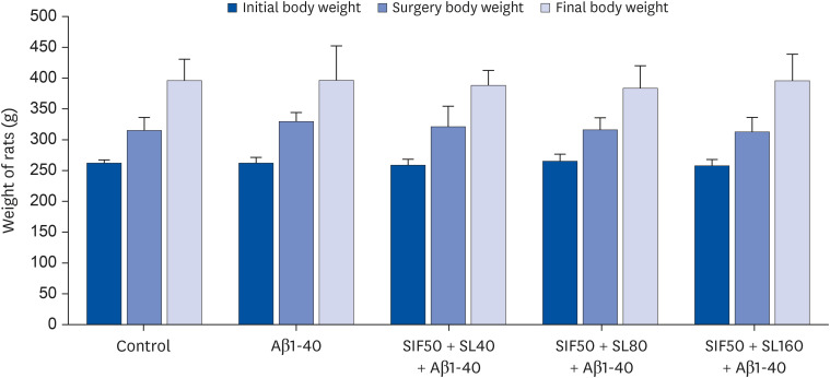
Effects of soybean isoflavones on Aβ1-42 mediated body weight in rats
The weight analysis results before, during, and after the animal experiment showed no statistical difference in the initial and surgery body weight of rats in each group (P > 0.05). There was no significant difference in the final body weight of rats in each group after preventive gavage, lateral ventricle injection, and postoperative gavage until the end of the experiment (P > 0.05). Repeated measure ANOVA was conducted to determine the weight changes in the experimental rats in each group two weeks before the Aβ1-40 injection and the weight changes among the groups affected by drugs after and during surgery. There was no statistical significance in the weight difference among the groups before SIF by intragastric administration, and there was no significant difference in body weight between the groups of Aβ toxicity and SIF protection after and during surgery (P > 0.05) (Fig. 1).
In vivo study
The protection of SIF + SL on the cognitive ability of rats
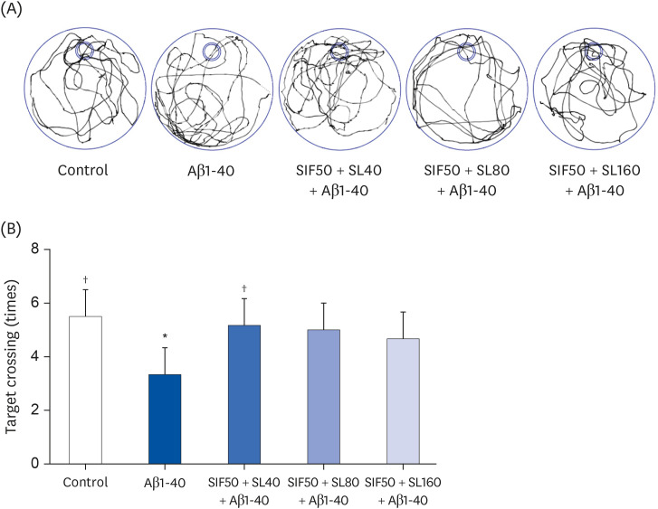
The learning and memory ability of rats was assessed using the Morris water maze test to determine the protective effect of 50 mg/kg·bw SIF combined with what level of SL on Aβ1-42-induced cognitive impairment of rats. The escape latency of rats in the Aβ1-40 group (modal group) was prolonged than that in the control group. On the other hand, a downtrend of escape latency was observed in three SIF+SL pretreatment groups (Fig. 2A). The target crossing times of the rats in the SIF50 + SL40, SIF50 + SL80, and SIF50 + SL160 groups were significantly longer than that in the Aβ1-40 group (Fig. 2B).
Fig. 2
Neuroprotective effect of SIF combined with SL on (A) swimming trace and (B) target crossing of learning and memory impairment rats induced by Aβ1-40. The control group was the rats treated with 0.5% CMC-Na and injected with normal saline; the Aβ1-40 group was rats treated with 0.5% CMC-Na and injected with Aβ1-40; the SIF50 + SL40 + Aβ1-40 group was the rats treated with SIF (25 mg/kg·d) + SL (40 mg/kg·d) and injected with Aβ1-40; the SIF50 + SL80 + Aβ1-40 group and the SIF50 + SL160 + Aβ1-40 group were the same as that shown above (n = 6 per group). All the data are reported as mean ± SD.
SIF, soy isoflavone; SL, soy lecithin; Aβ, β-amyloid.
*P < 0.05 compared with the control group; †P < 0.05 compared with the Aβ1-40 group.
The regulation of SIF + SL on CBF of rats
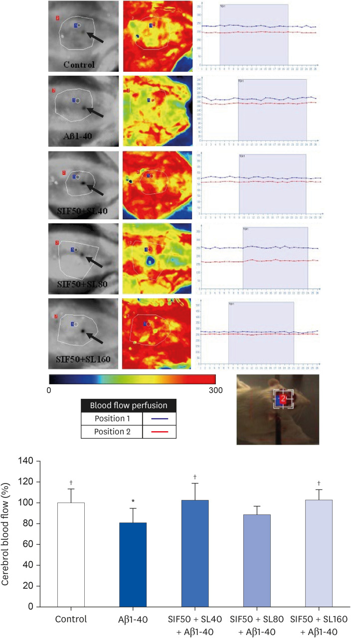
LSCI, which provides an index of blood flow based on speckle contrast analysis [14], was used to detect the CBF in the parietal cortex of rats in the present study and determine if the reduction of CBF existed in Aβ-induced cognitive impairment rats and if SIF + SL had positive effects on CBF. The result was presented in Fig. 3. The CBF of rats in the Aβ1-40 group was uncommonly lower than that in the control group. On the other hand, an enhancement of the CBF in the SIF+SL pretreatment groups and significant differences in the SIF50 + SL40 and SIF50 + SL160 groups were found compared to the Aβ1-40 group.
Fig. 3
Neuroprotective effect of SIF combined with SL on the CBF (%) of learning and memory impairment rats induced by Aβ1-40. The 3 pictures of every group are detected position image, CBF perfusion color image, and blood flow time window (TOI is 60 seconds detection time). Position 1 is the target of CBF detection. → showed marked bregma of rats. The control group was the rats treated with 0.5% CMC-Na and injected with normal saline; the Aβ1-40 group was the rats treated with 0.5% CMC-Na and injected with Aβ1-40; the SIF50 + SL40 + Aβ1-40 group was the rats treated with SIF (50 mg/kg·d) + SL (40 mg/kg·d) and injected with Aβ1-40; SIF50 + SL80 + Aβ1-40 group and the SIF50 + SL160 + Aβ1-40 group were the same as that shown above (n = 5 per group). All the data are reported as mean ± SD.
SIF, soy isoflavone; SL, soy lecithin; CBF, cerebral blood flow; Aβ, β-amyloid; TOI, time of interest.
*P < 0.05 compared with control group; †P < 0.05 compared with Aβ1-40 group.
The prevention of SIF + SL on cerebrovascular morphologic injury of rats
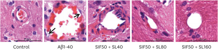
Cerebrovascular injury plays a vital role in abnormal CBF and nervous system damage. The result showed the attenuation of the endothelium in cerebral vessels, atrophy of cerebrovascular smooth muscle cells, aberrations in the capillary wall, narrowing of the capillary diameter, and even the reduction of cerebral microvessels in the rats of the Aβ1-40 group. On the other hand, these pathological changes were much less in the rats in the SIF+SL pretreatment groups, particularly in the SIF50 + SL40 and SIF50 + SL160 groups (Fig. 4).
Fig. 4
Neuroprotective effect of SIF combined with SL on brain tissue and cerebrovascular learning and memory impairment rats induced by Aβ1-40. The control group was the rats treated with 0.5% CMC-Na and injected with normal saline; the Aβ1-40 group was the rats treated with 0.5% CMC-Na and injected with Aβ1-40; the SIF50 + SL40 + Aβ1-40 group was the rats treated with SIF (50 mg/kg·d) + SL (40 mg/kg·d) and injected with Aβ1-40; SIF50 + SL80 + Aβ1-40 and SIF50 + SL160 + Aβ1-40 group were the same as that shown above (n = 3 per group). The staff gauge showed 50 μm at the right side of the bottom of every picture.
SIF, soy isoflavone; SL, soy lecithin; Aβ, β-amyloid.
Inhibition of SIF + SL on oxidative damage and anti-oxidative status of rats
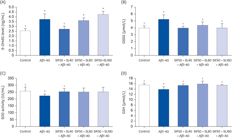
The 8-OhdG, which is the oxidative damage production marker of mRNA and GSSG, was tested to determine the effect of SIF + SL on preventing oxidative damage induced by Aβ, 8. In Fig. 5A and B, significant up-regulation of the 8-OhdG and GSSG levels was found in the serum of rats in the Aβ1-40 group compared with that in the control group. The results of SIF+SL pretreatment showed that the 8-OhdG level of the SIF50 + SL40 group was significantly lower than in the Aβ1-40 group. The GSSG showed a significant decrease in the SIF50 + SL40, SIF50 + SL80, and SIF50 + SL160 groups compared to the Aβ1-40 group. To understand how SIF + SL inhibits the oxidative damage of rats, SOD and GSH, which are the markers of two anti-oxidation pathways, were measured in Fig. 5C and D. The SOD level and GSH in the rats of the Aβ1-40 group were remarkably lower than that in the control group. On the other hand, the SIF+SL pretreatment results showed that the SOD level in the SIF50 + SL40 group was much higher than that in the Aβ1-40 group. Similarly, higher SOD levels appeared in the SIF50 + SL160 group than in the Aβ1-40 group, but it was not significant (P = 0.052). The GSH showed a significant increase in SIF50 + SL40, SIF50 + SL80, and SIF50 + SL160 groups compared to the Aβ1-40 group.
Fig. 5
Neuroprotective effect of SIF combined with SL on RNA oxidative damage maker 8-OHdG (A), GSSG (B), SOD (C), and GSH (D) levels in the plasma of learning and memory impairment rats induced by Aβ1-40. The Control group was the rats treated with 0.5% CMC-Na and injected with normal saline; the Aβ1-40 group was the rats treated with 0.5% CMC-Na and injected with Aβ1-40; the SIF50 + SL40 + Aβ1-40 group was the rats treated with SIF (50 mg/kg·d) + SL (40 mg/kg·d) and injected with Aβ1-40; the SIF50 + SL80 + Aβ1-40 group and SIF50 + SL160 + Aβ1-40 group were the same as that shown above (n = 5 per group). All the data are reported as mean ± SD.
SIF, soy isoflavone; SL, soy lecithin; 8-OHdG, 8-hydroxy-2′-deoxyguanosine; GSSG, oxidized glutathione; SOD, superoxide dismutase; GSH, glutathione; Aβ, β-amyloid.
*P < 0.05 compared with control group; †P < 0.05 compared with Aβ1-40 group.
In vitro study
Screening SL pretreatment dose and time in bEND.3 cells
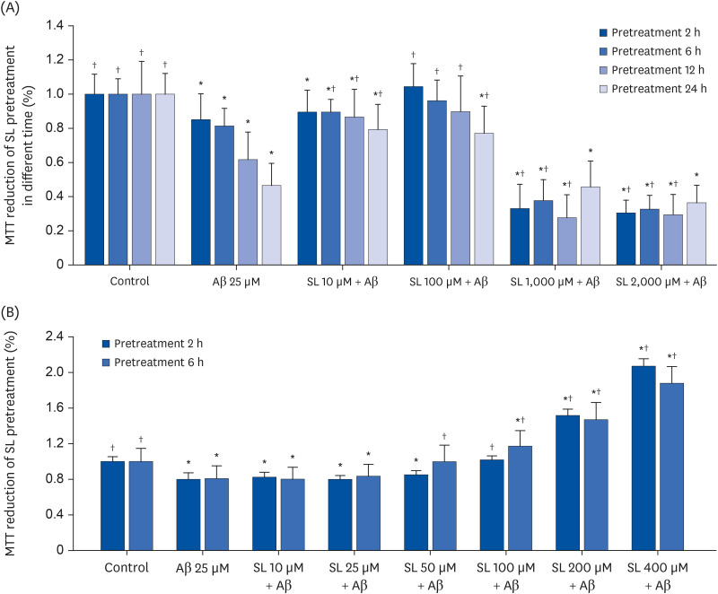
The viability of bEND.3 cells was examined to assess the SL pretreatment dose and time in bEND.3 cells. The 25 μM Aβ25-35 (Aβ25 group) showed a significant decrease in the cell viability in a time-dependent manner (Fig. 6A). The SL intervention showed that a 2 hours pretreatment of SL was sufficient to prevent the toxicity of Aβ25-35 on the cell viability (Fig. 6). The first rough estimation showed the effective dose of SL might be more than 10 μM and less than 1,000 μM (Fig. 6A). The test of the appropriate pretreatment dose showed that 50 and 100 μM of SL could reverse the decreased cell viability induced by Aβ25-35 alone (Fig. 6B). Therefore, 25, 50, and 100 μM were chosen as the low, medium, and high dose of the combination.
Fig. 6
Cell viability of bEND.3 cells in different pretreatment doses of SL or Gen at different times. (A) Cell viability of bEND.3 cells from untreated cells (control group), cells exposed to 25 μM Aβ25-35 (Aβ25 group), and cells exposed to 10/100/1,000/2,000 μM SL 2/6/12/24 hours before 25 μM Aβ25-35 was added (SL10/100/1,000/2,000 μM + Aβ groups). (B) Cell viability of bEND.3 cells from untreated cells (control group), cells exposed to 25 μM Aβ25-35 (Aβ25 group), and cells exposed to 10/25/50/100/200/400 μM SL 2/6 hours before 25 μM Aβ25-35 was added (SL10/25/50/100/200/400 μM + Aβ groups). All data are reported as the mean ± SD.
SL, soy lecithin; Gen, genistein; Aβ, β-amyloid; MTT, 3-(4,5-dimethylthiazol-2-yl)-2,5-diphenyltetrazolium bromide.
*P < 0.05 compared to the control group; †P < 0.05 compared to the Aβ25 group.
Rectification of SIF + SL on oxidative damage and anti-oxidative status of bEND.3 cells: the second proof of the in vivo study
As the second proof of SIF+SL on the redox system of health found the in vivo study, the oxidative damage (8-OhdG and GSSG) and anti-oxidative status (SOD and GSH) in Fig. 7 of bEND.3 cells were tested. In the Aβ25 group, the levels of 8-OhdG and GSSG were significantly higher than that in the control group. On the other hand, the SOD levels and GSH in the Aβ25 group were much lower than those in the control group.
Fig. 7
Neuroprotective effect of SIF combined with SL on RNA oxidative damage maker 8-OHdG (A), GSSG (B), SOD (C), and GSH (D) levels in bEND.3 cell. The control group was untreated cells; the Aβ25 group was the cells exposed to 25 μM Aβ25-35; the Gen50 + SL25 + Aβ group was the cell exposed to 25 μM Aβ25-35 and treated with Gen (50 μM) + SL (25 μM); the Gen50 + SL50 + Aβ group and Gen50 + SL100 + Aβ group were the same as that shown above. All data were shown as mean ± SD.
SIF, soy isoflavone; SL, soy lecithin; 8-OHdG, 8-hydroxy-2′-deoxyguanosine; GSSG, oxidized glutathione; SOD, superoxide dismutase; GSH, glutathione; Aβ, β-amyloid.
*P < 0.05 compared with control group; †P < 0.05 compared with Aβ1-40 group.
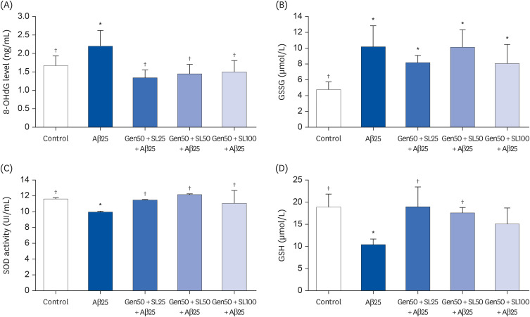
The Gen + SL pretreatment results showed that the 8-OhdG levels in Gen50 + SL25 (50 μM Gen + 25 μM SL), Gen50 + SL50 (50 μM Gen + 50 μM SL), and Gen50 + SL100 (50 μM Gen + 100 μM SL) groups were lower than that in the Aβ25 group. The GSSG showed a similar trend. The SOD levels in the Gen50+SL25, Gen50+SL50, and Gen50+SL100 groups showed a significant increase compared to that in the Aβ25 group. The GSH of the cells in the Gen50 + SL25 and Gen50 + SL50 groups showed an upward adjustment compared to the Aβ25 group.
DISCUSSION
Cognitive impairment is the main clinical manifestation in a series of events in the development of AD. The vascular factor is one of the vital pathogenesis of AD [413]. Evidence shows that more than 90% of AD patients have cerebral amyloid angiopathy, and up to 60% have ischemic white matter damage [15]. Moreover, some researchers declare that a dysfunction of the vascular system might be an initial factor for AD development. Increased atherosclerotic plaque accumulation is found in the main cerebral artery of AD patients [16]. In addition, Aβ could lead to an injury to the brain vessel wall and oxidative damage to cerebrovascular endothelial cells in vivo and in vitro [417]. These cerebrovascular abnormalities cause the progressive consequence of decreased CBF. Ischemia is the defining pathogenesis of vascular dementia, but evidence suggests that it contributes to the development of AD [15]. In recent years, phytotherapy research has confirmed that many biological components of plants, such as soy isoflavone, lycopene, lecithin, and turmeric, have beneficial effects on atherosclerotic, cardiovascular disease, ischemia, and AD [51218]. A previous study showed that 50 mg/kg·d SIF or Gen supplementation could benefit the impaired brain function or cells [1920]. On the other hand, there is little evidence showing the effect of soy isoflavone plus soy lecithin on the impairment of cognition and abnormal CBF. This study suggested that SIF + SL could prevent hypoperfusion and cognitive defects. In addition, these beneficial effects might be attributed to its antioxidant activity for protecting cerebrovascular, which was verified both in vivo and in vitro studies. The administration of 50 mg/kg·d SIF or Gen and a low dose of SL was the optimal combination in the rat experiments and cell tests.
Learning and memory impairment is present from the extremely early pre-clinical phase of AD, which is called mild cognitive impairment (MCI), and persists for every progressive stage with increased impairment in the development of AD [21]. Given the irreversible loss of cognition in AD patients, preventive methods need to be explored, such as introducing functional food to the daily diet of elderly and susceptible people. There is evidence to show that 6 months of soybean-derived phosphatidylserine (Soy-PS) treatment in both supplement doses of 100 and 300 mg/day could enhance the memory functions of the elderly with memory complaints (MCI). Researchers have declared that Soy-PS is a safe food ingredient [22]. Previous studies have shown that a single SIF treatment (80 mg/kg·d) could protect learning and memory ability, brain tissue, and the cerebrovascular wall of rats damaged by Aβ1-42 [1013]. The present study focused on the combined effects of SIF and SL on cognitive impairment. The results showed that a SIF + SL pretreatment could significantly increase the target crossing times and shorten the total distance of rats compared to the Aβ1-40-induced impairment rat model. These results suggest that a combination treatment with a lower dose of SIF and SL could prevent Aβ1-40-induced impairment of spatial learning and memory capability. In addition, there were no growth side effects (body weight) and vital signs during SIF + SL treatment during the experiment. This is similar to the experiment showing that quercetin plus alpha-tocopherol could prevent the accumulation of lipids and glycoprotein components in rats with isoproterenol-induced myocardial infarction through anti-lipid peroxidation. Furthermore, a combined pretreatment was better than a single pretreatment [23].
Cerebrovascular abnormalities are common in AD [15], and over time, these pathological changes could lead to catastrophic neuronal damage, dysfunction, and progressive degeneration, which explain the cognitive impairment. In research on the cerebrovascular-related pathogenesis of AD, it was reported that in animal studies, a decrease in CBF may precede the development of dementia [24] and manifest well before behavioral or pathological changes [2526]. Furthermore, disrupted perfusion occurs in MCI patients and in healthy people who have a high risk of AD [27]. It is unclear if CBF is an effective earlier therapeutic target for Aβ-induced cognitive impairment. There is evidence that the administration of lecithin (300 mg/kg) and alpha-tocopherol (200 mg/kg) alone or in combination could alleviate ischemia/reperfusion injury in rat brains through their antioxidant action [28]. In a previous study, 0.7 mg/kg folic acid (FA) + 160 mg/kg SIF supplementation resulted in a lower incidence of cyclophosphamide-induced post-neural tube closure defects in rodents [29]. In the present study, Aβ1-40 could lead to a decrease in CBF in the parietal cortex of rats, but the SIF + SL could prevent the toxicity of Aβ1-40 on CBF. Hence, the administration of SIF + SL could alleviate the reduction of CBF induced by the accumulation of Aβ and might be one target for SIF + SL to protect the cognitive ability of rats.
The 8-OHdG and GSSG, reflecting the chronic oxidative damage of subjects, were detected to explore how SIF + SL inhibits the morphological injury induced by Aβ1-40. In addition, with the production of RNA and GSH oxidation, lower levels of 8-OHdG and GSSG were observed in the SIF + SL pretreatment groups. The results suggested that SIF + SL might protect the cells of cerebrovascular tissue through its anti-oxidative capability. Different levels of SOD and GSH in the serum of animal models and SIF + SL groups confirmed this speculation. This judgment is similar to a previous study, showing that FA+SIF supplementation during pregnancy could decrease the damage of the CPA, such as damaged nuclear DNA, early apoptotic morphological changes, and Bax and P53 expression on embryo brains [29]. In addition, the analogous evidence showed that SL + vitamin E could restore CAT activity with a slight tendency to normalize SOD activity in the rat brain during ischemia/reperfusion [28]. Moreover, supplementation of soy isoflavone protein (12.95 mg) + SIF (50 mg) in the sample of healthy postmenopausal women could increase the high-density lipoprotein/low-density lipoprotein ratio significantly [12].
As the second proof of the results in vivo study, the cerebrovascular protective effect of SIF + SL was confirmed using an immortalized mouse brain endothelial cell line (bEND.3 cell). First, the appropriate dose of cell treatment was selected. The results showed that 50 and 100 μM of SL could protect the viability of cells separately. Thus, 100 μM of SL was chosen as the dose of the high combination, 50 μM as the medium dose and 25 μM as the lower dose. A Gen + SL pretreatment was performed for 2 hours before Aβ25-35 was added. The results of 8-OHdG and GSSG indicated that the treatment of Gen + SL could effectively inhibit the oxidative damage induced by Aβ25-35, which resulted in cerebrovascular endothelium injury. The anti-oxidative capability of cells indicated by SOD and GSH could be increased significantly by the treatment of Gen + SL. A review showed genistein could exert an anti-oxidant effect in Aβ-induced oxidative damage in PC12 cells, which could serve as a model for Alzheimer disease [30]. The results of the in vitro study dramatically verified the deduction of in vivo study that Gen + SL could prevent the damage of cerebrovascular endothelial cells induced by Aβ by upregulating the antioxidant activity of cells. This cerebrovascular protection might be the mechanism through which Gen + SL maintains the CBF and improves the cognitive ability of rats.
The aim of this study was to provide essential data for the next exploration of middle cerebral artery functional protection of SIF + SL and the further detection of SIF + SL supplementation to AD high-risk people or MCI patients. Thus, the optimal combination of SIF and SL was investigated in both in vivo and in vitro studies. The results showed that SIF50 + SL40 in the rat experiment and Gen50 + SL25 in the cell test were the best joint doses for alleviating cognitive impairment and regulating CBF by protecting the cerebrovascular tissue through its antioxidant activity. These effective doses of SIF/Gen and SL are much lower than the single effective treatment dose of SIF or SL reported elsewhere. For example, 100 and 300 mg/day supplementation of soybean-derived phosphatidylserine could protect the memory of the elderly [22]. The administration of lecithin (300 mg/kg) and alpha-tocopherol (200 mg/kg) alone or in combination could alleviate ischemia/reperfusion insults in rat brains [28]. Combination treatment studies showed that SL (500 mg/kg), SL (500 mg/kg) + vitamin B complex and tocopheryl acetate (80 mg/kg) could maintain the normal level of aspartate transaminase and alanine aminotransferase activities, SOD activity, GSH content, and thiobarbituric acid reactive substances level, which are disordered by ethanol feeding in BALB/c mice [31]. Evidence shows that the combination of SIF and SL could be used to prevent osteoporosis in mice [32]. Furthermore, a previous study showed that SIF (80 mg/kg) could protect neurons and cerebral vessels in rats with cognitive impairment induced by Aβ [1013]. Hence, SIF combined with SL could protect cognition and CBF in a lower and much safer dose. The combined treatment was better than single supplementation. Moreover, the lack of growth side effects (body weight) and vital signs during the SIF + SL treatment suggested that SIF + SL might allow long-term supplementation in the elderly, even in middle-aged people with a high risk of AD. This deduction needs more evidence and human exploration.
In conclusion, SIF + SL could significantly prevent the cognitive defects induced by Aβ by regulating CBF, which might be attributed to its antioxidant activity in protecting cerebral vessels. In this study SIF50 + SL40 in the rat experiment and Gen50 + SL25 in the cell test were the optimal combination for alleviating cognitive impairment and regulating CBF by prohibiting cerebrovascular tissue oxidative damage induced by Aβ.
 XML Download