This article has been corrected. See "Erratum: Replantation of autotransplanted mature third molar in anterior open bite patient: case report" in Volume 39 on page 104.
Abstract
Autotransplantation of third molars with completely formed roots is known to be effective and provide a high long-term success rate. However, in case of severe mobility or unexpectedly extraction is observed during the monitoring period after surgery, it is generally considered as a failure. This case report describes successful replantation of autotransplanted mature third molar into surgically created molar socket. 1 year follow up of transplanted tooth showed clinically normal periodontal pocket depth and tooth mobility. Root resorption or bone loss were not observed. Provided that there is no apparent sign of inflammation, re-insertion into socket is a viable alternative to immediate determination of extraction.
초록
성숙된 치근을 갖는 제3대구치를 이용한 자가치아이식술은 효과적이며, 장기간 높은 성공률 보여주는 것으로 알려져 있다. 자가이식술 이후 생착 여부에 대해 경과 관찰하는 기간동안 치아가 심한 동요도를 보이는 경우, 이를 실패로 간주하고 발치를 하는 것이 일반적이다. 하지만, 이번 증례에서는, 자가치아 이식술 후 치아가 고정이 되지 않아, 구강 외로 탈락되었으나, 시기 적절히 재식을 시도하여 다시 생착을 획득하였고, 1년 이후까지도 성공적인 임상 결과를 보였다. 재식하였지만, 치근 흡수나 골소실도 보이지 않았다. 자가치아이식 후 교합적 문제로 고정을 보이지 않으나, 염증소견이 없는 치아라면 치조와에 재식하는 것이 발치에 앞선 대안이 될 수 있다.
Optimal treatment options for a non-restorable tooth include 1) extraction and replacement either using a single-tooth implant or a fixed dental prosthesis 2) extraction without replacement and 3) transplantation. Among these options, tooth autotransplantation is defined as ‘transplantation of an unerupted or erupted tooth in the same individual, from one site to another or to a new surgically prepared socket’. The first recorded surgery with details about tooth bud transplantation was performed by the French dentist Ambroise Pare in 1564. Afterwards, a molar transplantation technique was described in 1956, the general guidelines of which remain very similar.1 When choosing autotransplantation for a patient with a non-restorable tooth, comprehensive case evaluation including surgeon’s skill and knowledge, patient selection, local inflammatory status, endodontic treatment, and availability of the periodontal ligament (PDL) in both the donor and recipient sites is important.
Autotransplantation has shown higher success rates with recent advancements in technologies including cone-beam computerized tomography (CBCT) and better biological understanding. According to one meta-analysis, cases with 1- and 5-year follow‑ups have been reported with a success rate of 81 - 98%.2 Tooth autotransplantation with complete root formation is a favorable treatment with rare failure.3 Even with this reported high success rate, a small percentage of failures leading to extraction of the transplanted tooth exists for several reasons. Among a total of 182 cases, extraction was conducted in nine failed cases (4.5%).4 According to another report, of 366 observed transplanted teeth, 10 failed and were lost due to unsuccessful PDL regeneration and persistent mobility grade III (or greater).5 Among the various failure criteria that leads to an inevitable extraction, stability loss is a primary one. However, successful immediate replantation of an autotransplanted tooth that exhibited noticeable mobility and fell out of the socket in an open bite patient has never been reported. Therefore, this case report is aimed at demonstrating the one-year follow-up of successful replantation of an autotransplanted maxillary third molar into a mandibular first molar extraction site.
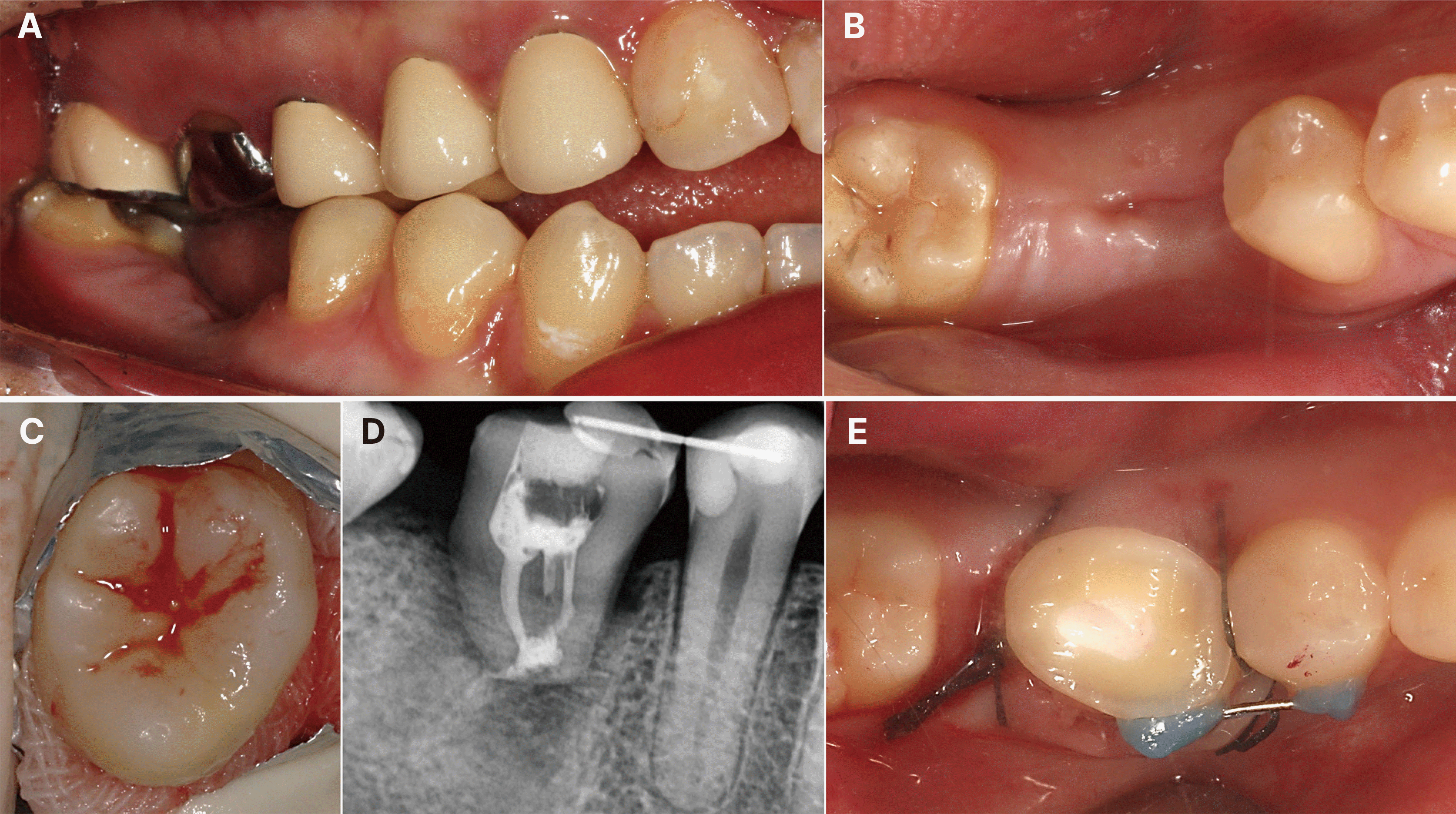
This case report was written according to the Preferred Reporting Items for Case reports in Endodontics (PRICE) 2020 guidelines. A 22-year-old Asian female presented for evaluation of her right mandibular first molar (#46) that suffered a crown fracture due to extensive caries and infection of the root canal system with chronic apical periodontitis (Fig. 1A and 1B). The patient was classified as ASA I with no history of smoking. The tooth did not exhibit pain on percussion or mobility. Fortunately, she had three third molars, which seemed to have favorable root forms and development (Fig. 1A), as potential donor teeth for autotransplantation. The patient’s compromised first mandibular molar with poor potential endodontic and restorative treatment represents typical indications for autotransplantation. Therefore, treatment options that aim to replace the severely compromised tooth were proposed as 1) Extraction without replacement 2) Extraction and replacement using a single-tooth implant and 3) Autotransplantation using a third molar. After discussion, the patient chose extraction only and wished to postpone the replacement decision. Therefore, after performing an inferior alveolar nerve block with local anesthesia using three ampules of 2% lidocaine with 1:100,000 epinephrine, luxation and extraction of the roots of #46 were performed followed by delicate curettage to remove the periapical granulation tissue (Fig. 1C). After extraction, the need for orthodontic treatment was emphasized. Consultation with an orthodontist was strongly recommended to the patient prior to restoring her posterior occlusion.
Two months later, the patient suddenly presented to the clinic with an intention to undergo autotransplantation surgery (Fig. 2A, 2B). She brought a consultation paper from her orthodontist stating that tooth #48 was planned to be included in the future orthodontic treatment. Therefore, tooth #48 was excluded as a candidate for autotransplantation. Tooth #38 was also excluded due to its mesial impaction with unfavorable prognosis during the extraction procedure. Information on the risks and benefits of autotransplantation using tooth #18 with a relatively conical shape were provided to the patient. Small-field of view preoperative CBCT was performed to create a resin replica of the donor tooth with three-dimensional (3D) information about the anatomy of the tooth and its surrounding structures.
To decrease the intraoral bacterial load, the patient underwent an oral hygiene session including supragingival scaling and chlorhexidine (0.12%) use one week prior to surgery. The patient consented to the autotransplantation procedure before the surgery. The surgical sites were disinfected using chlorhexidine saturated cotton balls (0.12%) to maintain a clean field during the procedure. A horizontal incision on the healed ridge was made. The recipient site was prepared with a low-speed handpiece (20,000 rpm) with a carbide round bur under copious irrigation. Try-in of the handmade resin replica in the recipient socket was performed, but it could not be placed into the appropriate position. Due to this incompatibility between the receiving alveolus and the replica, additional alveoloplasty was performed to reduce the inter-radicular septum and widen the receiving bed. After recipient site preparation, an intrasulcular incision was created at tooth #18 to interrupt the circular fibers of the ligament. Luxation and extraction were performed, avoiding contact with the radicular surface to prevent damaging the PDL cells. The donor tooth was wrapped with gauze soaked with a sterile saline solution to preserve the vitality of the PDL cells (Fig. 2C). After extraoral endodontic treatment and apicoectomy using MTA, the donor tooth was placed in the recipient site as soon as possible. The total duration from extraction to stabilization (extraoral time) was less than 13 minutes. Since analysis of the CBCT revealed the oval-shaped crown form of #18, the transplant was rotated 90° due to the narrow recipient ridge (Fig. 2E). Single mesial and distal stitches were made with black silk 4/0 sutures in an interrupted manner. Once hemostasis was under control, the operative field was dried. Since the transplanted tooth exhibited slight mobility, semirigid splinting using composite and steel wire (diameter 0.3 - 0.4 mm) was applied. To prevent excessive trauma, selective adjustment was performed on its occlusal surface until minimum sub-occlusion was obtained. A periapical postoperative radiograph was taken to corroborate the position of the tooth in the socket (Fig. 2D). Antibiotic and antiseptic therapy was prescribed after the surgery. Medical prescriptions included the antibiotic amoxicillin 500 mg (one capsule three times per day for three days), the non-steroidal anti-inflammatory drug dexibuprofen 400 mg (one tablet three times per day for three days), and mouthwash with chlorhexidine gluconate 0.12% (twice a day for seven days).
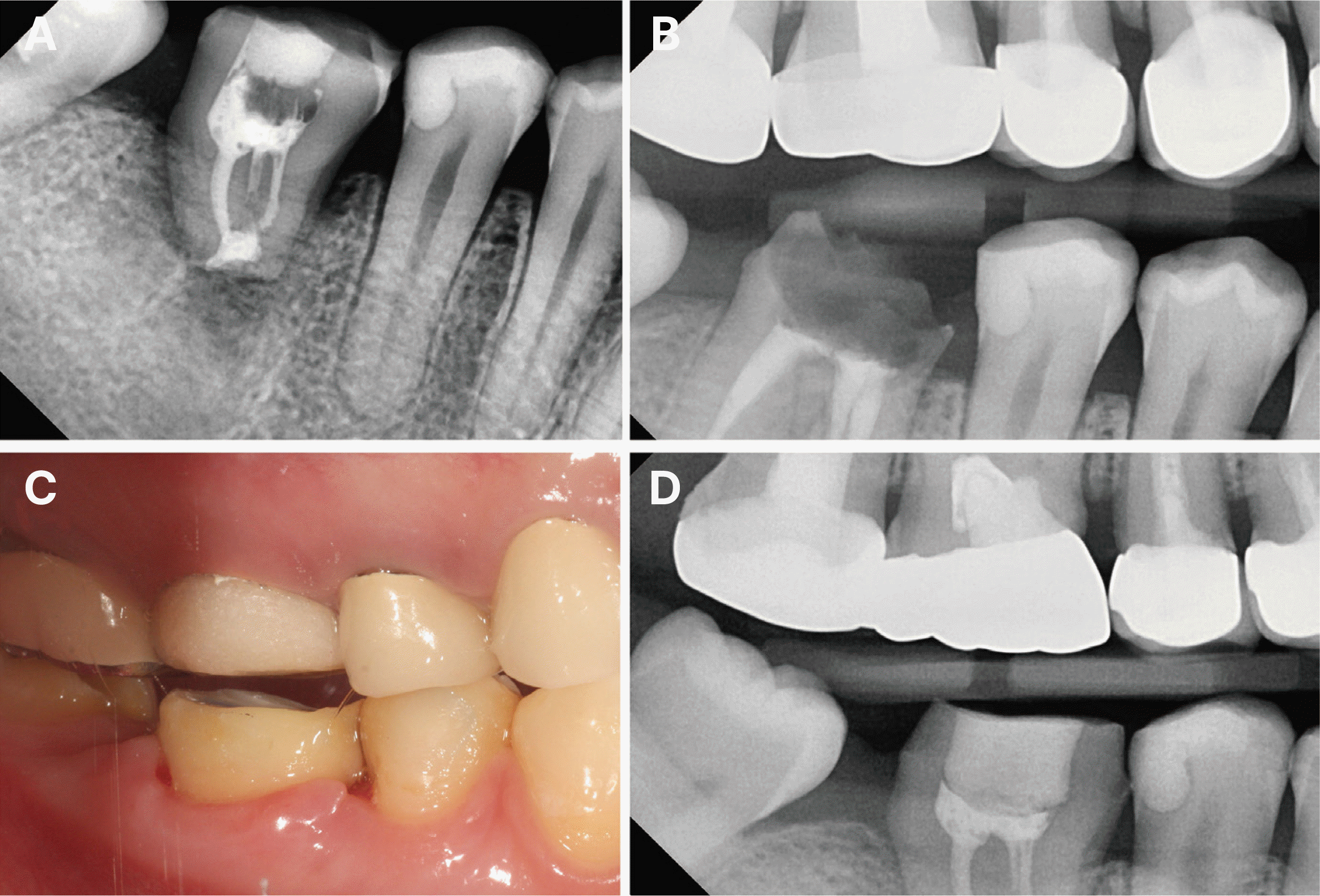
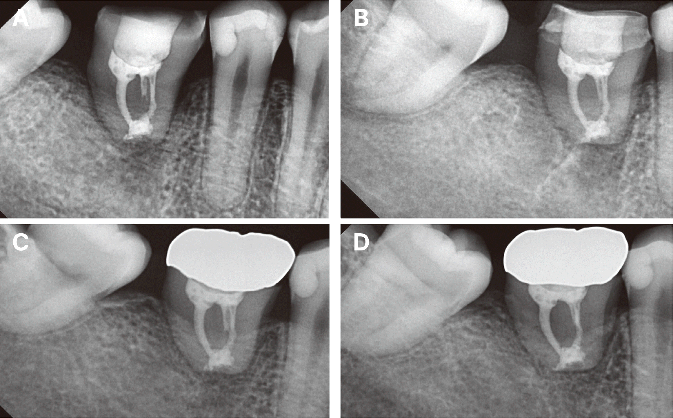
Twenty days after the surgery, the patient presented to the clinic for splint removal and did not report any significant discomfort. To assess tooth’s mobility, splint removal was initiated. Immediately, however, the transplanted tooth was unexpectedly slipping out of the socket. The tooth was immediately washed with sterile saline and reinserted in the socket with digital pressure (Fig. 3A). When occlusion was analyzed, the opposing maxillary first molar restored with a metal crown had no occlusal contact. Nevertheless, the old crown was removed and replaced with a provisional resin crown to ensure definitive disocclusion (Fig. 3C). After splinting for two more weeks, stability was confirmed on the autotransplanted tooth. During the second splinting period, a new crown was fabricated and placed on the opposing first molar (Fig. 3D). Next, the splint was removed (Fig. 4A). Another two weeks of monitoring of the provisionally crowned replanted tooth (#46) demonstrated acceptable stability under occlusal function with the opposing tooth (Fig. 4B). At 3.5 months after surgery, the patient stated that she was comfortable with chewing (Table 1). After confirming painless biting with the transplanted tooth and favorable bone healing, final placement of a gold crown was performed (Fig. 4C). Since then, the patient has exhibited improved oral hygiene care and high compliance with the regular recall program. The radiograph revealed no pathological features at the one-year follow‑up (Fig. 4D). The transplanted tooth has been functioning without any symptoms (Fig. 5).
In the event of tooth extraction, procedures such as decoronation and orthodontic extrusion may be useful to preserve the hard and soft tissues for future dental implant placement. Another preservation option is dental autotransplantation (DAT), which provides a chance to retain the natural tooth in the dentition for a longer period of time. In adult patients, autotransplantation of third molars with completely formed roots is effective in both surgically created and fresh extraction sockets and provides a high long-term success rate.6 Factors that can improve the likelihood of success and survival of the transplanted tooth include reduction of extra-oral exposure time of the donor tooth, non-rigid splinting to allow physiological mobility of the donor tooth, and the application of bite force during the initial post-treatment period to prevent ankylosis.7,8 Primary stability was ideal with one-degree mobility in the present case. However, a flexible minimum length splint with a steel wire was applied for two weeks to account for the open bite malocclusion (Fig. 2E). The replanted tooth was planned to undergo monthly checks for the first three months due to the possibility of infection-related resorption caused by damage to a small part of the PDL and resulting in subsequent pulp infection.9 Transplanted teeth with good initial stability exhibited better initial healing compared to those with poor initial stability.4 In the present case, the patient did not mention any discomfort until the first follow-up appointment. However, the transplanted tooth lost stability over a period of time, and the tooth was displaced from the socket during removal of the splint, showing complete stability loss in 20 days. It is assumed that minimum subocclusion and the short splint length were not enough to maintain stability in the presence of an open bite. Therefore, the transplanted tooth was re-inserted after a brief explanation provided to the patient about the risk of failure. A same-length splint was applied for another two weeks. Application of a longer splint reaching the adjacent teeth would have been challenging due to the limited bonding surface. Tsukiboshi suggested that, in cases where the mobility of the transplant is high, rigid splinting with wire and resin should be applied for another three weeks after removing the suture splint.10 Typically, the splinting period ranges from two weeks to two months depending upon the mobility of the transplant.
Verifying the status of the opposing dentition also is important in DAT. Extrusion of the opposing maxillary first molar into the interocclusal space was suspected as a cause of stability loss. One report showed that 83 percent of unopposed teeth are likely to overerupt, some to a substantial extent.11 Therefore, any occlusal interference should be removed immediately to stabilize the autotransplanted tooth. On the other hand, many previous findings suggest that immediate treatment may not be critical in patients with a missing single posterior tooth. Extrusion of the opposing tooth was ≤ 1 mm in 99 percent of the cases (median follow-up period of 6.9 years) in a previous study.12 In the present case, it was unclear whether extrusion caused the issue because subocclusion was maintained during the monitoring period. When the occlusal plane was re-assessed, the open bite was very linear, with only the molars exhibiting full contact. The second premolars had shallow contact, and the open bite increased progressively from the posterior to the anterior teeth, as typically seen in anterior open bite patients. In addition, the mandibular second molar was missing. Bite force concentration on the first molar was assumed to be the main cause of stability loss. Therefore, the old metal crown restoration of the opposing maxillary first molar exhibited compromised crown margins and was removed. Maximum subocclusion was achieved by decreasing the height of the provisional crown during the monitoring period after replantation (Fig. 3C).
As in this case, patients with malocclusion require special care throughout the DAT procedure. In severe Class III malocclusion patients, autotransplantation of impacted maxillary third molar to maxillary second molar extraction sites has been reported.13 However, the transplanted maxillary molar had no opposing teeth due to the protruded mandibular arch position, which merits less consideration for occlusal interference. On the other hand, when the tooth is autotransplanted in the posterior area, especially in anterior open bite patients similar to our case, occlusion verification should be performed more meticulously. Anterior open bite results from the combined influences of skeletal, dental, functional, and habitual factors. According to one study involving patients whose mean age of treatment initiation was 23.7 years, 80 percent of the total relapses of the orthodontically intruded maxillary first molars occurred during the first year.14 This tendency is also importantly applicable in anterior open bite patients during and after DAT splinting. Another contributing factor that negatively impacts transplant stability is anterior tongue position at rest, which is reported to have a large impact on tooth position.15 As seen in many open bite patients, tongue thrusting habits may result in increased forces toward the lingual side of the tooth. In such a case, double splinting (both vestibular and lingual aspects) could be an option to prevent harmful force. After providing maximum subocclusion, the present transplant regained stability. According to a case report of occlusal adjustment (overbite decrease greater than 2 mm) to correct an open bite in relapsed orthodontic patients, anterior open bite correction was reasonably stable, and the overbite did not return to the initial value.16
The absence of primary stability is known to contribute to a large number of complications during healing. Contrary to this, the stability of the replanted transplant has been maintained with a favorable outcome for one year. Healing was confirmed with a single standard radiograph to avoid unwarranted radiation exposure (Fig. 4D). The presumed success factor is survival of the PDL. The most critical factor for success of DAT is presence of a viable PDL on the root surface of the transplanted teeth (donor teeth), regardless of whether the transplanted teeth are immature or mature.17 The transplant was re-inserted promptly without curettage. Many studies have shown that granulation tissues containing mesenchymal stem cells help to heal the socket.18 The use of maxillary third molar was another contributing factor because extraction in the maxilla is less traumatic than that in the mandible. In the case of a radiographically formed apex, it is advisable to perform root canal treatment before surgery to reduce the extraoral time. However, accessibility was potentially difficult to achieve due to distally tilted upper third molars (Fig. 1A). Therefore, occlusal reduction before extraction and fast extraoral one-visit root canal treatment was performed to avoid compromise of the periodontal cells. Moreover, during the extraoral time, the tooth was immersed in sterile saline. In addition, the use of a donor tooth replica reduced the extraoral time and the number of fitting attempts, minimizing iatrogenic mechanical damage to the PDL and complications such as root resorption. Good patient cooperation with a soft diet, strict control of infection, no smoking, and being of young age (younger than 30 years) improved the overall result.19
This reattachment of an autotransplanted tooth is comparable with an intentional tooth reimplantation, which is an alternative treatment for periodontally hopeless teeth (PHT) with secondary occlusal trauma.20 Ankylosis is an important reason for the decrease of PHT mobility since thorough debridement during surgery led to direct contact of the root surface and the alveolar socket. Another similar situation is clinical use of a tooth cryopreserved for up to three years, even though the risk of replacement root resorption has been reported.21 While periodontally diseased teeth typically have considerable amounts of PDL surface loss, the PDL on the transplant’s root surface in the present case was speculated to have been survived under wet and bloody intra-alveolar socket conditions. Moreover, mobility was caused only by traumatic occlusal interference in a non-infectious environment. With a help of PDL preservation in the present case, attachment presumably occurred between the bone and the transplant PDL.
An alternative and valid method is the placement of dental implants. However, this patient was 22 years old and was expected to undergo changes in the jaws and teeth with aging. When completion time was evaluated as the time from the beginning of treatment until regained function, implant treatment might have needed a longer completion time than DAT; the patient’s transplant started to function within three months of replantation (Table 1). A recent DAT case report suggested reconsidering the traditional standards in immediate transplantation.22 With the context of these new suggestions, the present case also pose a question to aforementioned failure criteria. Moreover, this case report provides initial exploration of re-insertion of highly mobile autotransplanted teeth and demonstrates the possibility for reattachment. For a more convincing conclusion, long-term results are necessary to determine if any failures occur due to replacement resorption (i.e., ankylosis-related resorption) or secondary attachment loss.
Failure signs of an autotransplanted tooth are frustrating but common. In conditions of minimal inflammation and transplant mobility due to occlusal or lateral overload, replantation of the transplanted tooth can be considered. When performing autotransplantation in a patient with open bite, more frequent and meticulous occlusal verifications are essential to ensure relief from occlusal contact.
References
1. Ravi Kumar P, Jyothi M, Sirisha K, Racca K, Uma C. 2012; Autotransplantation of mandibular third molar: a case report. Case Rep Dent. 2012:629180. DOI: 10.1155/2012/629180. PMID: 23346422. PMCID: PMC3533607. PMID: 0a3dd264e3fc4c36b6fe245a1b3035ad.

2. Singh AK, Khanal N, Acharya N, Hasan MR, Saito T. 2022; What Are the Complications, Success and Survival Rates for Autotransplanted Teeth? An Overview of Systematic Reviews and Metanalyses. Healthcare. 10:835. DOI: 10.3390/healthcare10050835. PMID: 35627972. PMCID: PMC9141500. PMID: 036b2ce97aaf41d5b2dfaefa6ca9ce0c.
3. Chung WC, Tu YK, Lin YH, Lu HK. 2014; Outcomes of autotransplanted teeth with complete root formation: a systematic review and meta-analysis. J Clin Periodontol. 41:412–23. DOI: 10.1111/jcpe.12228. PMID: 24393101.
4. Kim E, Jung JY, Cha IH, Kum KY, Lee SJ. 2005; Evaluation of the prognosis and causes of failure in 182 cases of autogenous tooth transplantation. Oral Surg Oral Med Oral Pathol Oral Radiol Endod. 100:112–9. DOI: 10.1016/j.tripleo.2004.09.007. PMID: 15953925.
5. Abela S, Murtadha L, Bister D, Andiappan M, Kwok J. 2019; Survival probability of dental autotransplantation of 366 teeth over 34 years within a hospital setting in the United Kingdom. Eur J Orthod. 41:551–6. DOI: 10.1093/ejo/cjz012. PMID: 31144709.
6. Yu HJ, Jia P, Lv Z, Qiu LX. 2017; Autotransplantation of third molars with completely formed roots into surgically created sockets and fresh extraction sockets: a 10-year comparative study. Int J Oral Maxillofac Surg. 46:531–8. DOI: 10.1016/j.ijom.2016.12.007. PMID: 28062250.
7. Andreasen J, Paulsen HU, Yu Z, Schwartz O. 1990; A long-term study of 370 autotransplanted premolars. Part III. Periodontal healing subsequent to transplantation. Eur J Orthod. 12:25–37. DOI: 10.1093/ejo/12.1.25. PMID: 2318260.
8. Bauss O, Schilke R, Fenske C, Engelke W, Kiliaridis S. 2002; Autotransplantation of immature third molars: influence of different splinting methods and fixation periods. Dent Traumatol. 18:322–8. DOI: 10.1034/j.1600-9657.2002.00147.x. PMID: 12656866.
9. Andreasen JO. 1981; Relationship between surface and inflammatory resorption and changes in the pulp after replantation of permanent incisors in monkeys. J Endod. 7:294–301. DOI: 10.1016/S0099-2399(81)80095-7. PMID: 6942086.
10. Tsukiboshi M, Yamauchi N, Tsukiboshi Y. 2019; Long-term outcomes of autotransplantation of teeth: A case series. Dent Traumatol. 35:358–67. DOI: 10.1111/edt.12495. PMID: 31127697.
11. Craddock HL, Youngson CC. 2004; A study of the incidence of overeruption and occlusal interferences in unopposed posterior teeth. Br Dent J. 196:341–8. DOI: 10.1038/sj.bdj.4811082. PMID: 15044991.
12. Shugars DA, Bader JD, Phillips SW Jr, White BA, Brantley CF. 2000; The consequences of not replacing a missing posterior tooth. J Am Dent Assoc. 131:1317–23. DOI: 10.14219/jada.archive.2000.0385. PMID: 10986832.
13. Kitahara T, Nakasima A, Shiratsuchi Y. 2009; Orthognathic treatment with autotransplantation of impacted maxillary third molar. Angle Orthod. 79:401–6. DOI: 10.2319/022008-103.1. PMID: 19216595.

14. Baek MS, Choi YJ, Yu HS, Lee KJ, Kwak J, Park YC. 2010; Long-term stability of anterior open-bite treatment by intrusion of maxillary posterior teeth. Am J Orthod Dentofacial Orthop. 138:396. DOI: 10.1016/j.ajodo.2010.05.006.

15. Proffit WR, Field HW, Sarver DM. 2013. Contemporary Orthodontics. 5th ed. Mosby;St. Louis: DOI: 10.1093/ejo/cjs071.
16. Janson G, Crepaldi MV, Freitas KM, de Freitas MR, Janson W. 2010; Stability of anterior open-bite treatment with occlusal adjustment. Am J Orthod Dentofacial Orthop. 138:14. DOI: 10.1016/j.ajodo.2010.03.002.

17. Andreasen JO. 1987; Experimental dental traumatology: development of a model for external root resorption. Endod Dent Traumatol. 3:269–87. DOI: 10.1111/j.1600-9657.1987.tb00636.x. PMID: 2894300.

18. Seo BM, Miura M, Gronthos S, Bartold PM, Batouli S, Brahim J, Young M, Robey PG, Wang CY, Shi S. 2004; Investigation of multipotent postnatal stem cells from human periodontal ligament. Lancet. 364:149–55. DOI: 10.1016/S0140-6736(04)16627-0. PMID: 15246727.

19. Tsukiboshi M, Yamauchi N, Tsukiboshi Y. 2019; Long-term outcomes of autotransplantation of teeth: A case series. Dent Traumatol. 35:358–67. DOI: 10.1111/edt.12495. PMID: 31127697.

20. Zhang J, Luo N, Miao D, Ying X, Chen Y. 2020; Intentional replantation of periodontally involved hopeless teeth: a case series study. Clin Oral Investig. 24:1769–77. DOI: 10.1007/s00784-019-03039-z. PMID: 31410671.

21. Yoshizawa M, Koyama T, Izumi N, Niimi K, Ono Y, Ajima H, Funayama A, Mikami T, Kobayashi T, Ono K, Takagi R, Saito C. 2014; Autotransplantation or replantation of cryopreserved teeth: a case series and literature review. Dent Traumatol. 30:71–5. DOI: 10.1111/edt.12039. PMID: 23480134.

22. Al-Khanati NM, Kara Beit Z. 2022; Reconsidering some standards in immediate autotransplantation of teeth: Case report with 2-year follow-up. Ann Med Surg (Lond). 75:103470. DOI: 10.1016/j.amsu.2022.103470. PMID: 35386797. PMCID: PMC8978091.
Fig. 1
(A) Pre-operative panoramic view showing the presence of third molars for transplantation, (B) Radiograph showing a non-restorable mandibular first molar with extensive caries and an apical radiolucency with previously treated canals, (C) Extraction of #46.

Fig. 2
Vertical bone resorption at the extraction site. (A) The opposing maxillary first molar showing slight extrusion. Tongue thrusting in open bite occlusion is also noted, (B) Healed ridge two months after extraction, (C) Extraction of #18 as a donor tooth, (D) Postoperative radiograph after transplantation of the third molar into the socket of the non-restorable mandibular first molar, (E) Autotransplantation of the upper third molar to the mandibular first molar extraction site.

Fig. 3
Complete loss of stability of the transplant was observed after 20 days. (A) Radiograph after immediate re-insertion into the socket, (B) Previous opposing old crown (#16), (C) Maximum occlusal clearance was achieved with the temporary crown on the upper opposing maxillary first molar, (D) Fabrication of the new crown on #16.

Fig. 4
Radiographs after replantation of the transplanted tooth. (A) Splint removal was performed two weeks after replantation, (B) Provisional restoration in function for two weeks after splint removal. Note the regeneration of the mesial bone, (C) Final crown setting 3.5 months after autotransplantation, (D) One-year postoperative radiograph showing excellent results.

Fig. 5
Clinical view after one year. (A) A definite vertical stop was established only on #16. Shallow occlusion was seen on the buccal cusp of #15. Note that healthy attached gingiva was maintained, (B) Decreased size of occlusal table was designed for prevention of occlusal overload.

Table 1
Follow-up results


 PDF
PDF Citation
 Print
Print


 XML Download