This article has been
cited by other articles in ScienceCentral.
Abstract
A three-dimensional (3D) segmentation and model reconstruction is a specialized tool to reveal spatial interrelationship between multiple internal organs by generating images without overlapping structures. This technique can also be applicable to mummy studies, but related reports have so far been very rare. In this study, we applied 3D segmentation and model reconstruction to computed tomography images of a Korean mummy with congenital diaphragmatic hernia. As originally revealed by the autopsy in 2013, the current 3D reconstruction reveals that the mummy’s heart is shifted to the left due to the liver pushing up to thoracic cavity thorough diaphragmatic hernial defect. We can generate 3D images by calling up the data exclusively from mummy’s target organs, thus minimizing the confusion of diagnosis that could be caused by overlapping organs.
Keywords: Computed tomography, Congenital diaphragmatic hernia, Image reconstruction, Korea, Joseon dynasty
Introduction
Recently, destructive investigations like autopsy are often no longer possible for most mummy studies, so radiological analysis becomes more popular among concerned researchers. Radiological research is very rapid in development and new sophisticated techniques to reveal the status of internal cavities without any destruction are appearing one after another. Many researchers note that three-dimensional (3D) reconstruction technique can be useful in studies of mummies worldwide. For the past decades, computed tomography (CT) 3D reconstruction was tried for the studies on ancient Egyptian [
1-
6] or Croatian mummies [
7].
A 3D segmentation and model reconstruction is a specialized tool to generate images without overlapping structures, by calling up the radiological data exclusively from target organs. Since it can provide considerable help to clinicians by visualizing the spatial interrelationship between multiple organs and by estimating the volume of internal structures very easily, this technique can be applicable to mummy studies as well. Nevertheless, the related reports have so far been very rare from mummy studies. In this report, we thus applied 3D segmentation and model reconstruction technique to the extant CT images of a Joseon period Korean mummy with a congenital diaphragmatic hernia (CDH). At the time of the first investigation on this case, an autopsy should have been performed for final confirmation because the 2D radiological diagnosis was very confusing at that time [
8]. Considering the technical potential of 3D segmentation and model reconstruction technique, we would like to revisit this case for estimating what extent the CDH can be diagnosed by this novel radiological tool.
Case Report
The male mummy was discovered in 2013, at the Joseon period grave of Andong city, South Korea. The estimated date of mummy was 230±30 BP by radiocarbon dating. Originally, our investigation in 2013 was authorized by the Institutional Review Board (IRB), Seoul National University Hospital (H-1108-049-120) [
8], and further for the current 3D segmentation and model reconstruction, the same review board confirmed that they could be exempt from board review (IRB No. 2017–001). CT was conducted using 64 multidetector computed tomography (64 MDCT) scanner (VCT; GE Healthcare, Chicago, IL, USA). The CT scan intervals were 0.625 mm from head to toe. Autopsy was performed for this case too. The 2D CT axial images and autopsy data were reported in our previous report [
8]. In brief, we observed that a part of liver was extended into right thoracic cavity through the posterolateral defect (12 cm×8 cm) of the diaphragm (
Supplementary Figs. 1, 2). We also found a mediastinal shift to the left during autopsy (
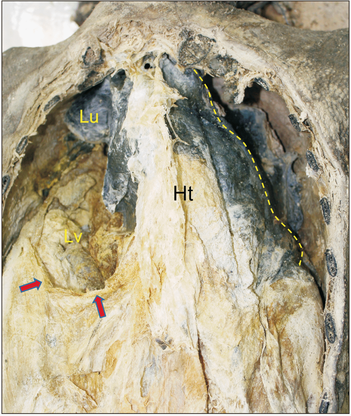
Fig. 1). Taken together this Andong case was diagnosed as Bochdalek-type CDH [
8].
Fig. 1
Autopsy view in 2013. Mediastinal shift to the left. Left border of heart (Ht) marked by a yellow broken line. Liver (Lv) is found in right thoracic cavity. Posterolateral defect of diaphragm marked by arrows. Lu, lung.

In this report, we newly performed 3D segmentation and model reconstruction technique on the CT data of Andong mummy’s heart and liver. The mummy’s DICOM file is loaded into the MEDIP PRO (
https://medicalip.com/Medip; MEDIP PRO, version 2.3.0; MEDICALIP Co., Ltd., Seoul, Korea). We then conducted an image segmentation task that separates the target structure from its surroundings. Semi-automatic techniques were mostly used to find the boundary between the target and surrounding structure. The segmented data are used to implement a 3D structure using an MEDIP program (3D rendering). The complete 3D image was adjusted to an angle that was easy to observe the mummified organs, and the color was allocated for each organ to facilitate researcher’s easy reading.
The results of 3D segmentation and model reconstruction are summarized in
Figs. 2–
5.
Fig. 2 is 3D reconstructed heart viewed from various angles. The shape of the heart was generally maintained but contracted and displaced a lot (
Fig. 2). We can see that the autopsy view of mummified heart in 2013 (
Supplementary Figs. 1, 2) could be similarly reproduced in the current data of 3D segmentation and model reconstruction (
Fig. 3A–C). In case of Andong mummy’s liver, the shape is generally maintained though the degree of contraction is severe (
Fig. 2). Judging from its location, mummy’s liver looks located above the diaphragm, mostly in the right thorax. In the figure viewed from the caudal, the mummified liver was flat and placed upon ribs and vertebral column in parts (
Fig. 3B–D). This is almost the same view as seen in autopsy in 2013 (
Supplementary Figs. 1, 2). At the time of autopsy in 2013, the mummy’s right lung was placed on the liver (
Fig. 4A), and when it was lifted, the liver was found on the bottom of the right thorax.
Fig. 2
Three-dimensional reconstructed heart (Ht) and liver (Lv) viewed from various angles. In brief, columns: (A) the view from the front; (B) the view from the left 45-degree angle; (C) the view from the left 90-degree angle; and (D) the view from the back.
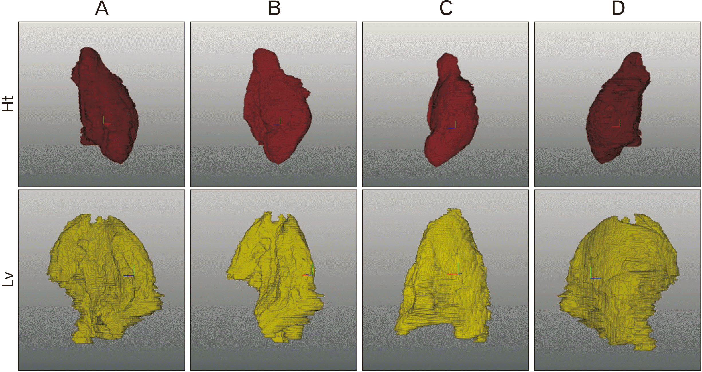

Fig. 3
Three-dimensional segmentation and model reconstruction of heart (A, C) and liver (B, D). The views from the front (A, B) and caudal (C, D). Mediastinal shift to the left could be seen (A, C). Liver can be found in right thoracic cavity due to diaphragmatic hernia (B, D).
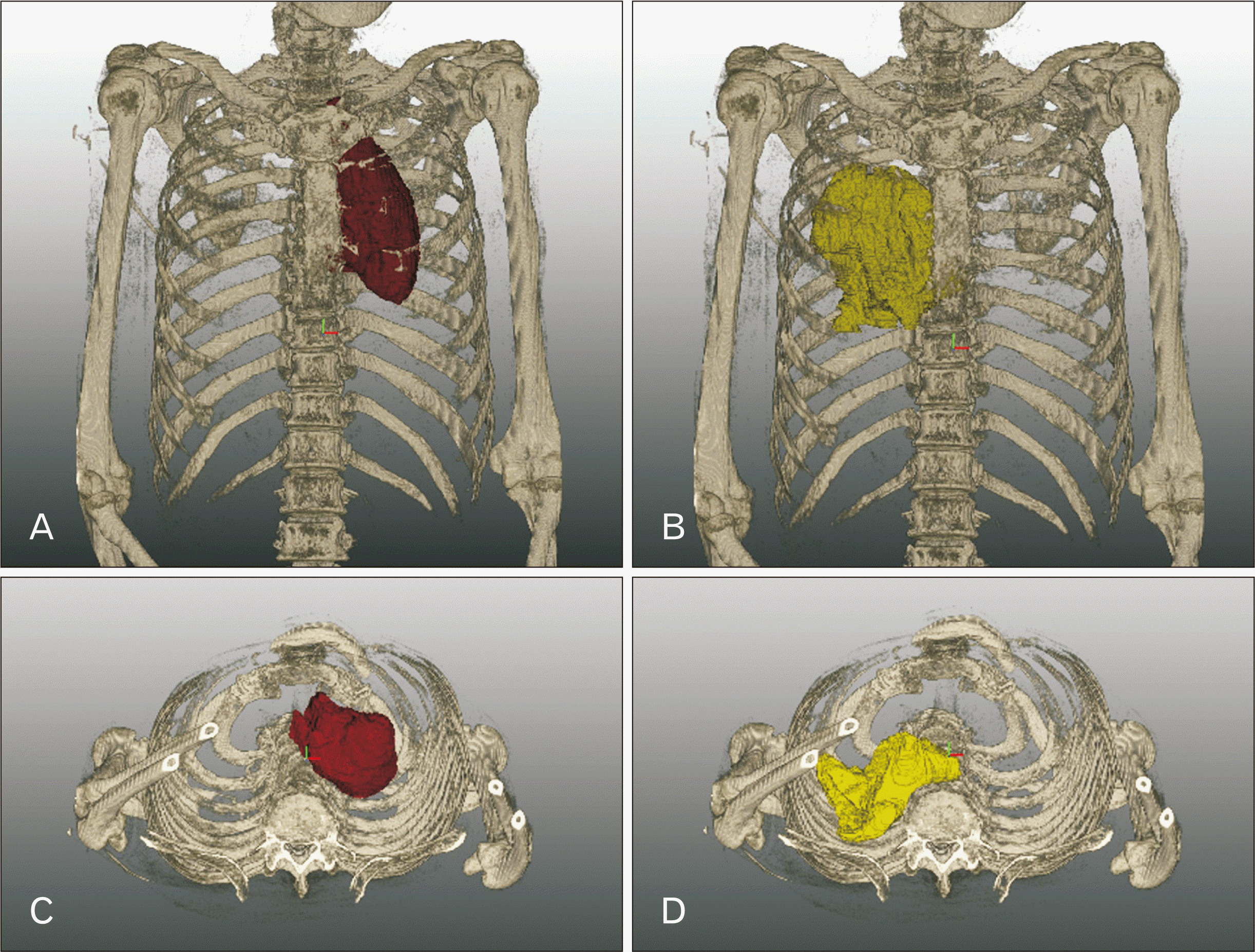

Fig. 4
(A) Autopsy (2013) and (B) three-dimensional segmentation and model reconstruction view of Andong mummy’s thoracic cavity. Liver is present below the right lung. Heart is encapsulated by pericardium. (B) Heart and liver is visible without overlapping organs by calling up the data exclusively from the target organs.
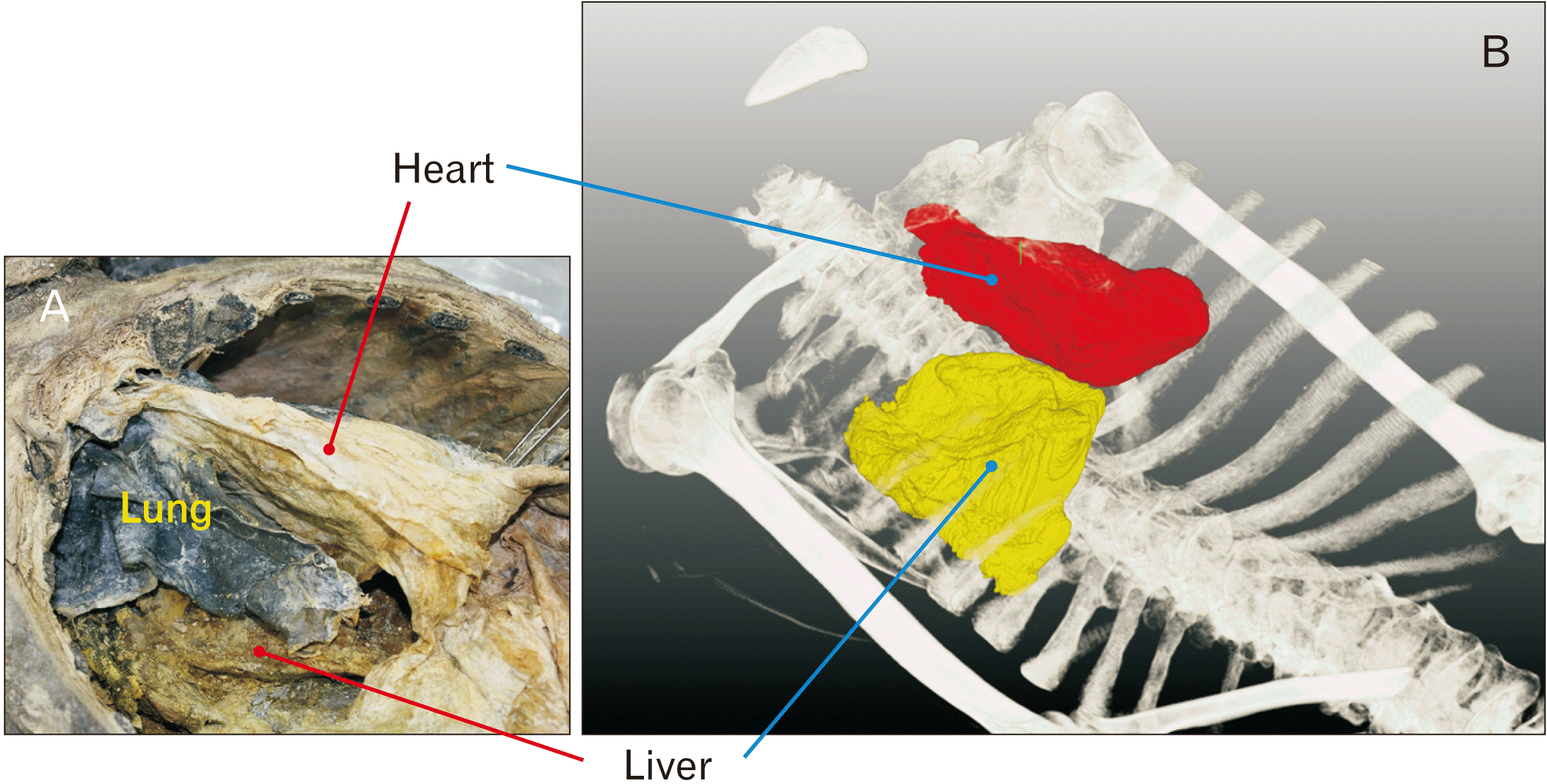

Fig. 5
Heart (red) and liver (yellow) in the thorax are viewed from different angles. (A) the view from the front; (B) the view from the left 45-degree angle; (C) the view from the left 90-degree angle; and (D) the view from the back.
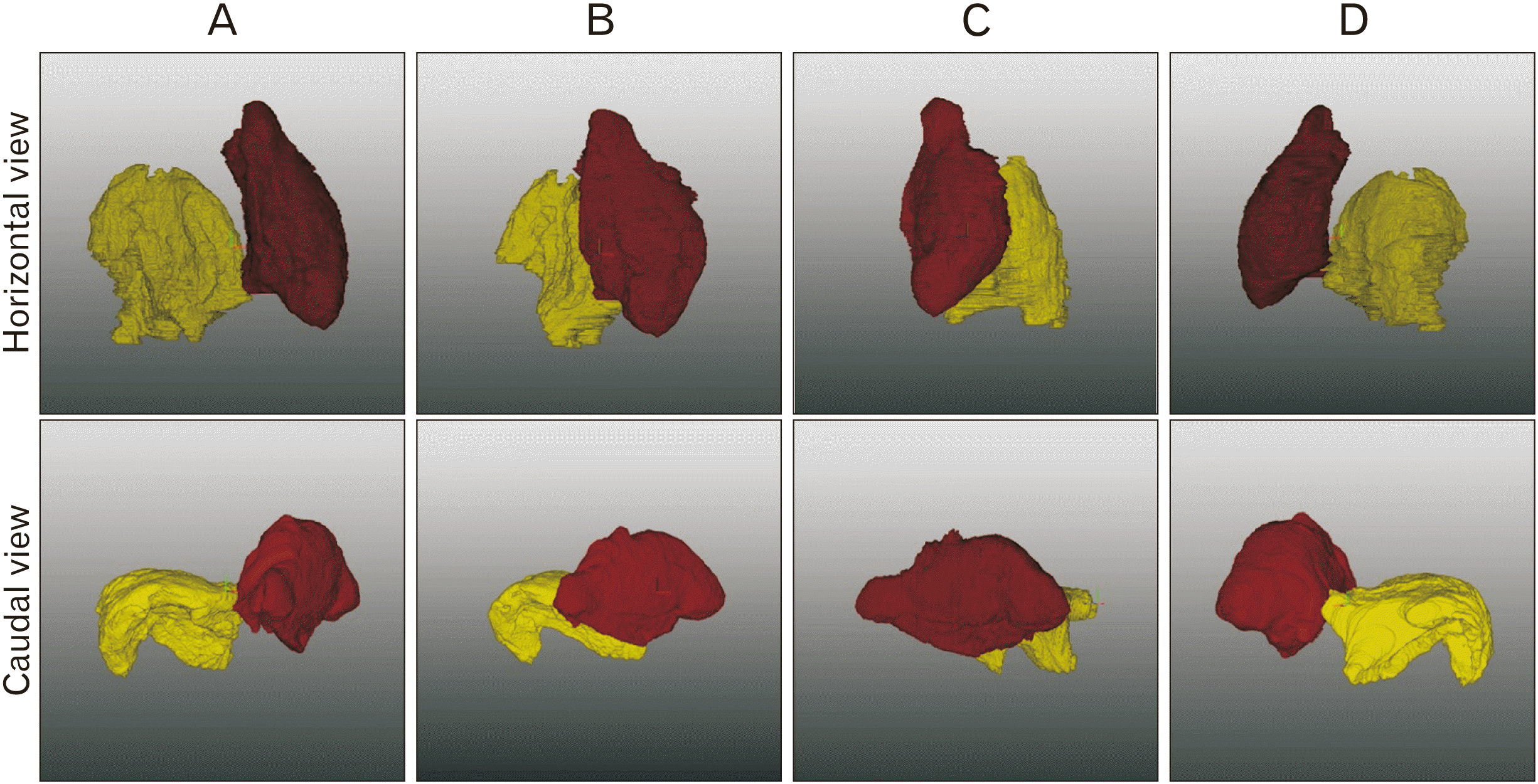

In this report, it is possible to confirm how the mummified organs (
i.e., liver and heart) are arranged in the thorax by implementing those segmented structures along with the other bony structures (
Fig. 4B) or from the rotational views of the organs (
Fig. 5). In the normal living people’s body, the base and apex of heart are generally located at the level of thoracic vertebrae (TV) 4 to 9. As seen in
Fig. 4B, the base of Andong mummy’s heart is located at TV4 and the apex at TV8, which means that its position does not change significantly compared to the normal body. On the other hand, normal individuals’ livers range from TV8–9 to TV9–lumvar vertebra 1 (LV1) [
9]. The Andong mummy’s liver is found at TV4 to TV8, indicating that it has moved up a lot compared to the living body (
Fig. 4B), which confirms the possibility of diaphragmatic hernia. In this report, we can also measure the volume of 3D reconstructed, mummified target organs. Mesh volume, voxel volume, maximum 2D diameter (slice), maximum 2D diameter (column), and maximum 2D diameter (row) can be measured for the Andong mummy’s 3D reconstructed heart and liver. The results are summarized in
Table 1.
Table 1
Volume and length measurements of Andong mummy’s heart and liver
|
Measurement |
Heart |
Liver |
|
Mesh volume (ml) |
242.95 |
188.39 |
|
Voxel volume (ml) |
243.00 |
188.52 |
|
Maximum 3D axis diameter (cm) |
14.93 |
12.27 |
|
Maximum 2D (slice) diameter (cm) |
8.39 |
9.63 |
|
Maximum 2D diameter (column) (cm) |
14.91 |
10.90 |
|
Maximum 2D diameter (row) (cm) |
12.04 |
11.69 |

Discussion
The Andong mummy case of the Joseon dynasty was originally investigated in 2013. The CDH diagnosed by 2D radiological analysis could be confirmed by an autopsy at the time [
8]. Nonetheless, since museum curators have recently argued that unnecessary destruction of mummy should be improved, we could not regard this case an ideal study in that the autopsy caused destructive damages on the Andong mummy. In fact, invasive techniques such as autopsy are more rarely used since 2013; and the dependence on CT radiographic has become a stronger trend in mummy research today. In this sense, it is meaningful to clarify to what extent reliable information can be attained from 3D segmentation and model reconstruction technique on the mummy’s CDH.
As originally confirmed by the autopsy in 2013, the current 3D segmentation and reconstruction images can reveal that the heart was pushed to the left due to the liver pushing up to the right thorax thorough hernial defect occurring in diaphragm. By the segmentation and reconstruction technique, we can see the spatial interrelationship between mummified liver and heart from various angles, estimating biometric volumetric measurements, and minimizing confusion due to overlapping organs by calling up the data exclusively from mummy’s target organs. In brief, when 3D segmentation technique is applied to Andong mummy’s CDH case, the obtained results are almost like those from the autopsy; and only based on the data, there was no difficulty in diagnosing this case of CDH. Since the latest trend in mummy research is to study them in more non-destructive way, it is meaningful to confirm how close it can be to the confirmed diagnosis of CDH by applying 3D segmentation and model reconstruction technique to this case. Based on this report, it is expected that the use of this technique, when similar congenital diseases are identified in mummies in the future, will produce results that are quite close to accurate diagnosis, even if autopsy is not possible for the case.
Acknowledgements
This work was supported by the National Research Foundation of Korea (NRF) grant funded by the Korea government (MSIT) (No. 2020R1A2C1010708) and Korea Ministry of Education (2020R1I1A1A01073501).
References
1. Hughes SW. Higgins T, Main P, Lang J, editors. 1996. Three-dimensional reconstruction of an ancient Egyptian mummy. Imaging the Past: Electronic Imaging and Computer Graphics in Museums and Archaeology. British Museum;London: p. 211–26.
3. Gerloni A, Cavalli F, Costantinides F, Costantinides F, Bonetti S, Paganelli C. 2009; Dental status of three Egyptian mummies: radiological investigation by multislice computerized tomography. Oral Surg Oral Med Oral Pathol Oral Radiol Endod. 107:e58–64. DOI:
10.1016/j.tripleo.2009.02.031. PMID:
19464646.
5. Cesarani F, Martina MC, Ferraris A, Grilletto R, Boano R, Marochetti EF, Donadoni AM, Gandini G. 2003; Whole-body three-dimensional multidetector CT of 13 Egyptian human mummies. AJR Am J Roentgenol. 180:597–606. DOI:
10.2214/ajr.180.3.1800597. PMID:
12591661.
6. Pelo S, Correra P, Danza FM, Amenta A, Gasparini G, Marianetti TM, Moro A. 2012; Evaluation of the dentoskeletal characteristics of an Egyptian mummy with three-dimensional computer analysis. J Craniofac Surg. 23:1159–62. DOI:
10.1097/SCS.0b013e31824e25c2. PMID:
22801114.
7. Cavka M, Janković I, Sikanjić PR, Ticinović N, Rados S, Ivanac G, Brkljacić B. 2010; Insights into a mummy: a paleoradiological analysis. Coll Antropol. 34:797–802. PMID:
20977064.
8. Kim YS, Lee IS, Jung GU, Kim MJ, Oh CS, Yoo DS, Lee WJ, Lee E, Cha SC, Shin DH. 2014; Radiological diagnosis of congenital diaphragmatic hernia in 17th century Korean mummy. PLoS One. 9:e99779. DOI:
10.1371/journal.pone.0099779. PMID:
24988465. PMCID:
PMC4079512.


