Journal List > Ann Lab Med > v.43(2) > 1516080062

Lee, Heo, Kim, Lee, Hwang, Park, Sung, Chang, Park, and Lee: Comprehensive Evaluation of the NeoBase 2 Non-derivatized MSMS Assay and Exploration of Analytes With Significantly Different Concentrations Between Term and Preterm Neonates

Abstract

Background

Despite the popularity of the NeoBase 2 Non-derivatized MSMS assay (PerkinElmer, Turku, Finland), there are no reports of its comprehensive evaluation, including the ability to distinguish transient tyrosinemia of the newborn (TTN) from tyrosinemia type 1 (TYR 1) using succinylacetone (SUAC). No newborn screening (NBS) cutoffs for preterm neonates in the Korean population have been suggested. We evaluated the NeoBase 2 assay and identified analytes requiring different cutoffs in preterm neonates.

Methods

Residual NBS dried blood spot samples and proficiency testing (PT) materials of the Newborn Screening Quality Assurance Program and the Korean Association of External Quality Assessment Service were used. Precision, accuracy, limit of detection (LOD), lower limit of quantification (LLOQ), linearity, recovery, carryover, and performance of SUAC were evaluated. Cutoffs were determined, and analytes requiring different cutoffs in preterm neonates were investigated.

Results

Mean CVs for within-run and between-day precision were within 15%. Accuracy analysis indicated high agreement with in-house derivatized assay results and results of other PT participants. All analytes demonstrated acceptable LOD, LLOQ, and linearity. Recoveries were acceptable, except for SUAC. Carryover was negligible. Cutoffs were established for all analytes; Tyr, adenosine, and C200-lysophosphatidylcholine required different cutoffs in preterm neonates. Differential diagnosis of TYR 1 and TTN was successful with simultaneous Tyr and SUAC measurement.

Conclusions

The NeoBase 2 assay demonstrated satisfactory performance. The additional analytes provide a wider diagnostic coverage, and the simultaneous measurement of Tyr and SUAC is efficient in excluding TYR 1. The new cutoffs for preterm neonates may decrease false-positive rates, without compromising diagnostic sensitivity.

INTRODUCTION

Newborn screening (NBS) allows early detection of inborn errors of metabolism (IEM) in the presymptomatic stage, permitting prompt intervention, which can possibly change the natural course of the disease. Traditional NBS assays relying on a few enzymes can detect only a limited number of diseases. The introduction of tandem mass spectrometry (MS/MS) has allowed a significant expansion of disease coverage through rapid detection and quantification of a wide range of metabolites, which has increased the detection rate and allowed earlier intervention [1, 2].
Samsung Medical Center, Seoul, Korea, has been using liquid chromatography (LC)-MS/MS with an in-house developed derivatized assay for NBS. In recent years, the number of laboratories using non-derivatized assays has steadily increased, whereas the number of laboratories using derivatized assays has decreased. The NeoBase 2 Non-derivatized MSMS kit (NeoBase 2; PerkinElmer, Turku, Finland) has been adopted in multiple institutes. According to the Newborn Screening Quality Assurance Program (NSQAP) QC report by the Centers for Disease Control and Prevention (CDC), approximately 65% of participants used a non-derivatized assay, and the NeoBase 2 assay accounted for approximately 21% of the non-derivatized assays [3]. Among 15 organizations participating in a program for metabolite testing provided by the Korean Association of External Quality Assessment Service (KEQAS), nine used the NeoBase 2 assay as of 2020.
Despite the popularity of the NeoBase 2 assay revealed in proficiency testing (PT), only one study has comparatively analyzed the evaluation parameters of linearity, precision, and carryover for the NeoBase 2 assay [4]. Although a verification report on the prior version—the NeoBase Non-derivatized MSMS kit (NeoBase; PerkinElmer)—has been published [5], there is no validation report on the analytical performance of the NeoBase 2 assay other than that from the manufacturer itself. The only published report regarding the performance of the NeoBase 2 assay is a premarket notification from the manufacturer for US Food and Drug Administration 510(k) clearance (510(k) No. K173568) [6]. Compared with the NeoBase assay, the NeoBase 2 assay measures additional analytes, expanding the diagnostic coverage, and has improved succinylacetone (SUAC) recovery and a shorter SUAC sample preparation time. In addition, despite SUAC being one of the target analytes in the NeoBase 2 assay, its performance in measuring SUAC and its ability to distinguish transient tyrosinemia of the newborn (TTN) from tyrosinemia type 1 (TYR 1) have not been reported.
The incidence of TTN ranges from 0.29% to 1.80% [7, 8]. The prevalence of TYR 1 is estimated to be 1 in 100,000 globally, and its prevalence in Korea is deemed to be lower, with only a few cases reported to date [9, 10]. Because of the difference in the prevalence of these two conditions, institutes without a SUAC assay set up are forced to repeat NBS in duplicate until the Tyr concentration returns to the normal range, unless the case is highly suspicious of TYR 1. As the NeoBase 2 assay allows for the simultaneous measurement of Tyr and SUAC, it would benefit laboratories not assaying SUAC. Furthermore, although the requirement for different preterm cutoffs has been suggested in the literature [11-13], there is no consensus on preterm cutoffs, and no cutoffs have been reported for the Korean population. Our aim was to carry out a comprehensive evaluation to determine the feasibility of adopting the NeoBase 2 assay and to establish new cutoffs in preterm neonates for analytes that require different cutoffs in the Korean population.

MATERIALS AND METHODS

Samples

Residual dried blood spot (DBS) samples submitted for NBS were stored at –20°C in sealed plastic bags with desiccant. The samples were collected on DBS cards (Honeywell Burdick & Jackson, Morristown, NJ, USA) and were punched with a DBS puncher (PerkinElmer) to a disk of 3.2 mm in diameter. PT materials were obtained from the NSQAP (seven amino acids, 21 acylcarnitines, and SUAC) and KEQAS (13 amino acids and 14 acylcarnitines). This study was approved by the Institutional Review Board (IRB) of Samsung Medical Center (IRB Nos. 2019-01-127 and 2021-02-078). The need for informed consent was waived because residual DBS samples were utilized.

Analytical method

The NeoBase 2 Non-derivatized MSMS kit was used following the manufacturer’s guidelines. The assay was conducted using a Waters ACQUITY H-Class ultraperformance liquid chromatography (UPLC) system coupled with a Waters XEVO tandem triple-quadrupole mass spectrometer (Waters Corporation, Milford, MA, USA). Analytes were quantified using the MassLynx 4.2 software (Waters Corporation). The mobile phase consisted of 84% acetonitrile (100% HPLC-grade acetonitrile diluted with 20% HPLC-grade distilled water) mixed with 0.1% formic acid. The optimal UPLC gradient was obtained at a flow rate of 130 µL/min (Supplemental Data Table S1). The MS analysis was conducted under the following conditions: capillary voltage, 3.0 kV; source temperature, 120°C; desolvation temperature, 350°C; desolvation gas flow, 800 L/hr; and cone gas flow, 70 L/hr. Detailed information on the MS operation parameters is presented in Supplemental Data Table S2. The in-house derivatized assay was performed as described previously [14].

Method validation

We comprehensively evaluated the NeoBase 2 assay, considering the CLSI NBS04, CLSI C62-A, and other guidelines for the validation of LC-MS/MS [15-17]. The evaluation parameters included limit of detection (LOD), lower limit of quantification (LLOQ), linearity, recovery, accuracy, precision, and carryover. The LOD and LLOQ were determined using blank DBS samples. Linearity and extraction recovery were assessed using AAAC Multilevel DBS (Lot No. 676609; PerkinElmer), which comprises 25 analytes at six dilution levels starting from the non-spiked endogenous concentration. The AACC Multilevel DBS samples were run in duplicates. Spike recoveries of the other five levels were calculated relative to the non-spiked endogenous concentration. Accuracy was evaluated using two methods: extraction recovery and method comparison. For method comparison, 20 NSQAP PT materials and 54 KEQAS PT materials were utilized. In addition, the NeoBase 2 assay was compared with the in-house derivatized assay in parallel for two weeks, using 64 DBS samples submitted for NBS.
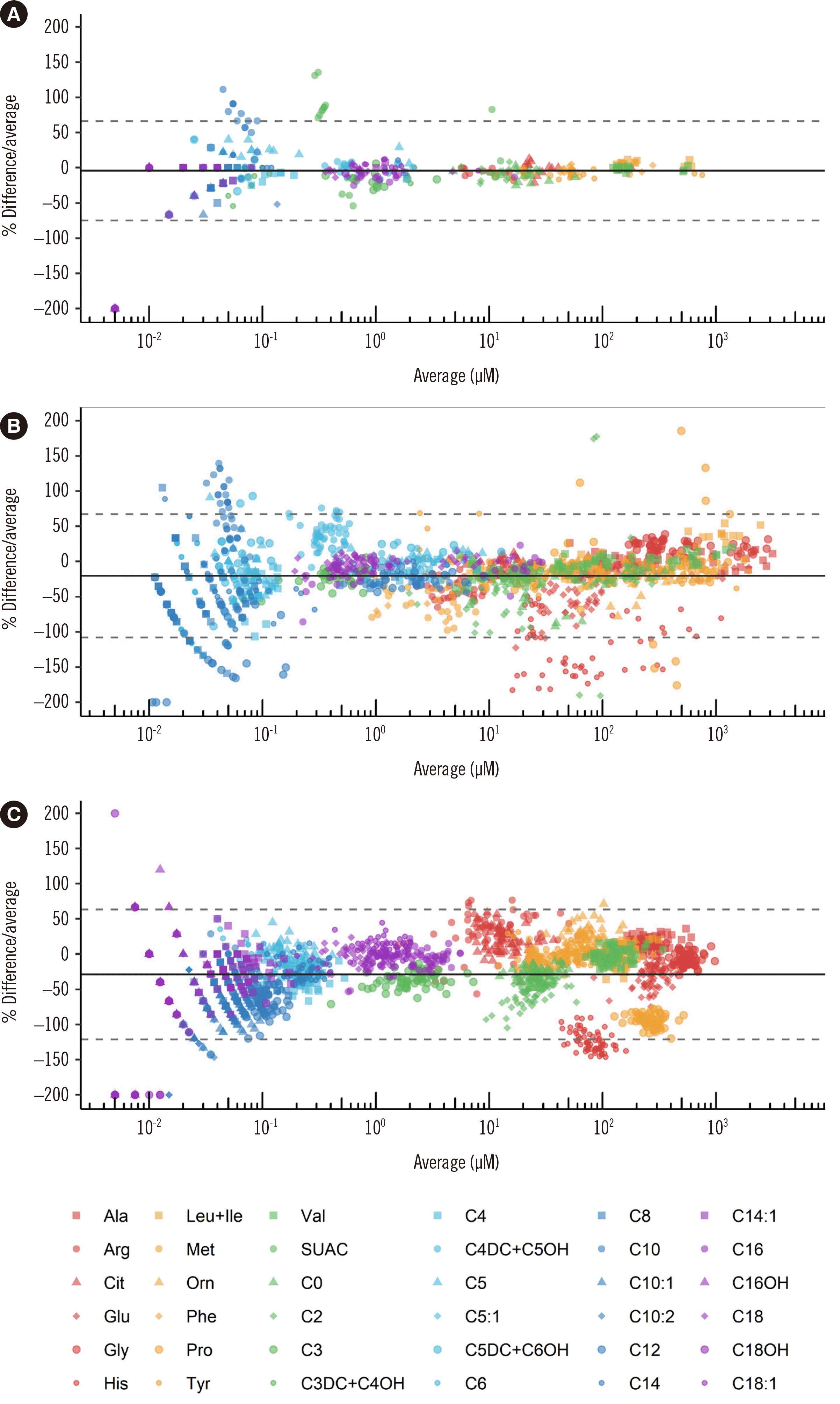
In total, 29, 27, and 32 analytes were compared using the NSQAP PT materials, KEQAS PT materials, and in-house derivatized assay, respectively. In a qualitative evaluation, the classification of a measured value as normal or abnormal was compared. For comparisons with the in-house derivatized assay, the agreement of the results was assessed based on the cutoffs for each method. For the PT samples, the agreement with the intended response was evaluated. PT samples that were not graded because of poor peer agreement (consensus <80%) were excluded from the qualitative analysis. For quantitative comparison, the results of 15 NSQAP PT materials were compared with the mean of the results for other participants using the NeoBase 2 assay. As there were at most 16 participants in the KEQAS PT, and kit-specific mean or median values of each analyte were not reported, the mean of all participants was imputed. As non-derivatized assays cannot distinguish certain analytes with similar properties, only analytes that are exclusively measured were compared with the in-house derivatized assay. The percent deviations were calculated, and Bland–Altman plots were generated.
Precision was evaluated using QC materials of the NeoBase 2 assay (PerkinElmer, Lot No. 687285) and the NSQAP (CDC, Lot Nos. A1915, B1915, C1915, and D1915). For five days, a run was performed in triplicate per day to assess within-run precision and between-day precision. To determine carryover, a blank DBS card (Honeywell Burdick & Jackson) was assayed directly after a high-level NeoBase 2 QC material was assayed, and this was repeated eight times. Throughout the study, data analyses were performed with R 4.0.2 (R Foundation for Statistical Computing, Vienna, Austria). All plots were generated using the ggplot2 3.3.5 package [18] in R 4.0.2 (R Foundation for Statistical Computing).

Cutoff determination

In total, 351 DBS samples from term neonates without definite medical problems submitted for NBS between September 2020 and February 2021 were evaluated. As only presumptively healthy neonates were included in the analysis, maximum or 99.5 percentile values were presumed to be the upper limits and minimum or 0.5 percentile values the lower limits. The presumed cutoffs were compared with cutoffs used in other institutes, and peer percentiles were retrieved from Collaborative Laboratory Integrated Reports (CLIR) (https://clir.mayo.edu). Cutoffs were established according to the CLSI NBS04 and CLSI EP28-A3 guidelines [15, 19]. Precision near the cutoff concentration was evaluated using QC materials having a concentration proximate to the cutoff of each analyte.
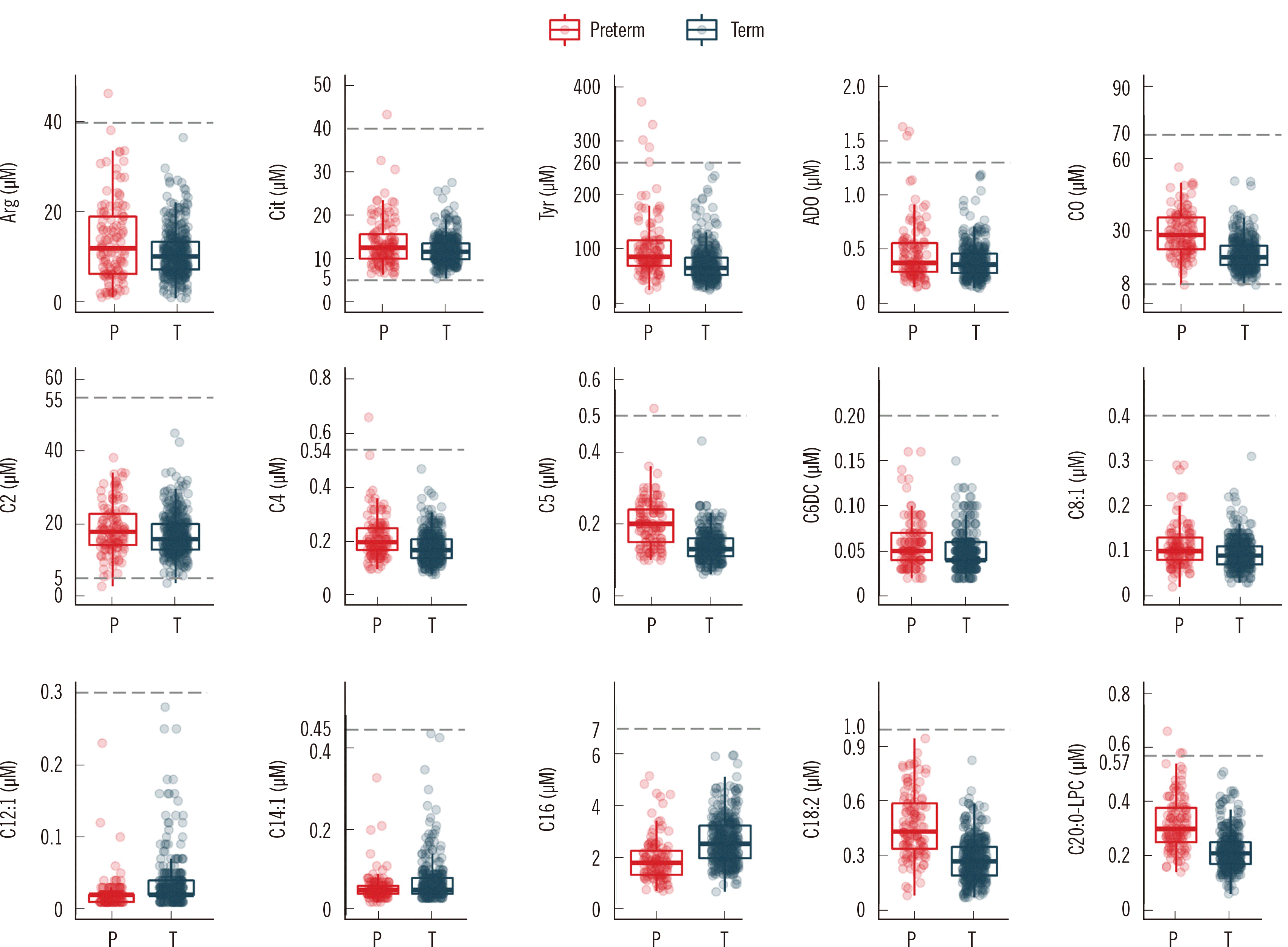
Cutoffs for preterm neonates (<37 weeks of gestation) were investigated using samples collected within 10 days after birth. In total, 118 preterm DBS samples were obtained between September 2020 and February 2021, and none of the neonates were diagnosed as having IEM. The distribution and 99.5 percentile of preterm and term neonates were determined for all analytes. If the difference of the 99.5 percentile (0.5 percentile for the lower cutoff) between preterm and term neonates was greater than the SD in term neonates, the requirement for a separate cutoff for the corresponding analyte was further investigated using box-and-whisker plots, which visually demonstrate the distribution and degree of false classification. If the degree of false classification was non-negligible (i.e., the assay was unable to classify abnormality properly), different cutoffs that better represent the distribution in preterm neonates were suggested. False classification of one neonate was considered acceptable. For analytes requiring a different cutoff for preterm neonates, the 99.5 percentile (0.5 percentile for the lower cutoff) value rounded to two significant figures was adopted.

In-depth evaluation of Tyr and SUAC

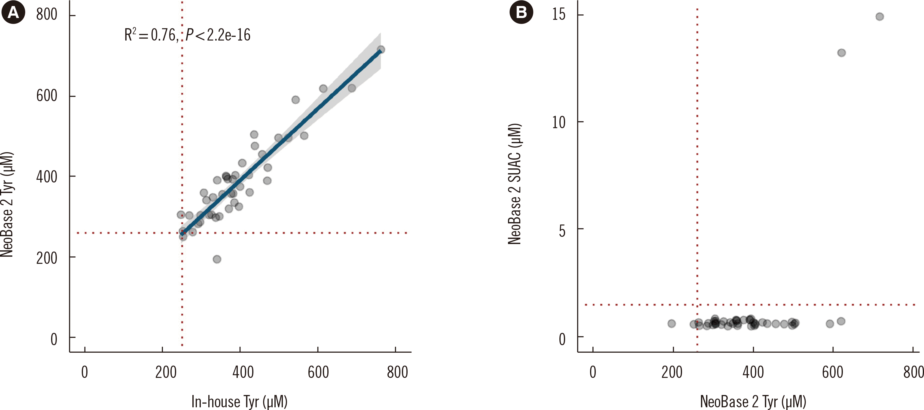
In total, 78 stored DBS samples from neonates with an increased Tyr concentration of >250 µM collected between March 2010 and December 2020 were used. Since there have been no TYR 1 patients in our institute, two NSQAP PT materials with increased Tyr and SUAC concentrations were also included in the analysis. To identify samples with a significant change in analyte concentrations due to long-term storage, the samples were retested using the in-house derivatized assay. Samples with a percent difference between the previous and retested concentrations >30% were excluded from the analysis. Tyr concentrations measured using the NeoBase 2 assay were compared with those measured using the in-house derivatized assay. The capability of the NeoBase 2 assay to simultaneously detect increases in Tyr and SUAC concentrations was evaluated.

RESULTS

Performance of the NeoBase 2 assay

The results of the performance evaluation are summarized in Table 1. Although the LOD and LLOQ of most amino acids were obtained, they were indeterminate for some analytes, including most of the acylcarnitines, as no definite peak was observed in repeated measurement of blank materials. For all analytes, the mean within-run precision and between-day precision CVs were within 15%. In the linearity evaluation, all analytes demonstrated a high coefficient of determination (R2)>0.99 (Supplemental Data Fig. S1). The mean recoveries of amino acids and acylcarnitines were 74.6%–96.2% and 83.3%–101.1%, respectively. SUAC had a low mean recovery rate (61.0%) compared with other analytes. Carryover was acceptable for all analytes. Assays of blank materials in the carryover evaluation did not reveal any interfering peaks.
Detailed results of the qualitative accuracy evaluation are presented in Table 2. The results for the NSQAP PT materials showed 100% agreement with the intended responses in all comparisons. However, among the 1,428 comparisons made using the KEQAS PT materials, three results were inconsistent with the intended responses (Supplemental Data Table S3). In addition, among 2,048 comparisons made with the in-house derivatized assay, seven results were discordant with the judgments made based on our in-house derivatized assay and cutoffs. The samples with discordant results all had concentrations near the cutoffs.
The results of the quantitative accuracy analysis are presented in Fig. 1. The mean percent difference between the NeoBase 2 and the NSQAP participants, KEQAS participants, and in-house derivatized assay was –4.3%, –20.4%, and –28.8%, respectively. In the quantitative comparison with NSQAP peer participants using the NeoBase 2 assay, decanoylcarnitine (C10) and SUAC showed a positive deviation. In the quantitative comparison with KEQAS peer participants, C10 showed a positive deviation, whereas dodecanoylcarnitine (C12) and histidine (His) showed negative deviations. In the comparison with the in-house derivatized assay, His and proline (Pro) showed negative deviations. Although the percent difference was large for analytes with low concentrations, the magnitude of the absolute difference in concentrations was insignificant (Supplemental Data Fig. S2).

Determined cutoffs and precision near the cutoff

The determined cutoffs are presented in Table 3, along with the 50 percentile cutoff of peers in the CLIR database, mean cutoffs provided in the NSQAP 2020 annual summary report [20] and KEQAS 2020 report [21], and the cutoffs determined by Cho, et al. [22], which is the only study that published cutoffs for the Korean population. For all analytes, precision near the cutoff was acceptable, with CVs within 15%.

Investigation of preterm cutoffs

Fifteen analytes showed a difference in the 99.5 percentile between preterm and term neonates greater than the SD in term neonates (Fig. 2). Among these, Tyr, adenosine (ADO), and C20:0-LPC exhibited at least two false abnormalities with the cutoffs determined based on term neonates. For these analytes, preterm cutoffs were designated as 350, 1.6, and 0.61 µM (260, 1.3, and 0.57 µM in term neonates), respectively. When we applied these new preterm cutoffs, only one neonate was classified as abnormal for each analyte.

Analysis of Tyr and SUAC

In total, 44 samples (42 residual samples and two NSQAP PT materials) with an increase in the Tyr concentration were analyzed. The results demonstrated a good correlation between the NeoBase 2 assay and the in-house derivatized assay, with a Spearman correlation coefficient of 0.87 (P<0.01). All 42 residual samples with an increase in the Tyr concentration exhibited a low SUAC concentration. The NeoBase 2 assay revealed increases in both the Tyr and SUAC concentrations in the two NSQAP PT materials, which supports the feasibility of using the NeoBase 2 assay in diagnosing TYR 1 (Fig. 3).

DISCUSSION

We demonstrated the satisfactory performance of the NeoBase 2 assay based on various validation parameters, listed cutoffs for all analytes, and suggested preterm cutoffs for three analytes for the first time in Korea. MS/MS is widely used for measuring various analytes [23-25], and several guidelines for the evaluation of MS/MS and NBS have been published [17]. However, there is no consensus on which parameters to assess and the degree of validation [17, 23]. The current comprehensive evaluation will aid future studies that aim to assess the performance of an MS/MS method in measuring a number of target analytes. While our study was mostly in line with previous reports on MS/MS validation in terms of the process used, our quantitative accuracy evaluation deserves attention. Although quantitative analysis based on percent differences and visualization in Bland–Altman plots provides intuitive result interpretation, it is not widely used in the evaluation of MS/MS methods as these are usually semi-quantitative and often include too many measurands. As this is a commonly used method in head-to-head comparison of quantitative chemistry analyzers, adopting this method in MS/MS method validation would be effortless for major clinical laboratories. This approach not only verifies the quantitative agreement but also reveals analytes with a systematic bias, which justifies the use of significantly different cutoffs among laboratories.
In quantitative comparison, there were few analytes with a systematic difference. The positive deviation of SUAC was due to low concentrations reported by NSQAP participants using the NeoBase 2 assay. Our SUAC results were consistent with those of peers who used an assay other than the NeoBase 2 assay. The positive deviation of C10 was mainly due to low analyte concentrations. While C4DC+C5OH showed a positive deviation throughout the measured range, the positively skewed results were mainly due to comparisons with KEQAS peer participants, including derivatized assays that solely measured C5OH. This finding is supported by the NSQAP PT and QC reports, which revealed higher concentrations for the NeoBase 2 assay than for other derivatized assays [3, 20]. While His showed a negative deviation from the mean of the KEQAS participants, 7 out of 10 participants submitted concentrations similar to ours, whereas the other three submitted approximately five-fold higher concentrations, causing a positively skewed mean value. Considering this, the results of the quantitative accuracy evaluation were considered acceptable.
A few analytes (Gln+Lys, Glu, and His) showed significant differences in cutoffs among institutes. The primary role of Gln and Glu in NBS is to support the diagnosis of disorders related to low citrulline, such as ornithine transcarbamylase deficiency and carbamoyl phosphate synthetase 1 deficiency [26], which are accompanied by an increase in Ala and a decrease in Arg concentrations [27]. His is no longer included as a screening item in NBS in many countries as histidinemia is currently regarded as a benign condition [28, 29]. Considering the nature of the related disorders and the qualitative agreement with other laboratories, we believe the discordant cutoffs would not cause clinical misclassification. The differences in cutoffs suggest that each laboratory should establish their own cutoffs due to various assay types used and/or population differences in analyte concentrations among ethnicities. NBS using MS/MS is generally regarded a semi-quantitative assay as some analytes are isobaric and QC materials are not available for certain analytes, which is another reason for the discrepancy in the quantitative results and cutoffs among laboratories [30]. To reduce assay-related biases, harmonization of quantitative results, allowing head-to-head comparison among laboratories, would be ideal [31]. In the qualitative accuracy evaluation using KEQAS PT materials, there was only one sample for each analyte with disagreement. All these samples had concentrations near the cutoffs, and 6%–19% of the peer participants submitted an unintended response identical to ours. Moreover, there were few qualitative disagreements between the NeoBase 2 assay and our previous in-house derivatized assay using clinical samples. Nonetheless, as there were no IEM patients during the study period and all samples with discordant results had concentrations near the cutoffs, this would not affect diagnostic sensitivity.
Differences in amino acid and acylcarnitine concentrations have been reported between preterm and term neonates, which complicate the interpretation of NBS results [32]. Acylcarnitine and amino acid concentrations are generally lower in preterm neonates [12, 13]. However, acylcarnitines derived from branched-chain amino acids showed an opposite trend [12]. The probability of TTN is higher in preterm than in term neonates [8]. The median ADO concentration is significantly higher in neonates with low birth weight [33]. In addition, the timing of blood collection affects analyte concentrations and thus the performance of NBS [11, 34]. These results complicate the interpretation of metabolic status in preterm neonates and substantiate the requirement for a different cutoff for IEM screening. The metabolic profile of preterm neonates in this study was in agreement with those in previous reports. We determined different cutoffs for three analytes (Tyr, ADO, and C20:0-LPC) that showed significant differences between preterm and term neonates and are screening markers of TYR 1, adenosine deaminase severe combined immunodeficiency (ADA-SCID), and X-linked adrenoleukodystrophy (X-ALD), respectively. The suggested preterm cutoffs may overlap with disease-related concentrations. However, as TYR 1, ADA-SCID, and X-ALD all have other means to support the diagnosis, a higher preterm cutoff would be beneficial in reducing the number of false positives. The pathognomonic marker for TYR 1 is SUAC [35]. The diagnosis of ADA-SCID can be supported by lymphopenia and low Ig concentrations [36]. The C20:0-LPC concentration does not significantly differ between healthy controls and ALD [37, 38], and increases in C24:0-LPC, C26:0-LPC, and the ratio of these markers to C20:0-LPC or C22:0-LPC are used for diagnosis [37-39].
There are a few limitations to our study. First, as a relatively small number of samples from preterm neonates were included in the analysis, we were unable to suggest cutoffs for subcategories of preterm neonates based on gestational age. Second, samples from confirmed TYR 1 patients were not included in our evaluation of Tyr and SUAC, primarily because of the low incidence of TYR 1. Our evaluation still showed that the NeoBase 2 assay can detect concurrent increases in Tyr and SUAC concentrations. Although SUAC had a low recovery rate, this phenomenon has also been observed in previous studies, which suggests that low recovery does not affect clinical sensitivity in distinguishing TYR 1 [35, 40].
In conclusion, this study confirmed the performance of the NeoBase 2 assay in various measures. As the NeoBase 2 assay provides a wide diagnostic coverage with numerous analytes, we suggest that the introduction of the NeoBase 2 assay in clinical laboratories may benefit patients, pediatricians, and laboratory personnel, including clinical pathologists and medical technologists. The cutoffs determined for the Korean population using the NeoBase 2 assay will be relevant for other institutes in Korea willing to introduce the NeoBase 2 assay in their laboratories. By applying different cutoffs for certain analytes in preterm neonates, false-positive rates are expected to decrease, without compromising diagnostic sensitivity.

ACKNOWLEDGEMENTS

We thank Kim DJ, Oh H, and Kim HK for performing the assays. We also thank Bio-Medical Science (BMS) for supplying the NeoBase 2 Non-derivatized MSMS kits.

Notes

AUTHOR CONTRIBUTIONS

Conceptualization: Lee B, Hwang N, Park HD, and Lee SY. Data curation: Lee B, Heo WY, Lee HS, and Hwang N. Formal analysis: Lee B, Heo WY, Kim JA, Lee HS, and Lee SY. Investigation: Lee B, Heo WY, Lee HS, Hwang N, and Lee SY. Methodology: Lee B, Heo WY, Hwang N, Park HD, and Lee SY. Project administration: Lee SY. Resources: Hwang N, Park HD, Sung SI, Chang YS, Park WS, and Lee SY. Supervision: Park HD, Sung SI, Chang YS, Park WS, and Lee SY. Validation: Lee B, Heo WY, Kim JA, Lee HS, and Lee SY. Visualization: Lee B. Writing — original draft: Lee B. Writing — review and editing: Lee B and Lee SY. All authors accept responsibility for the entire content of this manuscript and approve its submission.

CONFLICTS OF INTEREST

The authors declare no conflicts of interest.

RESEARCH FUNDING

None declared.

REFERENCES

1. Schulze A, Lindner M, Kohlmüller D, Olgemöller K, Mayatepek E, Hoffmann GF. 2003; Expanded newborn screening for inborn errors of metabolism by electrospray ionization-tandem mass spectrometry: results, outcome, and implications. Pediatrics. 111:1399–406. DOI: 10.1542/peds.111.6.1399. PMID: 12777559.
crossref
2. Wilcken B, Wiley V, Hammond J, Carpenter K. 2003; Screening newborns for inborn errors of metabolism by tandem mass spectrometry. N Engl J Med. 348:2304–12. DOI: 10.1056/NEJMoa025225. PMID: 12788994.
crossref
3. Centers for Disease Control and Prevention (CDC). Newborn Screening Quality Assurance Program (NSQAP) 2020 Quality control program report. https://www.cdc.gov/labstandards/pdf/nsqap/QC_Report_S1_2020-508.pdf. Updated on Jun 2020.
4. Cicalini I, Valentinuzzi S, Pieragostino D, Consalvo A, Zucchelli M, Donzelli S, et al. 2021; Analytical evaluation of the ideal strategy for high-throughput flow injection analysis by tandem mass spectrometry in routine newborn screening. Metabolites. 11:473. DOI: 10.3390/metabo11080473. PMID: 34436414. PMCID: PMC8399422.
crossref
5. Zheng Y, Chen Y, Qiu X, Chen W, Lin Q, Zeng Y, et al. 2019; A verification of the application of the non-derivatized mass spectrometry method in newborns screening of metabolic disorders. Medicine (Baltimore). 98:e15500. DOI: 10.1097/MD.0000000000015500. PMID: 31083189. PMCID: PMC6531236.
crossref
6. U.S. Food and Drug Administration. 510(k) Premarket notification (K17 3568). https://www.accessdata.fda.gov/cdrh_docs/reviews/K173568.pdf. Updated on Sept 2018.
7. Levy HL, Shih VE, Madigan PM, MacCready RA. 1969; Transient tyrosinemia in full-term infants. JAMA. 209:249–50. DOI: 10.1001/jama.1969.03160150035008. PMID: 5819230.
crossref
8. Zea-Rey AV, Cruz-Camino H, Vazquez-Cantu DL, Gutiérrez-García VM, Santos-Guzmán J, Cantú-Reyna C. 2017; The incidence of transient neonatal tyrosinemia within a Mexican population. J Inborn Errors Metab Screen. 5:e170016. DOI: 10.1177/2326409817744230.
crossref
9. Park HD, Lee DH, Choi TY, Lee YK, Kim JW, Ki CS, et al. 2009; Clinical, biochemical, and genetic analysis of a Korean neonate with hereditary tyrosinemia type 1. Clin Chem Lab Med. 47:930–3. DOI: 10.1515/CCLM.2009.223. PMID: 19569981.
crossref
10. Choi HJ, Bang HI, Ki CS, Lee SY, Kim JW, Song J, et al. 2014; Two novel FAH gene mutations in a patient with hereditary tyrosinemia type I. Ann Clin Lab Sci. 44:317–23.
11. Clark RH, Kelleher AS, Chace DH, Spitzer AR. 2014; Gestational age and age at sampling influence metabolic profiles in premature infants. Pediatrics. 134:e37–46. DOI: 10.1542/peds.2014-0329. PMID: 24913786.
crossref
12. Gucciardi A, Zaramella P, Costa I, Pirillo P, Nardo D, Naturale M, et al. 2015; Analysis and interpretation of acylcarnitine profiles in dried blood spot and plasma of preterm and full-term newborns. Pediatr Res. 77:36–47. DOI: 10.1038/pr.2014.142. PMID: 25268144.
crossref
13. Liu Q, Wu J, Shen W, Wei R, Jiang J, Liang J, et al. 2017; Analysis of amino acids and acyl carnitine profiles in low birth weight, preterm, and small for gestational age neonates. J Matern Fetal Neonatal Med. 30:2697–704. DOI: 10.1080/14767058.2016.1261395. PMID: 27844490.
crossref
14. Lee HS. 2021; Arginine, as a key indicator for real-time stability monitoring of quality control in the newborn screening test using dried blood spot. Separations. 8:201. DOI: 10.3390/separations8110201.
crossref
15. CLSI. 2017. Newborn screening by tandem mass spectrometry. CLSI NBS04. Clinical and Laboratory Standards Institute;Wayne, PA:
16. CLSI. 2014. Liquid chromatography-mass spectrometry methods; approved guideline. CLSI C62-A. Clinical and Laboratory Standards Institute;Wayne, PA:
17. Moon SY, Choi HJ, Kim S, Lee K, Lee SG, Song SH, et al. 2020; Recommendations for liquid chromatography-mass spectrometry in the clinical laboratory: part II. Method validation. Lab Med Online. 10:95–108. DOI: 10.3343/lmo.2020.10.2.95.
crossref
18. Wickham H. ggplot2: elegant graphics for data analysis. Use R;2016.
19. CLSI. 2010. Defining, establishing, and verifying reference intervals in the clinical laboratory; approved guideline-third edition. CLSI EP28-A3C. Clinical and Laboratory Standards Institute;Wayne, PA:
20. Centers for Disease Control and Prevention (CDC). Newborn Screening Quality Assurance Program (NSQAP) 2020 Annual Summary Report. https://www.cdc.gov/labstandards/pdf/nsqap/NSQAP_Annual_Summary_2020-508.pdf. Updated on Aug 2020.
21. Lee SY. 2020; Report of the Korean Association of External Quality Assessment Service on metabolite testing (2018-2019). Lab Med Qual Assur. 42:10–25. DOI: 10.15263/jlmqa.2020.42.1.10.
crossref
22. Cho SE, Park EJ, Seo DH, Lee IB, Lee HJ, Cho DY, et al. 2015; Neonatal screening tests for inherited metabolic disorders using tandem mass spectrometry: experience of a clinical laboratory in Korea. Lab Med Online. 5:196–203. DOI: 10.3343/lmo.2015.5.4.196.
crossref
23. Chae H, Cho SE, Park HD, Chun S, Lee YW, Yun YM, et al. 2019; Use of liquid chromatography-tandem mass spectrometry for clinical testing in Korean laboratories: a questionnaire survey. Ann Lab Med. 39:447–53. DOI: 10.3343/alm.2019.39.5.447. PMID: 31037863. PMCID: PMC6502944.
crossref
24. Choi R, Chun MR, Park J, Lee JW, Ju HY, Cho HW, et al. 2021; Quantification of thioguanine in DNA using liquid chromatography-tandem mass spectrometry for routine thiopurine drug monitoring in patients with pediatric acute lymphoblastic leukemia. Ann Lab Med. 41:145–54. DOI: 10.3343/alm.2021.41.2.145. PMID: 33063676. PMCID: PMC7591283.
crossref
25. Choi R, Park HD, Oh HJ, Lee K, Song J, Lee SY. 2019; Dried blood spot multiplexed steroid profiling using liquid chromatography tandem mass spectrometry in Korean neonates. Ann Lab Med. 39:263–70. DOI: 10.3343/alm.2019.39.3.263. PMID: 30623618. PMCID: PMC6340850.
crossref
26. Hall PL, Marquardt G, McHugh DM, Currier RJ, Tang H, Stoway SD, et al. 2014; Postanalytical tools improve performance of newborn screening by tandem mass spectrometry. Genet Med. 16:889–95. DOI: 10.1038/gim.2014.62. PMID: 24875301. PMCID: PMC4262759.
crossref
27. Saudubray J-M, van den Berghe G, Walter JHW. 2012. Inborn metabolic diseases: diagnosis and treatment. 5th ed. Springer;Berlin: p. 656.
28. Levy HL, Shih VE, Madigan PM. 1974; Routine newborn screening for histidinemia. Clinical and biochemical results. N Engl J Med. 291:1214–9. DOI: 10.1056/NEJM197412052912303. PMID: 4421298.
29. Lam WK, Cleary MA, Wraith JE, Walter JH. 1996; Histidinaemia: a benign metabolic disorder. Arch Dis Child. 74:343–6. DOI: 10.1136/adc.74.4.343. PMID: 8669938. PMCID: PMC1511463.
crossref
30. Rappold BA. 2022; Review of the use of liquid chromatography-tandem mass spectrometry in clinical laboratories: part I-development. Ann Lab Med. 42:121–40. DOI: 10.3343/alm.2022.42.2.121. PMID: 34635606. PMCID: PMC8548246.
crossref
31. Pickens CA, Sternberg M, Seeterlin M, De Jesús VR, Morrissey M, Manning A, et al. 2020; Harmonizing newborn screening laboratory proficiency test results using the CDC NSQAP reference materials. Int J Neonatal Screen. 6:75. DOI: 10.3390/ijns6030075. PMID: 33123642. PMCID: PMC7570198.
crossref
32. CLSI. 2019. Newborn screening for preterm, low birth weight, and sick newborns; approved guideline. 2nd ed. CLSI NBS03-A. Clinical and Laboratory Standards Institute;Wayne, PA:
33. Panfoli I, Cassanello M, Bruschettini M, Colella M, Cerone R, Ravera S, et al. 2016; Why do premature newborn infants display elevated blood adenosine levels? Med Hypotheses. 90:53–6. DOI: 10.1016/j.mehy.2016.03.007. PMID: 27063086.
crossref
34. Peng G, Tang Y, Cowan TM, Zhao H, Scharfe C. 2020; Timing of newborn blood collection alters metabolic disease screening performance. Front Pediatr. 8:623184. DOI: 10.3389/fped.2020.623184. PMID: 33553077. PMCID: PMC7854909.
crossref
35. De Jesús VR, Adam BW, Mandel D, Cuthbert CD, Matern D. 2014; Succinylacetone as primary marker to detect tyrosinemia type I in newborns and its measurement by newborn screening programs. Mol Genet Metab. 113:67–75. DOI: 10.1016/j.ymgme.2014.07.010. PMID: 25066104. PMCID: PMC4533100.
crossref
36. Flinn AM, Gennery AR. 2018; Adenosine deaminase deficiency: a review. Orphanet J Rare Dis. 13:65. DOI: 10.1186/s13023-018-0807-5. PMID: 29690908. PMCID: PMC5916829.
crossref
37. Turgeon CT, Moser AB, Mørkrid L, Magera MJ, Gavrilov DK, Oglesbee D, et al. 2015; Streamlined determination of lysophosphatidylcholines in dried blood spots for newborn screening of X-linked adrenoleukodystrophy. Mol Genet Metab. 114:46–50. DOI: 10.1016/j.ymgme.2014.11.013. PMID: 25481105.
crossref
38. Mashima R, Tanaka M, Sakai E, Nakajima H, Kumagai T, Kosuga M, et al. 2016; A selective detection of lysophosphatidylcholine in dried blood spots for diagnosis of adrenoleukodystrophy by LC-MS/MS. Mol Genet Metab Rep. 7:16–9. DOI: 10.1016/j.ymgmr.2016.02.007. PMID: 27331004. PMCID: PMC4908058.
crossref
39. Lee S, Clinard K, Young SP, Rehder CW, Fan Z, Calikoglu AS, et al. 2020; Evaluation of X-linked adrenoleukodystrophy newborn screening in North Carolina. JAMA Netw Open. 3:e1920356. DOI: 10.1001/jamanetworkopen.2019.20356. PMID: 32003821. PMCID: PMC7042889.
crossref
40. Adam BW, Hall EM, Meredith NK, Lim TH, Haynes CA, De Jesus VR, et al. 2012; Performance of succinylacetone assays and their associated proficiency testing outcomes. Clin Biochem. 45:1658–63. DOI: 10.1016/j.clinbiochem.2012.08.007. PMID: 22906829. PMCID: PMC4557813.
crossref

Fig. 1
Bland–Altman plots comparing the NeoBase 2 non-derivatized assay results with those of (A) NSQAP participants using the Neo-Base 2 assay for amino acids, SUAC, and acylcarnitines, (B) KEQAS participants regardless of the assay for amino acids and acylcarnitines, and (C) the in-house derivatized assay.
Abbreviations: NSQAP, Newborn Screening Quality Assurance Program; SUAC, succinylacetone; KEQAS, Korean Association of External Quality Assessment Service; amino acids are abbreviated as three-letter symbols according to the International Union of Pure and Applied Chemistry (IUPAC) nomenclature.
alm-43-2-153-f1.tif
Fig. 2
Box-and-whisker plots of selective analytes that potentially require a different cutoff in preterm neonates. Red dots and blue dots represent the values measured in preterm neonates and term neonates, respectively, and the dashed gray line indicates the established cutoff for term neonates.
Abbreviations: P, Preterm; T, Term; ADO, adenosine; LPC, lysophosphatidylcholine; amino acids are abbreviated as three-letter symbols according to the International Union of Pure and Applied Chemistry (IUPAC) nomenclature.
alm-43-2-153-f2.tif
Fig. 3
Scatter plots for (A) comparison of Tyr concentrations between the in-house derivatized assay and the NeoBase 2 assay and (B) Tyr and SUAC measured using the NeoBase 2 assay. The samples used in the analysis are composed of 42 residual clinical samples with an increased Tyr concentration and two NSQAP PT materials with an increase in both Tyr and SUAC concentrations.
Abbreviations: Tyr, tyrosine; SUAC, succinylacetone; NSQAP, Newborn Screening Quality Assurance Program; PT, proficiency testing.
alm-43-2-153-f3.tif
Table 1
Summary of the analytical performance characteristics
Analyte LOD (μM) LLOQ (μM) Precision* (%CV) Linearity Recovery* (%) Carryover (μM)


Within-run Between-day Range (μM) R2
Ala 1.13 2.26 5.06 ± 1.13 7.44 ± 2.90 398.40–2,710.77 0.9976 80.2 ± 13.2 3.45
Arg 0.17 0.33 3.46 ± 0.94 1.72 ± 1.59 6.03–1,311.86 0.9997 90.7 ± 3.8 0.65
Cit 0.21 0.42 5.10 ± 0.91 1.80 ± 2.06 23.44–917.48 0.9978 92.5 ± 7.4 0.23
Gln + Lys 0.46 0.92 4.50 ± 1.39 1.99 ± 1.68 2.57
Glu 0.17 0.33 4.56 ± 1.11 4.79 ± 1.99 0.14
Gly 2.16 4.31 4.27 ± 1.46 2.24 ± 1.86 424.46–2,791.61 0.9990 96.2 ± 13.6 8.42
His 0.18 0.35 5.16 ± 1.14 7.34 ± 1.80 0.63
Leu + Ile 0.21 0.43 4.14 ± 1.06 2.66 ± 1.60 242.94–1,224.43 0.9962 88.4 ± 16.1 1.74
Met 0.50 1.00 4.40 ± 1.27 5.56 ± 1.94 15.67–466.28 0.9981 81.5 ± 6.2 0.53
Orn 0.66 1.33 3.72 ± 1.05 2.52 ± 1.64 114.72–1,317.90 0.9984 79.1 ± 6.9 9.90
Phe 5.30 10.59 4.14 ± 1.21 3.43 ± 2.32 74.78–1,000.80 0.9986 83.9 ± 8.4 0.23
Pro 0.20 0.39 4.20 ± 1.20 5.56 ± 2.08 212.29–1,408.03 0.9976 74.6 ± 10.6 1.96
Tyr 0.52 1.05 3.66 ± 1.39 3.21 ± 2.10 78.77–1,452.52 0.9990 79.6 ± 7.7 0.62
Val 0.15 0.30 4.01 ± 0.85 4.00 ± 1.73 227.75–1,188.23 0.9960 87.2 ± 16.5 1.36
ASA 10.96 ± 2.08 13.90 ± 11.28 0.64
SUAC 0.03 0.07 5.68 ± 1.98 3.62 ± 2.40 0.53–38.69 0.9995 61.0 ± 3.9 0.13
ADO 0.02 0.03 3.90 ± 0.14 4.13 ± 1.83 < 0.01
D-ADO < 0.01
C0 0.03 0.07 4.52 ± 1.14 3.37 ± 2.32 27.62–1,057.47 0.9995 83.6 ± 5.2 0.12
C2 0.03 0.07 4.40 ± 0.87 1.93 ± 1.52 9.51–333.59 0.9984 94.0 ± 6.9 0.07
C3 0.03 0.07 4.44 ± 0.85 1.85 ± 1.45 1.53–41.22 0.9990 93.3 ± 6.2 0.01
C3DC + C4OH 4.10 ± 0.71 4.29 ± 1.26 < 0.01
C4 4.96 ± 0.76 1.30 ± 1.45 0.26–19.79 0.9991 87.0 ± 4.6 0.01
C4DC + C5OH 4.29 ± 1.35 2.48 ± 2.12 0.01
C5 4.71 ± 1.02 4.01 ± 2.25 0.14–18.05 0.9992 101.1 ± 4.8 0.02
C5:1 5.97 ± 1.02 3.04 ± 2.54 < 0.01
C5DC + C6OH 5.81 ± 1.62 4.22 ± 1.18 0.06–39.85 0.9998 83.3 ± 2.4 < 0.01
C6 4.63 ± 1.04 2.15 ± 2.15 0.05–21.63 0.9994 88.5 ± 5.4 < 0.01
C6DC < 0.01
C8 4.85 ± 1.07 2.24 ± 2.08 0.04–17.51 0.9990 97.5 ± 6.3 < 0.01
C8:1 < 0.01
C10 5.71 ± 1.21 2.72 ± 0.81 0.03–17.04 0.9990 83.5 ± 5.1 0.04
C10:1 < 0.01
C10:2 < 0.01
C12 5.47 ± 1.07 3.21 ± 2.17 0.03–21.44 0.9986 96.0 ± 6.2 < 0.01
C12:1 < 0.01
C14 5.15 ± 0.77 2.47 ± 2.29 0.11–20.17 0.9987 96.5 ± 6.4 < 0.01
C14OH < 0.01
C14:1 0.02 0.03 5.32 ± 0.97 2.20 ± 2.10 < 0.01
C14:2 0.02 0.03 < 0.01
C16 0.02 0.03 5.05 ± 1.75 3.00 ± 2.21 1.13–54.88 0.9984 91.4 ± 7.2 0.01
C16OH 6.14 ± 2.82 4.43 ± 1.51 < 0.01
C16:1 < 0.01
C16:1OH 0.01
C18 5.10 ± 1.81 2.75 ± 2.40 0.80–37.70 0.9981 92.5 ± 7.5 0.01
C18OH 7.23 ± 2.09 2.16 ± 2.73 < 0.01
C18:1 5.93 ± 2.88 3.13 ± 3.21 < 0.01
C18:1OH < 0.01
C18:2 6.54 ± 3.03 3.20 ± 3.11 < 0.01
C18:2OH 0.04
C20 < 0.01
C22 0.01
C24 < 0.01
C26 4.82 ± 1.57 2.19 ± 3.09 0.01
C20:0-LPC 6.74 ± 2.87 4.13 ± 2.89 < 0.01
C22:0-LPC 6.41 ± 1.69 3.08 ± 2.92 < 0.01
C24:0-LPC 6.65 ± 1.88 1.59 ± 2.33 < 0.01
C26:0-LPC 9.07 ± 6.80 3.38 ± 2.98 0.02

*The results of precision and recovery analyses are presented as mean±SD; The mean values of the duplicate runs were used.

Abbreviations: LOD, limit of detection; LLOQ, lower limit of quantification; ASA, argininosuccinic acid; SUAC, succinylacetone; ADO, adenosine; D-ADO, 2´-deoxyadenosine; LPC, lysophosphatidylcholine; amino acids are abbreviated as three-letter symbols according to the International Union of Pure and Applied Chemistry (IUPAC) nomenclature.

Table 2
Summary of the accuracy results with the degree of qualitative agreement
Analyte Accuracy (% agreement [total agreement rate, abnormality agreement rate])

NSQAP PT* KEQAS PT* In-house derivatized assay
Ala 100% (52/52, 14/14) 100% (64/64, 0/0)
Arg 100% (20/20, 2/2) 100% (54/54, 11/11) 95.3% (61/64, 1/1)
Cit 100% (20/20, 3/3) 100% (54/54, 13/13) 100% (64/64, 1/1)
Glu 100% (49/49, 6/6) 100% (64/64, 0/0)
Gly 98.1% (52/53, 12/12) 100% (64/64, 2/2)
His 100% (49/49, 11/11) 98.4% (63/64, 1/1)
Leu + Ile 100% (19/19, 4/4) 100% (53/53, 20/20) 100% (64/64, 3/3)
Met 100% (20/20, 5/5) 100% (54/54, 21/21) 98.4% (63/64, 1/2)
Orn 100% (51/51, 15/15) 100% (64/64, 2/2)
Phe 100% (20/20, 3/3) 100% (54/54, 21/21) 100% (64/64, 1/1)
Pro 100% (54/54, 16/17) 100% (64/64, 0/0)
Tyr 100% (20/20, 2/2) 100% (53/53, 19/19) 100% (64/64, 1/1)
Val 100% (20/20, 5/5) 100% (52/52, 15/15) 100% (64/64, 2/2)
SUAC 100% (20/20, 2/2)
C0 100% (20/20, 1/1) 100% (49/49, 18/18) 100% (64/64, 1/1)
C2 100% (18/18, 0/0) 100% (54/54, 18/18) 98.4% (63/64, 0/1)
C3 100% (19/19, 1/1) 100% (53/53, 16/16) 100% (64/64, 2/2)
C3DC + C4OH 100% (20/20, 3/3)
C4 100% (20/20, 1/1) 100% (53/53, 12/12) 100% (64/64, 0/0)
C4DC + C5OH 100% (20/20, 2/2) 98.1% (53/54, 6/6)
C5 100% (20/20, 2/2) 98.1% (53/54, 16/16) 100% (64/64, 0/0)
C5:1 100% (20/20, 1/1) 100% (64/64, 0/0)
C5DC + C6OH 100% (19/19, 1/1) 100% (53/53, 17/17)
C6 100% (19/19, 1/1) 100% (54/54, 13/13) 100% (64/64, 0/0)
C8 100% (20/20, 1/1) 100% (54/54, 13/13) 100% (64/64, 0/0)
C10 100% (20/20, 1/1) 100% (54/54, 12/12) 100% (64/64, 0/0)
C10:1 100% (20/20, 1/1) 100% (64/64, 0/0)
C10:2 100% (20/20, 1/1) 100% (64/64, 0/0)
C12 100% (54/54, 13/13) 100% (64/64, 0/0)
C14 100% (20/20, 4/4) 100% (53/53, 13/13) 100% (64/64, 0/0)
C14:1 100% (19/19, 3/3) 100% (64/64, 0/0)
C16 100% (20/20, 1/1) 100% (54/54, 14/14) 100% (64/64, 0/0)
C16OH 100% (20/20, 2/2) 100% (64/64, 0/0)
C18 100% (20/20, 1/1) 100% (53/53, 13/13) 98.4% (63/64, 0/0)
C18OH 100% (20/20, 2/2) 100% (64/64, 0/0)
C18:1 100% (20/20, 1/1) 100% (64/64, 0/0)

*Blank results indicate that the corresponding analytes are not included in the PT program; The lower cutoff, upper cutoff, and both lower and upper cutoffs were applied for comparison with the NSQAP PT, KEQAS PT, and in-house derivatized assay, respectively.

Abbreviations: ASA, argininosuccinic acid; SUAC, succinylacetone; ADO, adenosine; D-ADO, 2´-deoxyadenosine; LPC, lysophosphatidylcholine; KEQAS, Korean Association of External Quality Assessment Service; NSQAP, Newborn Screening Quality Assurance Program; PT, proficiency testing; amino acids are abbreviated as three-letter symbols according to the International Union of Pure and Applied Chemistry (IUPAC) nomenclature.

Table 3
Determined cutoffs and distribution of analyte concentrations in term and preterm neonates
Analyte (μM) Cutoff Distribution (99.5 percentile [mean±SD])


Our lab CLIR 50 percentile NSQAP [20] KEQAS [21] Cho, et al. [22] Preterm Term
Ala 700 698.20 694 532.30 (261.79 ± 74.86) 603.96 (266.15 ± 75.50)
Arg 40 50 57.60 57.10 40 41.76 (13.84 ± 8.98) 28.33 (11.24 ± 5.30)
Cit 5 (L), 40 5 (L), 50 49.80 48.00 40 6.45*, 37.06 (13.67 ± 5.70) 6.24*, 25.61 (11.95 ± 3.19)
Gln + Lys 1,200 850 1,179.91 (600.04 ± 194.87) 1,164.96 (654.53 ± 148.29)
Glu 500 850 409.30 (239.99 ± 61.36) 432.26 (224.09 ± 55.85)
Gly 900 851 893.00 1,000 730.90 (389.19 ± 105.64) 777.01 (430.59 ± 92.15)
His 70 249.20 252 56.61 (25.77 ± 8.04) 54.26 (28.96 ± 7.36)
Leu + Ile 240 292 308.40 288.50 236 224.68 (135.37 ± 37.20) 250.77 (116.62 ± 30.92)
Met 45 51 55.00 58.60 60 42.32 (22.13 ± 6.13) 39.63 (20.58 ± 5.03)
Orn 300 228 280.20 300 131.79 (59.56 ± 21.45) 125.24 (59.14 ± 17.62)
Phe 100 100 130.20 118.20 99 72.15 (43.50 ± 8.89) 78.30 (42.56 ± 9.50)
Pro 350 401 362.30 354 280.47 (141.14 ± 39.85) 250.56 (138.33 ± 33.24)
Tyr 260 275 296.30 287.00 270 347.85 (103.28 ± 58.74) 231.79 (73.71 ± 33.91)
Val 250 250 270.20 269.60 211 208.96 (115.11 ± 31.75) 213.58 (100.71 ± 25.99)
ASA 1 0.61 (0.11 ± 0.13) 0.55 (0.10 ± 0.11)
SUAC 1.50 2 2.20 0.78 (0.63 ± 0.07) 0.81 (0.60 ± 0.07)
ADO 1.30 1.61 (0.46 ± 0.27) 1.17 (0.39 ± 0.16)
D-ADO 0.06 0.02 (0.01 ± 0.01) 0.03 (0.01 ± 0.01)
C0 8 (L), 70 8 (L), 65 8.37 (L) 62.47 8.20 (L), 60 10.42*, 71.61 (29.83 ± 10.88) 9.18*, 49.24 (20.34 ± 6.34)
C2 5 (L), 55 7 (L) 7.03 (L) 55.45 49.56 4.24*, 35.96 (18.72 ± 7.08) 5.64*, 43.14 (17.04 ± 6.30)
C3 5.50 5.20 5.31 5.45 7.18 3.96 (1.73 ± 0.78) 4.13 (1.76 ± 0.69)
C3DC + C4OH 0.30 0.42 0.49 0.57 0.20 (0.09 ± 0.03) 0.23 (0.08 ± 0.04)
C4 0.54 1.05 0.95 0.72 0.70 0.58 (0.22 ± 0.08) 0.38 (0.18 ± 0.06)
C4DC + C5OH 0.65 0.80 0.71 0.66 0.60 0.36 (0.18 ± 0.05) 0.39 (0.21 ± 0.05)
C5 0.50 0.63 0.67 0.67 0.47 0.43 (0.20 ± 0.07) 0.25 (0.14 ± 0.04)
C5:1 0.05 0.11 0.15 0.20 0.02 (0.01 ± 0.00) 0.02 (0.01 ± 0.00)
C5DC + C6OH 0.20 0.30 0.34 0.28 0.30 0.16 (0.09 ± 0.02) 0.17 (0.09 ± 0.02)
C6 0.23 0.24 0.27 0.28 0.15 0.12 (0.05 ± 0.02) 0.12 (0.05 ± 0.02)
C6DC 0.20 0.20 0.16 (0.06 ± 0.03) 0.12 (0.05 ± 0.02)
C8 0.30 0.31 0.34 0.33 0.50 0.16 (0.07 ± 0.03) 0.14 (0.04 ± 0.02)
C8:1 0.40 0.33 0.29 (0.11 ± 0.05) 0.22 (0.09 ± 0.03)
C10 0.35 0.35 0.36 0.37 0.40 0.23 (0.10 ± 0.04) 0.25 (0.10 ± 0.04)
C10:1 0.16 0.22 0.25 0.22 0.09 (0.04 ± 0.02) 0.08 (0.04 ± 0.01)
C10:2 0.05 0.11 0.15 0.12 0.03 (0.01 ± 0.00) 0.03 (0.01 ± 0.00)
C12 0.35 0.49 0.41 0.51 0.25 (0.06 ± 0.03) 0.30 (0.07 ± 0.04)
C12:1 0.30 0.35 0.17 (0.02 ± 0.02) 0.25 (0.03 ± 0.03)
C14 0.50 0.63 0.60 0.62 0.61 0.42 (0.19 ± 0.07) 0.44 (0.19 ± 0.06)
C14OH 0.05 0.06 0.14 0.03 (0.01 ± 0.01) 0.03 (0.01 ± 0.01)
C14:1 0.45 0.48 0.46 0.40 0.26 (0.06 ± 0.04) 0.37 (0.07 ± 0.05)
C14:2 0.06 0.13 0.04 (0.02 ± 0.01) 0.04 (0.02 ± 0.01)
C16 7 7 6.92 6.21 5.57 4.99 (1.95 ± 0.87) 5.94 (2.65 ± 0.90)
C16OH 0.08 0.10 0.11 0.15 0.03 (0.01 ± 0.00) 0.04 (0.01 ± 0.01)
C16:1 0.60 0.53 0.36 (0.12 ± 0.06) 0.43 (0.16 ± 0.08)
C16:1OH 0.10 0.13 0.08 (0.04 ± 0.01) 0.08 (0.04 ± 0.01)
C18 1.80 2 2.10 1.98 2.00 1.47 (0.76 ± 0.26) 1.59 (0.83 ± 0.25)
C18OH 0.05 0.09 0.08 0.08 0.02 (0.00 ± 0.01) 0.02 (0.01 ± 0.01)
C18:1 3 3 3.09 1.81 2.74 (1.41 ± 0.38) 2.54 (1.27 ± 0.36)
C18:1OH 0.05 0.09 0.18 0.03 (0.01 ± 0.01) 0.03 (0.01 ± 0.01)
C18:2 1 0.65 0.90 (0.47 ± 0.18) 0.63 (0.28 ± 0.12)
C18:2OH 0.15 0.10 0.09 (0.05 ± 0.02) 0.09 (0.05 ± 0.02)
C20 0.08 0.05 (0.02 ± 0.01) 0.06 (0.02 ± 0.01)
C22 0.03 0.01 (0.01 ± 0.00) 0.01 (0.01 ± 0.00)
C24 0.05 0.03 (0.01 ± 0.01) 0.03 (0.02 ± 0.01)
C26 0.03 0.02 (0.01 ± 0.00) 0.02 (0.01 ± 0.00)
C20:0-LPC 0.57 0.61 (0.32 ± 0.10) 0.45 (0.22 ± 0.07)
C22:0-LPC 0.56 0.47 (0.24 ± 0.08) 0.52 (0.20 ± 0.08)
C24:0-LPC 0.80 0.60 (0.36 ± 0.08) 0.57 (0.33 ± 0.08)
C26:0-LPC 0.40 0.28 (0.16 ± 0.04) 0.25 (0.15 ± 0.03)

*The 0.5 percentile value is also shown as the lower cutoff was applied.

Abbreviations: ASA, argininosuccinic acid; SUAC, succinylacetone; ADO, adenosine; D-ADO, 2´-deoxyadenosine; LPC, lysophosphatidylcholine; L, lower cutoff; CLIR, Collaborative Laboratory Integrated Reports; NSQAP, Newborn Screening Quality Assurance Program; amino acids are abbreviated as three-letter symbols according to the International Union of Pure and Applied Chemistry (IUPAC) nomenclature; (L), lower cutoff.

TOOLS
Similar articles