Journal List > J Korean Med Sci > v.37(40) > 1516079306

Kim, Park, Chong, Shim, and Jo: Neutrophil-Lymphocyte Ratio and Monocyte-Lymphocyte Ratio According to the Radiologic Severity of Mycobacterium avium Complex Pulmonary Disease

Abstract

Background

To date, no study has investigated whether the neutrophil-lymphocyte ratio (NLR) and monocyte-lymphocyte ratio (MLR) have a clinical value in Mycobacterium avium complex (MAC)-pulmonary disease (PD).

Methods

We aimed to assess whether the baseline NLR and MLR were different according to the severity of MAC-PD based on the radiologic classification by retrospectively analyzing 549 patients treated in a tertiary referral center in South Korea.

Results

Both NLR and MLR were significantly higher as 3.33 and 0.43 respectively in the fibrocavitary type, followed by 2.34 and 0.27 in the cavitary nodular bronchiectatic type and significantly lower as 1.88 and 0.23 in the non-cavitary nodular bronchiectatic type.

Conclusion

The baseline NLR and MLR showed a distinct difference in accordance with the radiologic severity of MAC-PD.

Graphical Abstract

jkms-37-e292-abf001.jpg

Introduction

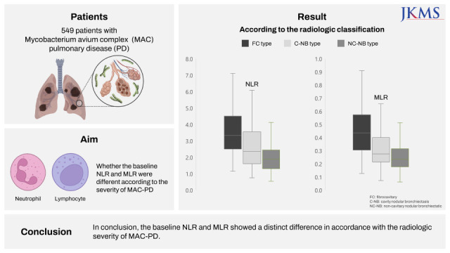
The neutrophil-lymphocyte ratio (NLR) and monocyte-lymphocyte ratio (MLR) are readily available laboratory markers calculated from a complete blood count (CBC). Previous studies have reported that both NLR and MLR were useful laboratory markers of the severity, treatment response, or recurrence of pulmonary or extrapulmonary tuberculosis.12345 Although the clinical characteristics of Mycobacterium avium complex (MAC)-pulmonary disease (PD) are similar to those of tuberculosis in many aspects, such as symptoms, radiologic findings, or treatment regimen,6 to date, no study has investigated the value of NLR and MLR in MAC-PD. Therefore, we aimed to assess this issue in patients with MAC-PD. Considering that, notably, NLR and MLR were associated with tuberculosis severity, we investigated whether the baseline NLR and MLR were different according to the severity of MAC-PD based on the radiologic classification.

Methods

Study subjects

Patients were enrolled in the Asan Medical Center in Seoul, South Korea. From 2001 to 2019, 1,148 patients with MAC-PD who had initiated treatment with a macrolide-containing regimen were identified. We excluded those 1) who did not undergo CBC examination within 90 days prior to treatment initiation (n = 384), 2) whose computed tomography (CT) findings were categorized as unclassifiable type (n = 150), and 3) who had comorbidities that could affect the results of white blood cell differential count, including liver disease (n = 24), infectious disease such as pneumonia (n = 21), were receiving calcineurin inhibitor (n = 7) or anti-cancer chemotherapy (n = 6), and others (n = 7). The medical records of the remaining patients were retrospectively analyzed in November 2021.

Radiologic classification and baseline NLR and MLR

Radiologic findings on chest CT were classified into one of the three major types: fibrocavitary (FC), cavitary nodular bronchiectatic (C-NB), and non-cavitary nodular bronchiectatic (NC-NB) types.7 The baseline NLR and MLR were compared according to each radiologic type. NLR and MLR were calculated from peripheral CBC. An automatic blood count device was employed for the analysis of CBC. NLR and MLR were determined as follows: NLR = Absolute Neutrophil Count/Absolute Lymphocyte Count, MLR = Absolute Monocyte Count/Absolute Lymphocyte Count.4

Statistical analysis

All data are presented as means ± standard deviation or as medians (interquartile range [IQR]) for continuous variables and number (%) for categorical variables. The baseline NLR and MLR values according to radiologic classification were compared using the Kruskal-Wallis test. For statistical analysis, an IBM SPSS Statistics for Windows, version 26.0 (IBM Corp., Armonk, NY, USA) software was used.

Ethics statement

The study protocol was approved by the Institutional Review Board (IRB) of the Asan Medical Center (IRB No. 2021-1210). The requirement for informed consent was waived by the board because of the retrospective nature of this study.

Results

Study participants

The eligibility screening identified 549 patients with MAC-PD, including 95, 176, and 278 patients with FC, C-NB, and NC-NB types, respectively. Their median age was 64.0 (IQR, 56.0–72.0) years, and women were predominant (61.4%). The mean body mass index was 20.3 ± 2.9 kg/m2. Sputum acid-fast bacillus (AFB) smear positivity was noted in 49.0% of the patients. As shown in Table 1, a statistically significant difference was noted in terms of age, gender, body mass index, smoking history, etiologic organism, and sputum AFB smear positivity.
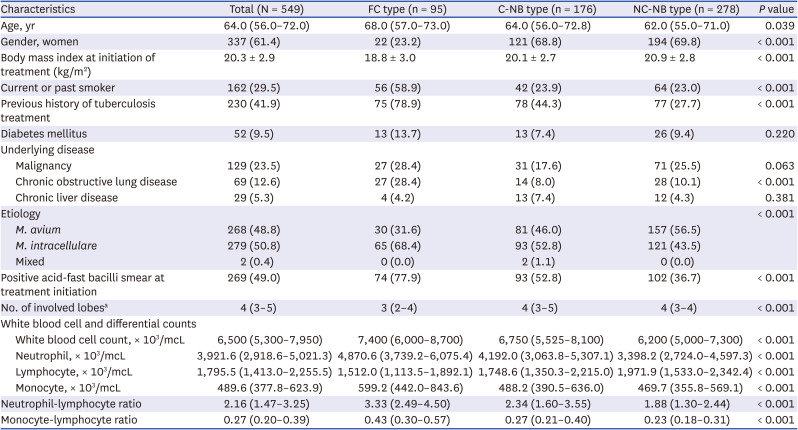
Table 1

Clinical characteristics of 549 patients with Mycobacterium avium complex pulmonary disease according to the radiologic classification

jkms-37-e292-i001
Characteristics Total (N = 549) FC type (n = 95) C-NB type (n = 176) NC-NB type (n = 278) P value
Age, yr 64.0 (56.0–72.0) 68.0 (57.0–73.0) 64.0 (56.0–72.8) 62.0 (55.0–71.0) 0.039
Gender, women 337 (61.4) 22 (23.2) 121 (68.8) 194 (69.8) < 0.001
Body mass index at initiation of treatment (kg/m2) 20.3 ± 2.9 18.8 ± 3.0 20.1 ± 2.7 20.9 ± 2.8 < 0.001
Current or past smoker 162 (29.5) 56 (58.9) 42 (23.9) 64 (23.0) < 0.001
Previous history of tuberculosis treatment 230 (41.9) 75 (78.9) 78 (44.3) 77 (27.7) < 0.001
Diabetes mellitus 52 (9.5) 13 (13.7) 13 (7.4) 26 (9.4) 0.220
Underlying disease
Malignancy 129 (23.5) 27 (28.4) 31 (17.6) 71 (25.5) 0.063
Chronic obstructive lung disease 69 (12.6) 27 (28.4) 14 (8.0) 28 (10.1) < 0.001
Chronic liver disease 29 (5.3) 4 (4.2) 13 (7.4) 12 (4.3) 0.381
Etiology < 0.001
M. avium 268 (48.8) 30 (31.6) 81 (46.0) 157 (56.5)
M. intracellulare 279 (50.8) 65 (68.4) 93 (52.8) 121 (43.5)
Mixed 2 (0.4) 0 (0.0) 2 (1.1) 0 (0.0)
Positive acid-fast bacilli smear at treatment initiation 269 (49.0) 74 (77.9) 93 (52.8) 102 (36.7) < 0.001
No. of involved lobesa 4 (3–5) 3 (2–4) 4 (3–5) 4 (3–4) < 0.001
White blood cell and differential counts
White blood cell count, × 103/mcL 6,500 (5,300–7,950) 7,400 (6,000–8,700) 6,750 (5,525–8,100) 6,200 (5,000–7,300) < 0.001
Neutrophil, × 103/mcL 3,921.6 (2,918.6–5,021.3) 4,870.6 (3,739.2–6,075.4) 4,192.0 (3,063.8–5,307.1) 3,398.2 (2,724.0–4,597.3) < 0.001
Lymphocyte, × 103/mcL 1,795.5 (1,413.0–2,255.5) 1,512.0 (1,113.5–1,892.1) 1,748.6 (1,350.3–2,215.0) 1,971.9 (1,533.0–2,342.4) < 0.001
Monocyte, × 103/mcL 489.6 (377.8–623.9) 599.2 (442.0–843.6) 488.2 (390.5–636.0) 469.7 (355.8–569.1) < 0.001
Neutrophil-lymphocyte ratio 2.16 (1.47–3.25) 3.33 (2.49–4.50) 2.34 (1.60–3.55) 1.88 (1.30–2.44) < 0.001
Monocyte-lymphocyte ratio 0.27 (0.20–0.39) 0.43 (0.30–0.57) 0.27 (0.21–0.40) 0.23 (0.18–0.31) < 0.001
Data are presented as mean ± standard deviation, median (interquartile range), or frequencies (%).
FC = fibrocavitary, C-NB = cavitary nodular bronchiectatic, NC-NB = non-cavitary nodular bronchiectatic.
aThe extent of the involved lobe was assessed according to how many of the six lung lobes were involved, considering the lingular segment to be a separate lobe.

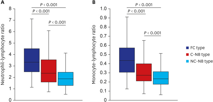
Baseline NLR and MLR according to radiologic classification

The baseline NLR of the 549 patients was 2.16 (IQR, 1.47–3.25). As shown in Fig. 1A, NLR was significantly higher in the FC type (3.33 [IQR, 2.49–4.50]), followed by that in the C-NB type (2.34 [IQR, 1.60–3.55]), and significantly lower in the NC-NB type (1.88 [IQR, 1.30–2.44]) (P < 0.001). In addition, the baseline MLR of the 549 patients was 0.27 (IQR, 0.20–0.39), and it was significantly higher in the FC type (0.43 [IQR, 0.30–0.57]), followed by that in the C-NB type (0.27 [IQR, 0.21–0.40]), and NC-NB type (0.23 [IQR, 0.18–0.31]) (P < 0.001), as shown in Fig. 1B.
Fig. 1

Neutrophil-lymphocyte ratio (A) and monocyte-lymphocyte ratio (B) according to the radiologic classification of 549 patients with Mycobacterium avium complex pulmonary disease.

FC = fibrocavitary, C-NB = cavitary nodular bronchiectatic, NC-NB = non-cavitary nodular bronchiectatic.
jkms-37-e292-g001

Discussion

Although previous studies have shown that NLR and MLR are related to the treatment outcomes or severity of tuberculosis,1245 whether these two laboratory markers would also have a clinical value in MAC-PD has not been assessed, so far. This study investigated this aspect by retrospectively analyzing 549 patients with MAC-PD in a tertiary referral center in South Korea. We found a distinct difference in the baseline NLR and MLR according to the radiologic severity of MAC-PD; both NLR and MLR showed higher values when the radiologic findings of MAC-PD were more severe.
Radiologic classification has vital importance in MAC-PD regarding various aspects including the treatment regimen, outcome, and prognosis. In terms of drug regimen composition, an intermittent oral-drug therapy is recommended for patients with NC-NB type, whereas an aminoglycoside-containing daily regimen is recommended for those with cavitary type (i.e., FC and C-NB types).6 Notably, the treatment success rate for cavitary MAC-PD was reported to be significantly lower than that for NC-NB type despite receiving standard treatment.7 In addition, significant difference in the mortality rate was noted according to the radiologic type of MAC-PD. A recent study reported that the overall survival rates were higher in patients with NC-NB type than in those with a cavitary disease, and those patients with C-NB type showed mortality rates intermediate between those of NC-NB and FC forms.8
These clinical differences according to radiologic classification could be explained by cavitation, which indicates a higher mycobacterial burden in MAC-PD.9 Therefore, it can be predicted that the presence of cavity is accompanied by a severe overall inflammatory response. In general, the immune responses of circulating leukocytes to various inflammatory events are characterized by an increased number of neutrophils in the peripheral blood.10 In the case of tuberculosis, extensive neutrophilic response is a sign of severity and has been particularly related to tissue destruction.11 Neutrophilia is independently associated with a higher risk of cavity formation.12 Additionally, an increase in matrix metalloproteinase-8-expressing neutrophil was related to cavitation in tuberculosis.9 Moreover, monocytes are one of the essential components of the innate immune responses and act as a link to the adaptive immune system.4 Therefore, as the disease progresses, the number of monocytes derived from the hematopoietic stem cells could increase to phagocytize and restrict mycobacterium.13 An ongoing immune response against the cavitary lesion could result in further increase in the percentage of blood monocytes.141516 Moreover, lymphopenia could result from the accumulation of lymphocytes at the infection site, leading to their decreased number in the peripheral blood.17 These theoretical changes in neutrophils, lymphocytes, and monocytes can reasonably explain why NLR and MLR were increased in accordance with the radiologic severity in MAC-PD.
Only those patients who received treatment were included in this study. This is because patients with cavitary form MAC-PD without treatment are rarely encountered, as immediate treatment initiation is recommended for this type.18 When we performed the same analysis in patients with MAC-PD who did not receive treatment using the data of the patients enrolled in our previous study,19 we found a statistical difference in NLR and MLR between the cavitary vs. non-cavitary type (Supplementary Fig. 1). However, the number of patients with cavitary type was too small to analyze.
There may be other laboratory test results that can be related to the radiologic severity of MAC-PD including erythrocyte sedimentation rate (ESR), C-reactive protein (CRP), or platelet count considering that previous studies have shown these inflammatory markers were associated with severity or the immune response of mycobacterial disease.202122 We compared the ESR, CRP, or platelet count according to the radiologic classification among a portion of patients in this study for whom these inflammatory markers were measured, the results of which are shown in Supplementary Fig. 2. As the Figure denotes, statistically significant differences in CRP and platelet count were noted among the three groups. Besides laboratory markers, the extent of radiologic involvement could also reflect the severity of MAC-PD. Additional analysis and results regarding whether the baseline NLR and MLR differ according to the number of involved lobes in each radiologic type, are summarized in Supplementary Fig. 3.
This study had some limitations; the most significant limitation being that it was conducted at a single referral center, and it had a retrospective design. Second, although NLR and MLR were measured in all the enrolled patients within 90 days of treatment initiation, the measured time points were slightly different for each patient. It was unclear whether the slightly different measurement time points for each patient affected the overall results of NLR and MLR. Finally, whether NLR and MLR could be used as a marker to predict treatment outcomes was not assessed. Further studies are needed to determine whether these values at baseline or during treatment are related to treatment outcomes in terms of treatment success or mortality.
In conclusion, this study showed that the baseline NLR and MLR had a distinct difference in accordance with the radiologic severity of MAC-PD.

Notes

Funding: This work was supported by the National Research Foundation of Korea grant funded by the Korea government (Ministry of Science and ICT; No. 2022R1A2C1002847) for Kyung-Wook Jo.

Disclosure: The authors have no potential conflicts of interest to disclose.

Author Contributions:

  • Conceptualization: Shim TS, Jo KW.

  • Data curation: Kim MA, Park YE, Chong YP.

  • Formal analysis: Kim MA, Shim TS, Jo KW.

  • Funding acquisition: Jo KW.

  • Investigation: Kim MA, Shim TS, Jo KW.

  • Writing-original draft: Kim MA, Jo KW.

  • Writing-review & editing: Shim TS, Jo KW.

References

1. Han Y, Kim SJ, Lee SH, Sim YS, Ryu YJ, Chang JH, et al. High blood neutrophil-lymphocyte ratio associated with poor outcomes in miliary tuberculosis. J Thorac Dis. 2018; 10(1):339–346. PMID: 29600065.
crossref
2. Yin Y, Kuai S, Liu J, Zhang Y, Shan Z, Gu L, et al. Pretreatment neutrophil-to-lymphocyte ratio in peripheral blood was associated with pulmonary tuberculosis retreatment. Arch Med Sci. 2017; 13(2):404–411. PMID: 28261295.
crossref
3. Iliaz S, Iliaz R, Ortakoylu G, Bahadir A, Bagci BA, Caglar E. Value of neutrophil/lymphocyte ratio in the differential diagnosis of sarcoidosis and tuberculosis. Ann Thorac Med. 2014; 9(4):232–235. PMID: 25276243.
crossref
4. Ngahane BH, Ebenezer AT, Eveline ND, Raïssa MN, Fernando KL, Lum CV, et al. Diagnostic value of leukocyte count abnormalities in newly diagnosed tuberculosis patients. Open J Respir Dis. 2019; 10(1):1–10.
crossref
5. Wang W, Wang LF, Liu YY, Yang F, Zhu L, Zhang XH. Value of the ratio of monocytes to lymphocytes for monitoring tuberculosis therapy. Can J Infect Dis Med Microbiol. 2019; 3270393. PMID: 31263513.
crossref
6. Daley CL, Iaccarino JM, Lange C, Cambau E, Wallace RJ Jr, Andrejak C, et al. Treatment of nontuberculous mycobacterial pulmonary disease: an official ATS/ERS/ESCMID/IDSA clinical practice guideline. Clin Infect Dis. 2020; 71(4):e1–36. PMID: 32628747.
crossref
7. Koh WJ, Moon SM, Kim SY, Woo MA, Kim S, Jhun BW, et al. Outcomes of Mycobacterium avium complex lung disease based on clinical phenotype. Eur Respir J. 2017; 50(3):1602503. PMID: 28954780.
8. Jhun BW, Moon SM, Jeon K, Kwon OJ, Yoo H, Carriere KC, et al. Prognostic factors associated with long-term mortality in 1445 patients with nontuberculous mycobacterial pulmonary disease: a 15-year follow-up study. Eur Respir J. 2020; 55(1):1900798. PMID: 31619468.
crossref
9. Ong CW, Elkington PT, Friedland JS. Tuberculosis, pulmonary cavitation, and matrix metalloproteinases. Am J Respir Crit Care Med. 2014; 190(1):9–18. PMID: 24713029.
crossref
10. Yoon NB, Son C, Um SJ. Role of the neutrophil-lymphocyte count ratio in the differential diagnosis between pulmonary tuberculosis and bacterial community-acquired pneumonia. Ann Lab Med. 2013; 33(2):105–110. PMID: 23482854.
crossref
11. Lyadova IV. Neutrophils in tuberculosis: heterogeneity shapes the way? Mediators Inflamm. 2017; 2017:8619307. PMID: 28626346.
crossref
12. de Melo MG, Mesquita ED, Oliveira MM, da Silva-Monteiro C, Silveira AK, Malaquias TS, et al. Imbalance of NET and alpha-1-antitrypsin in tuberculosis patients is related with hyper inflammation and severe lung tissue damage. Front Immunol. 2019; 9:3147. PMID: 30687336.
crossref
13. Fenton MJ, Vermeulen MW. Immunopathology of tuberculosis: roles of macrophages and monocytes. Infect Immun. 1996; 64(3):683–690. PMID: 8641767.
crossref
14. Lavalett L, Ortega H, Barrera LF. Infection of monocytes from tuberculosis patients with two virulent clinical isolates of Mycobacterium tuberculosis induces alterations in myeloid effector functions. Front Cell Infect Microbiol. 2020; 10:163. PMID: 32391286.
15. Rakotosamimanana N, Richard V, Raharimanga V, Gicquel B, Doherty TM, Zumla A, et al. Biomarkers for risk of developing active tuberculosis in contacts of TB patients: a prospective cohort study. Eur Respir J. 2015; 46(4):1095–1103. PMID: 26250497.
crossref
16. Urbanowski ME, Ordonez AA, Ruiz-Bedoya CA, Jain SK, Bishai WR. Cavitary tuberculosis: the gateway of disease transmission. Lancet Infect Dis. 2020; 20(6):e117–e128. PMID: 32482293.
crossref
17. Cohen R, Muzaffar S, Capellan J, Azar H, Chinikamwala M. The validity of classic symptoms and chest radiographic configuration in predicting pulmonary tuberculosis. Chest. 1996; 109(2):420–423. PMID: 8620716.
crossref
18. Kwon YS, Koh WJ. Diagnosis and treatment of nontuberculous mycobacterial lung disease. J Korean Med Sci. 2016; 31(5):649–659. PMID: 27134484.
crossref
19. Kwon BS, Lee JH, Koh Y, Kim WS, Song JW, Oh YM, et al. The natural history of non-cavitary nodular bronchiectatic Mycobacterium avium complex lung disease. Respir Med. 2019; 150:45–50. PMID: 30961950.
crossref
20. Kim HJ, Kwak N, Hong H, Kang N, Im Y, Jhun BW, et al. BACES score for predicting mortality in nontuberculous mycobacterial pulmonary disease. Am J Respir Crit Care Med. 2021; 203(2):230–236. PMID: 32721164.
crossref
21. Kwas H, Guermazi E, Zendah I, Ben Jemia E, Khattab A, Khouaja I, et al. C-reactive protein and pulmonary tuberculosis: what correlation with disease severity. Eur Respir J. 2015; 46:PA2751.
crossref
22. Kirwan DE, Chong DL, Friedland JS. Platelet activation and the immune response to tuberculosis. Front Immunol. 2021; 12:631696. PMID: 34093524.
crossref

SUPPLEMENTARY MATERIALS

Supplementary Fig. 1

NLR (A) and MLR (B) according to the radiologic classification of 107 patients with Mycobacterium avium complex pulmonary disease who did not receive treatment. NLR was higher in the FC type (3.96 [IQR, 1.69–6.78]), followed by that in the C-NB type (2.46 [IQR 1.60–4.20]), and significantly lower in the NC-NB type (1.57 [IQR, 1.29–2.06]). In addition, MLR was higher in the FC type (0.33 [IQR, 0.29–0.49]), followed by that in the C-NB type (0.27 [IQR, 0.23–0.39]), and NC-NB type (0.20 [IQR, 0.16–0.23]).
jkms-37-e292-s001.doc

Supplementary Fig. 2

ESR, CRP, and platelet count according to the radiologic classification of patients with Mycobacterium avium complex pulmonary disease. Notably, the number of patients was lower than 549 patients whose neutrophil-lymphocyte ratio and monocyte-lymphocyte ratio were assessed in the main analysis, because only some patients underwent laboratory tests for ESR and CRP. (A) No statistical difference in ESR was noted among the three radiologic types with 34.0 (20.5–90.8), 32.5 (16.0–66.6), and 26.0 (18.5–55.5) in the FC, C-NB, and NC-NB types, respectively. (B) CRP was significantly higher in the FC type (2.38 [IQR, 1.09–4.17]), followed by that in the C-NB type (0.43 [IQR, 0.12–1.68]), and significantly lower in the NC-NB type (0.16 [IQR, 0.10–0.54]). (C) Platelet count was significantly higher in the FC type (287,000 [IQR, 220,000–346,000]), followed by that in the C-NB type (245,000 [IQR, 202,000–302,000]), and significantly lower in the NC-NB type (229,000 [IQR, 193,000–275,000]).
jkms-37-e292-s002.doc

Supplementary Fig. 3

The comparison of the baseline NLR and MLR according to the involved lobe in each radiologic type. The extent of the involved lobe was assessed according to how many of the six lung lobes were involved, considering the lingular segment a separate lobe. After dividing into two groups according to the median number of involved lobes in each radiologic type, NLR and MLR were compared. As seen, there was no statistical difference in these ratios according to the number of involved lobes among all the three radiologic types.
jkms-37-e292-s003.doc
TOOLS
Similar articles