Journal List > Cancer Res Treat > v.54(3) > 1516078556

Choi, Yang, Lee, Lee, Kim, Yong, and Lee: Usefulness of Pulmonary Rehabilitation in Non–Small Cell Lung Cancer Patients Based on Pulmonary Function Tests and Muscle Analysis Using Computed Tomography Images

Abstract

Purpose

The usefulness of rehabilitation in patients with reduced lung function before lung surgery remains unclear, and there is no adequate method for evaluating the effect of rehabilitation. We aimed to evaluate the usefulness of rehabilitation in patients with non–small cell lung cancer (NSCLC) undergoing lung cancer surgery.

Materials and Methods

We retrospectively analyzed the medical records of NSCLC patients at Korea University Guro Hospital between 2018 and 2020. Patients were divided into two groups depending on whether they underwent rehabilitation. Pulmonary function test data and muscle determined using chest computed tomography images were analyzed. Because the baseline characteristics were different between the two groups, propensity score matching was performed.

Results

Of 325 patients, 75 (23.1%) and 250 (76.9%) were included in the rehabilitation and non-rehabilitation (control) groups, respectively. The rehabilitation group had a worse general condition at baseline. After propensity score matching, 45 patients remained in each group. Pulmonary function (forced expiratory volume in 1 second, %) (p=0.001) and the Hounsfield unit of erector spinae muscle (p=0.001) were better preserved in the rehabilitation group. Muscle loss of 3.4% and 0.6% was observed in the control and rehabilitation groups, respectively (p=0.003). In addition, the incidence of embolic events was lower in the rehabilitation group (p=0.044).

Conclusion

Pulmonary rehabilitation is useful in patients with NSCLC undergoing lung surgery. Pulmonary rehabilitation preserves lung function, muscle and reduces embolic events after surgery. Pulmonary rehabilitation is recommended for patients with NSCLC undergoing surgery.

Introduction

Rehabilitation is essential in various clinical situations and diseases [1]. The therapeutic efficacy of surgery or medicine can be improved through rehabilitation. Rehabilitation can maintain the quality of life of patients and prevent the worsening of chronic diseases. For example, rehabilitation can reduce the occurrence of post-intensive care syndrome in the intensive care unit [2]. Rehabilitation can improve sequelae and prevent recurrence in patients with coronary artery disease or cerebrovascular disease [3,4]. Similarly, the value of rehabilitation in various respiratory diseases is also attracting attention [5].
Pulmonary rehabilitation improves respiratory symptoms and complications in patients with chronic obstructive pulmonary disease (COPD) [6]. Early pulmonary rehabilitation in patients with severe acute exacerbation of COPD can decrease mortality and improve the health-related quality of life [7]. Moreover, pulmonary rehabilitation can improve exercise capacity, dyspnea, and quality of life in patients with idiopathic pulmonary fibrosis [8]. Various studies related to pulmonary rehabilitation in lung cancer and lung operation have been published [9]. Pulmonary rehabilitation can reduce postoperative complications and functional depletion after lung surgery [10]. Pulmonary rehabilitation enables patients with lung cancer with poor lung function to undergo surgery [11]. Furthermore, pulmonary rehabilitation is considered helpful in palliative care for patients with advanced lung cancer.
Pulmonary rehabilitation is known to be useful for treating various respiratory diseases. However, its value and benefits are overlooked in the clinical field [12,13], possibly owing to the lack of objective and specific data. Therefore, this study aimed to provide objective evidence about the usefulness of pulmonary rehabilitation through chest computed tomography (CT) and pulmonary function test (PFT) assessments in patients with non–small cell lung cancer (NSCLC) undergoing lung cancer surgery.

Materials and Methods

1. Patients

We retrospectively analyzed the medical records of patients with NSCLC who underwent lung surgery at Korea University Guro Hospital between January 2018 and December 2020. We included patients with histologically proven primary NSCLC according to the tumor-node-metastasis classification of the International Union Against Cancer, 8th edition. We included all types of lung operations (lobectomy, segmentectomy, wedge resection, and bilobectomy). The patients were divided into two groups depending on whether they underwent pulmonary rehabilitation. We included patients with preoperative and postoperative chest CT images or PFT data. In our hospital, PFT and chest CT are performed within a month from the date of discharge for all patients to check the degree of postoperative complications and deterioration of lung function. For this reason, all 325 patients included in this study had preoperative and postoperative PFT and chest CT data. Postoperative PFT and chest CT were mostly performed on the same day, and cases performed 2 months or later from the date of discharge were excluded from this study. Based on the day of discharge, postoperative PFT and chest CT were performed on 24.4±7.3 days later. For PFT-related analysis, we included only patients with forced expiratory volume in 1 second (FEV1), forced vital capacity (FVC), and carbon monoxide diffusion capacity (DLCO) data
We analyzed several clinical data to assess the effects of rehabilitation treatment on postoperative prognosis. For example, we analyzed the period from admission to discharge, high-flow nasal cannula (HFNC) application, duration of HFNC use, 90-day mortality and 180-day mortality. And major cardiovascular events, arrhythmia, pneumonia, pneumothorax, chest pain, dyspnea, pulmonary embolism, deep vein thrombosis, and pleural effusion were evaluated to evaluate major postoperative complications. ‘Major cardiovascular events, arrhythmias, pneumonia, pneumothorax, chest pain, dyspnea, deep vein thrombosis and pleural effusion’ were defined only for patients who visited the emergency room or readmitted within 1 month from the date of discharge. ‘Pulmonary embolism’ was defined as a case seen on postoperative CT.
Primary endpoints were changes in lung function parameters (FEV1, FVC, and DLCO) and muscle after surgery. Secondary endpoints were prognostic assessment during the hospital stay and major postoperative complications.

2. Pulmonary rehabilitation protocol

Pulmonary rehabilitation consisted of exercise and education programs. The exercise program was performed one to three times a week for 30–40 minutes per session. The education program was conducted repeatedly in the outpatient clinic and exercise therapy room. The exercise program consisted of aerobic (walking, bicycle ergometer, treadmill, and arm ergometer), strength (mainly upper-limb strengthening training), flexibility, and inspiratory muscle exercises. The education program consisted of smoking-cessation education, breathing re-training (pursed-lip breathing, diaphragmatic breathing, and segmental breathing), and secretion removal training (coughing exercise, huffing, assisted coughing, and postural drainage). The patients’ exercise intensity was determined according to metabolic equivalent, maximal oxygen consumption, and heart rate values.
Pulmonary rehabilitation was performed at least one to two times before surgery. We assessed the patients’ condition 2–3 weeks after the operation and resumed pulmonary rehabilitation. We provided educational materials (pamphlets, notes, and posters) to patients to enable them to perform daily rehabilitation exercises at home.

3. Muscle measurement

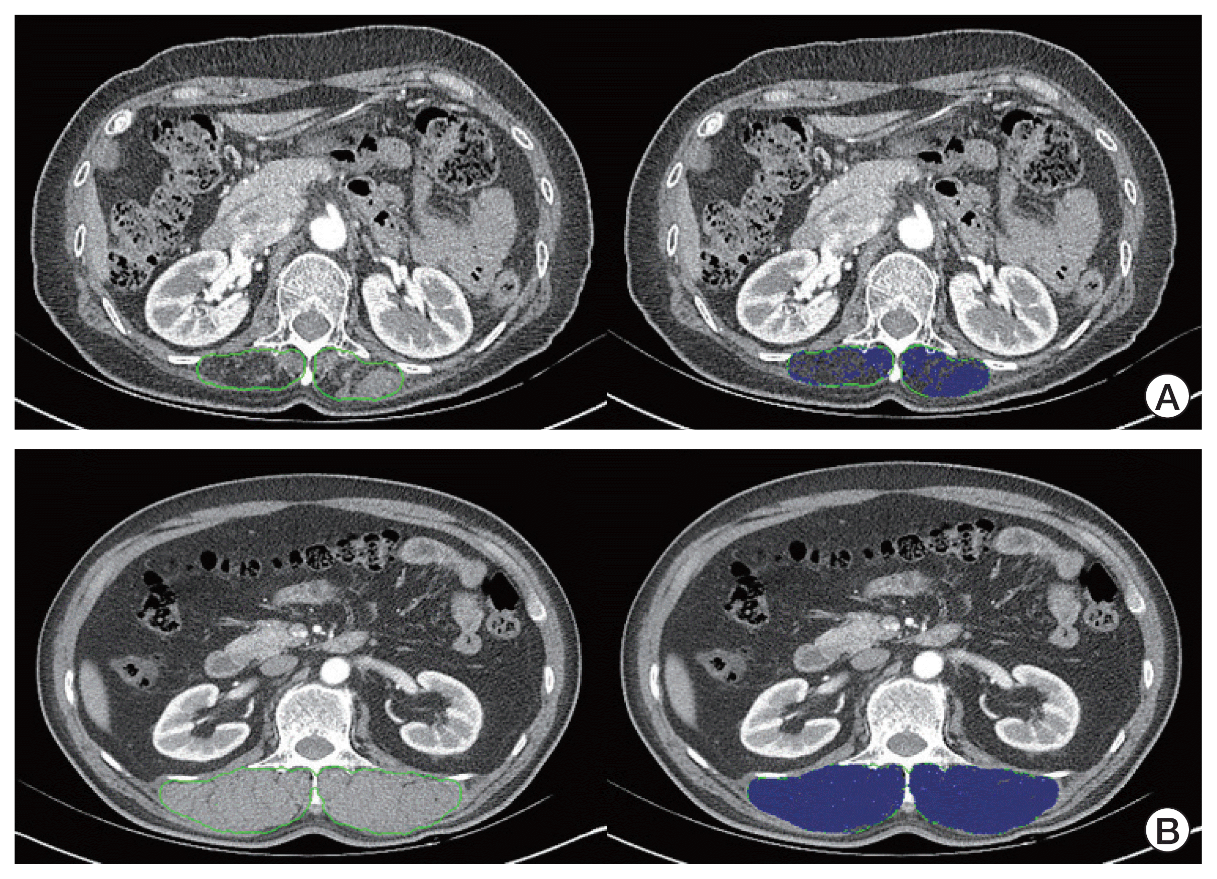
There are three muscles (pectoralis muscle, erector spinae muscle (thoracic level), erector spinae muscles (lumbar level)) we considered when measuring the muscle. Of these, the erector spinae muscles (first lumbar level) were chosen for three reasons. As a previous study, there is a study analyzing the prognosis of COPD patients using CT-based muscle measurement of the pectoralis muscle and erector spinae muscle (thoracic level) [14,15]. However, unlike the previous COPD study, this study was conducted on patients who underwent lung surgery and chest tube insertion or management, and it was thought that the pectoralis muscle, erector spinae muscle (thoracic level) may be inaccurate due to damage. Second, routine chest CT included only images up to first lumbar level. Third, in a study analyzing the prognosis of lung cancer patients, not patients with COPD, there was a study that erector spinae muscle (first lumbar level) was better than pectoralis muscle [16]. So, we set the erector spinae muscles (first lumbar level) as the standard for this study (Fig. 1A and B).
We used the Hounsfield unit (HU) average value measured within the patient’s spinae muscle area in the CT image to evaluate muscle mass. We defined and used ‘HUESMcsa’ as HU of average intensity value of erector spinae muscle in cross-sectional area in CT.
First, to overcome the variations in image quality based on patient size and scanning protocol, denoising was performed for all chest CT images using commercial software (ClariCT.ai, ClariPi, Seoul, Korea). Second, the HUESMcsa was manually calculated by one experienced clinician and two researcher using in-house developed software that can measure muscle and fat indices and semi-automatically calculate the HU range. The HUESMcsa measurement process was performed twice per person, and the average value was used to avoid user error and to check for repeatability.
The detailed mechanism and principle of measuring HUESMcsa are described below (Mechanism of in-house developed software):
We selected the region of interests (ROI) with in-house developed software, which could get the muscle area by clicking it on screen with mouse cursor from the cross-sectional area in CT image. Then, well-shown homogeneous muscle area and adipose tissue area from the whole area of CT were selected manually for ‘intensity calibration process’, for setting HU intensity range of muscle and adipose tissue for simpler way. The HU dividing points of fat and muscle were set to −30 at 120 kVp, according to previous studies [17,18]. Around the selected area, the average intensity HU values were measured, and validated via HU value calculation formula from the literatures of ‘National Institute of Standards and technology’, since the scanning process of the patients may have various conditions (such as different scanning protocols and parameters which may affect the average intensity value of muscle or adipose tissue value in unexpected range).
After that, with simple image processing techniques, ROI of demanded area was acquired with removing border line of muscle area. Modified flood fill technique was applied to get the ROI of demanded area, which could get the boundary area when relatively large intensity difference was met. This simple technique could get the erector spinae muscle area precisely, especially without boundary edge area which might include inhomogeneous intensity from partial volume area effects.
By getting the ROI from erector spinae muscle area and the intensity range of each target, the muscle and adipose tissue fracture distribution could be calculated by having the threshold value in the middle of muscle and adipose tissue intensity. The muscle and adipose intensity fracture distribution could be directly calculated as muscle density or ‘muscle index’ from the paper, since it could be interpreted as the mixture organ with pure muscle and fat.

4. Statistical analysis

Data were analyzed using SPSS ver. 20 software (IBM Corp., Armonk, NY). Continuous variables are reported as the mean±standard deviation, and categorical variables are reported as the number (%). Continuous variables were compared using the Student’s t test. Categorical variables were compared using the chi-square test or Fisher exact test. Fisher exact test was used when the expected number of events was less than five.
In general, pulmonary rehabilitation is recommended for patients with poor lung function before lung surgery. Therefore, differences in baseline characteristics and pulmonary function were observed between the two groups. To balance these differences, propensity score matching was performed. Propensity scores were calculated for each patient using multivariable logistic regression based on the following covariates: age, sex, height, weight, FEV1 (%), DLCO (%), comorbidities, inhaler use, cancer-related treatment (neoadjuvant chemotherapy and neoadjuvant concurrent chemoradiotherapy), type of surgery, and operation site. Matching was performed using the nearest-neighbor method to select the most similar propensity scores. We performed 1:1 matching and calculated the effect size of the standardized mean difference (d) to express the suitability of propensity score matchings.
In addition, to increase the reliability of the HUESMcsa, the correlation between the HUESMcsa and PFT values was analyzed. Correlation analysis was performed using Pearson correlation coefficients.

Results

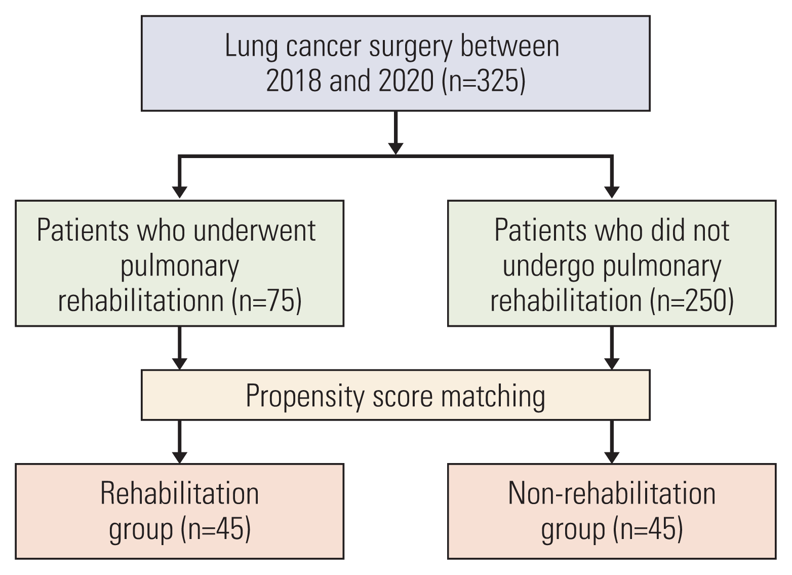
We retrospectively identified 325 patients with NSCLC who underwent lung surgery at our hospital between 2018 and 2020. Of these 325 patients, 75 (23.1%) were in the rehabilitation group and 250 (76.9%) were in the non-rehabilitation (control) group. After propensity score matching, 45 patients remained in each group (Fig. 2). The patients underwent 5.8±5.6 sessions of pulmonary rehabilitation on average.

1. Baseline characteristics

Table 1 shows the baseline characteristics of patients before and after propensity score matching. The rehabilitation group had a worse general condition than the control group. For example, those in the rehabilitation group were older, had a higher body mass index, and an increased prevalence of COPD. Accordingly, inhaler was used a lot in the rehabilitation group. With respect to the type of surgery, more patients in the rehabilitation group underwent bilobectomy or lobectomy with wedge resection. After propensity score matching, there were no significant differences between the two groups.

2. Pulmonary function test

Table 2 shows the preoperative PFT results before and after propensity score matching. Our hospital mainly recommends pulmonary rehabilitation for patients with poor lung function. Therefore, the baseline lung function was generally poor in the rehabilitation group. FEV1 (%), FVC (%), and DLCO (%) were all low in the rehabilitation group. We included FEV1 (%) and DLCO (%) as variables in propensity score matching. Therefore, there was no difference in lung function between the two groups after propensity score matching.
Table 3 shows the differences in lung function after surgery. Before and after propensity score matching, FEV1 (L, %) and FVC (L, %) were better preserved in the rehabilitation group. Even after propensity score matching, DLCO (L, %) did not show any statistically significant differences.

3. HUESMcsa

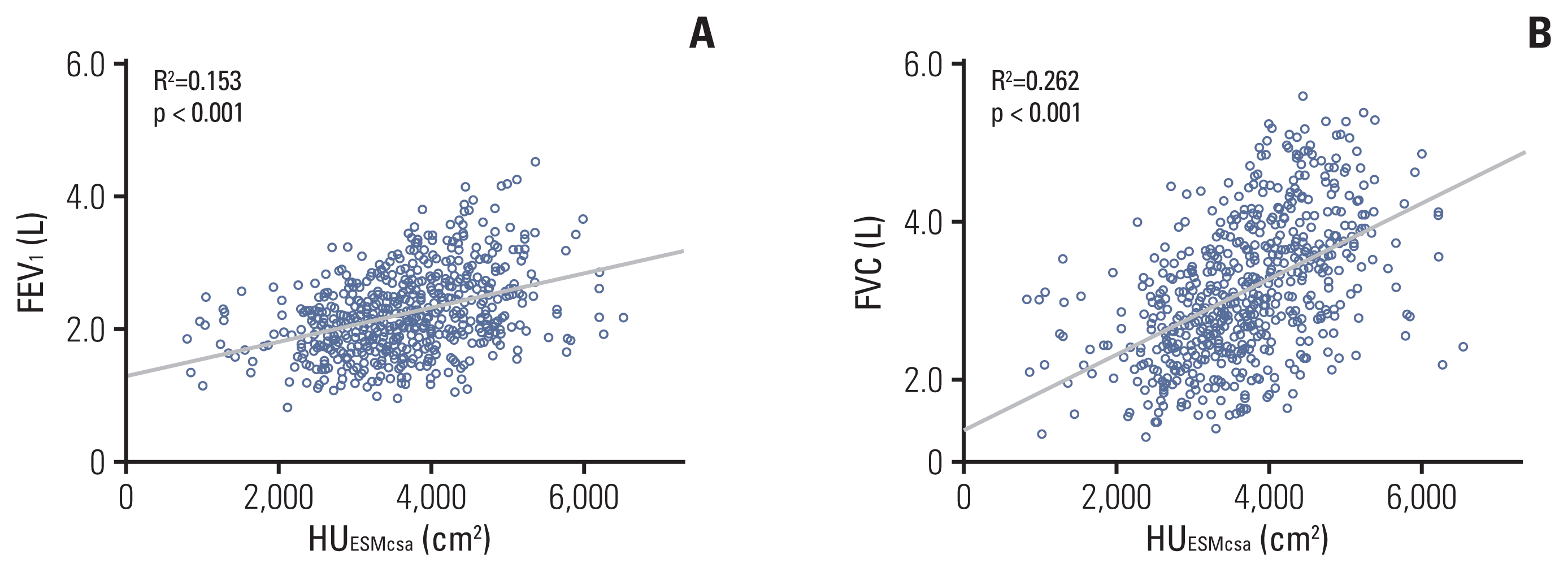
Before evaluating the HUESMcsa, we performed a correlation analysis between the HUESMcsa and PFT values (Fig. 3). FEV1 (L) and FVC (L) showed a clear positive correlation with the HUESMcsa (R2=0.153, p < 0.001 and R2=0.262, p < 0.001, respectively).
Before and after propensity score matching, the HUESMcsa was better preserved in the rehabilitation group (p=0.001 and p=0003, respectively) (Table 4). After propensity score matching analysis, there was 3.4% muscle loss in the control group and 0.6% muscle loss in the rehabilitation group (p=0.003).

4. Prognostic analysis

Before propensity score matching, the period from admission to discharge was longer in the rehabilitation group (p=0.029). After propensity score matching, the period from hospitalization to discharge was similar between the groups (p=0.926). With respect to HFNC application and the duration of HFNC, there were no differences between the two groups. No patients died within 30 days, and there was no difference between the two groups in terms of 90-day and 180-day mortality (Table 5).

5. Major postoperative complications

For major postoperative complications, there was no statistical difference in the analysis before propensity scoring matching, but after propensity scoring matching, there was a statistically significant decrease in major postoperative complications (p=0.027). In particular, the incidence of embolic events (pulmonary embolism or deep vein thrombosis) was lower in the rehabilitated patient group (p=0.044) (Table 6).

Discussion

This study is a first study to analyze the efficacy of lung rehabilitation by retrospectively analyzing the data of lung cancer patients who were operated in Korea. In this study, we attempted to demonstrate the usefulness of pulmonary rehabilitation using chest CT and PFT data. Despite the obvious necessity of pulmonary rehabilitation, it is not actively performed in the clinical setting, possibly because of the lack of specific and objective evidence. Therefore, we attempted to prove the necessity of pulmonary rehabilitation using numerical and objective data from CT and PFT assessments. According to our results, pulmonary rehabilitation preserves the FEV1, FVC, and HUESMcsa. Pulmonary rehabilitation should be actively recommended to patients with NSCLC scheduled for lung surgery.
It is commonly thought that pulmonary rehabilitation consists of only exercises that strengthen respiratory muscles. Ideally, pulmonary rehabilitation should be a combination of exercise and education programs [19]. Exercise programs include aerobic (walking, bicycle ergometer, treadmill, and arm ergometer), upper-limb strengthening training, flexibility, and inspiratory muscle exercises [20]. Education programs include smoking-cessation education, breathing re-training (pursed-lip breathing, diaphragmatic breathing, and segmental breathing), secretion removal training (coughing exercise, huffing, and assisted coughing), and postural drainage [21]. Furthermore, the addition of dietary education and emotional support programs would contribute to better outcomes [22,23]. The ideal form of pulmonary rehabilitation involves a harmonized combination of exercises and education aiming to correct the patients’ lifestyle and to encourage them to continuously engage in daily exercise. Therefore, we offered various educational programs in addition to the exercise programs to our patients. The patients in our study underwent fewer pulmonary rehabilitation sessions than those in other studies; however, this was compensated for through our education programs (S1 and S2 Figs.). Although the number of patients who underwent optimal rehabilitation treatments in this study was small, active respiratory rehabilitation was able to reduce the postoperative major complication rate in a small number of patients.
Pulmonary rehabilitation is useful for patients with NSCLC who are scheduled to undergo lung surgery because it can help preserve lung function and the respiratory muscles. Rehabilitation reduces postoperative complications [24] and enables the performance of lung surgery in patients with poor lung function [11]. Rehabilitation, whether performed only before surgery, only after surgery, or both before and after surgery, is helpful to patients [10]. As shown in our study, pulmonary rehabilitation preserved the patients’ FEV1 and FVC, although there was no clear evidence about the preservation of DLCO after propensity score matching. DLCO is mainly associated with structural changes in the lungs. Owing to the short follow-up period of our study, the DLCO results did not seem to indicate any significant improvement.
Previous pulmonary rehabilitation studies have used questionnaire scores or a 6-minute walk test. Questionnaire scores have limitations depending on the patient’s knowledge level and age. Application of the 6-minute walk test may not be possible in patients with poor physical condition. We attempted to prove the usefulness of rehabilitation through CT studies rather than conventional methods. Our study proved the usefulness of rehabilitation using objective data obtained by measuring the HUESMcsa. CT is considered a useful tool for overcoming the limitations of the existing methods.
Several studies have measured the muscle using chest CT in patients with respiratory diseases. For the measurement, the pectoralis major muscle or lumbar back muscle is typically selected [14,15]. In the case of the back muscle, some studies have measured and analyzed all muscle areas at the spine level (cross-sectional level) [25]. As one previous study reported that the lumbar back muscle had a good correlation with lung function, we selected the lumbar back muscle for analysis in our study [16]. The major problem in evaluating CT images is that the CT image quality differs depending on the CT machine used. We mainly evaluated chest CT images obtained at our hospital. By performing denoising and normalization, the CT image quality was equalized as much as possible. In addition, muscle and fat thresholds were set for each CT image.
Our study had several limitations. First, this was a retrospective study. Moreover, pulmonary rehabilitation is primarily recommended for patients with poor lung function at our hospital, which confers bias to this study. To overcome this problem, propensity score matching was performed. In propensity score matching, we tried to include as many factors as possible, including baseline characteristics and PFT parameters. Second, the number of pulmonary rehabilitation sessions was small. It is recommended to perform at least 10 rehabilitation sessions within 4 weeks by previous study [26]. However, center-based pulmonary rehabilitation is still a difficult issue in many countries like Korea [27,28]. To overcome this limitation, we focused on education programs and repeatedly implemented them in our outpatient clinic. The rehabilitation group continued to exercise at home when they did not visit the hospital for rehabilitation sessions. Third, we selected only the lumbar back muscle among several muscle groups. The usefulness of pulmonary rehabilitation can be evaluated more accurately by evaluating various respiratory muscles under various conditions. Fourth, assessments related to patients’ symptoms and quality of life are lacking. In rehabilitation group, dyspnea score, performance status, six-minute walk test, and maximal oxygen consumption (VO2 max) data could be collected. However, this study was a retrospective study, and the above data could not be collected in the non-rehabilitation group. Instead, we analyzed 90- and 180-day mortality and major postoperative complications (major cardiovascular events, arrhythmia, pneumonia, pneumothorax, pulmonary embolism, deep vein thrombosis, and pleural effusion).
In conclusion, we retrospectively analyzed 325 patients with NSCLC who underwent lung surgery. We analyzed the efficacy of pulmonary rehabilitation using chest CT and PFT data. Pulmonary function and the muscle were better preserved in the rehabilitation group. Pulmonary rehabilitation is overlooked in the clinical field because of the lack of objective data. This study objectively showed that pulmonary rehabilitation is beneficial for patients with lung cancer owing to its muscle preservation effect. Pulmonary rehabilitation should be actively recommended together with education programs for patients with NSCLC with poor lung function before lung surgery.

Electronic Supplementary Material

Supplementary materials are available at Cancer Research and Treatment website (https://www.e-crt.org).

Notes

Ethical Statement

This study was approved by the Institutional Review Board of Korea University Guro Hospital (approval no. 2017GR0037). Owing to the retrospective nature of this study and the low risk to the patients, the institutional review committee waived the need for signed informed consent forms.

Author Contributions

Conceived and designed the analysis: Choi J, Yong HS, Lee SY.

Collected the data: Choi J, Yang Z, Lee J, Lee JH, Kim HK.

Contributed data or analysis tools: Choi J, Yang Z.

Performed the analysis: Choi J, Yang Z.

Wrote the paper: Choi J

Interpretation and review and comment: Choi J, Yong HS, Lee SY.

Conficts of Interest

Confict of interest relevant to this article was not reported.

ACKNOWLEDGMENTS

This research was supported by Basic Science Research Program through the National Research Foundation of Korea (NRF) funded by the Ministry of Science and ICT (No.2017R1E1A1A01077436).

References

1. Mills JA, Marks E, Reynolds T, Cieza A. Rehabilitation: essential along the continuum of care. Jamison DT, Gel-band H, Horton S, Jha P, Laxminarayan R, Mock CN, editors. Disease control priorities: improving health and reducing poverty. 3rd ed. Washington, DC: The International Bank for Reconstruction and Development;2017. p. 285–95.
crossref
2. Colbenson GA, Johnson A, Wilson ME. Post-intensive care syndrome: impact, prevention, and management. Breathe (Sheff). 2019; 15:98–101.
crossref
3. Lui SK, Nguyen MH. Elderly stroke rehabilitation: overcoming the complications and its associated challenges. Curr Gerontol Geriatr Res. 2018; 2018:9853837.
crossref
4. Bellmann B, Lin T, Greissinger K, Rottner L, Rillig A, Zimmerling S. The beneficial effects of cardiac rehabilitation. Cardiol Ther. 2020; 9:35–44.
crossref
5. Al Moamary MS, Alorainy H, Al-Hajjaj MS. Pulmonary rehabilitation: a regional perspective evidenced-based review. Ann Thorac Med. 2014; 9:3–7.
crossref
6. Troosters T, Gayan-Ramirez G, Pitta F, Gosselin N, Gosselink R, Decramer M. Exercise effort training for COPD: physiological basis and results. Rev Mal Respir. 2004; 21:319–27.
7. Ryrso CK, Godtfredsen NS, Kofod LM, Lavesen M, Mogensen L, Tobberup R, et al. Lower mortality after early supervised pulmonary rehabilitation following COPD-exacerbations: a systematic review and meta-analysis. BMC Pulm Med. 2018; 18:154.
crossref
8. Dowman L, Hill CJ, Holland AE. Pulmonary rehabilitation for interstitial lung disease. Cochrane Database Syst Rev. 2014; (10):CD006322.
crossref
9. Tiep B, Sun V, Koczywas M, Kim J, Raz D, Hurria A, et al. Pulmonary rehabilitation and palliative care for the lung cancer patient. J Hosp Palliat Nurs. 2015; 17:462–8.
crossref
10. Bradley A, Marshall A, Stonehewer L, Reaper L, Parker K, Bevan-Smith E, et al. Pulmonary rehabilitation programme for patients undergoing curative lung cancer surgery. Eur J Cardiothorac Surg. 2013; 44:e266–71.
crossref
11. Vagvolgyi A, Rozgonyi Z, Kerti M, Agathou G, Vadasz P, Varga J. Effectiveness of pulmonary rehabilitation and correlations in between functional parameters, extent of thoracic surgery and severity of post-operative complications: randomized clinical trial. J Thorac Dis. 2018; 10:3519–31.
crossref
12. Pehlivan E, Balci A, Kilic L. Can functional inoperability in lung cancer patients be changed by pulmonary rehabilitation? Turk Gogus Kalp Damar Cerrahisi Derg. 2019; 27:212–8.
13. Arnold MT, Dolezal BA, Cooper CB. Pulmonary rehabilitation for chronic obstructive pulmonary disease: highly effective but often overlooked. Tuberc Respir Dis (Seoul). 2020; 83:257–67.
crossref
14. Bak SH, Kwon SO, Han SS, Kim WJ. Computed tomography-derived area and density of pectoralis muscle associated disease severity and longitudinal changes in chronic obstructive pulmonary disease: a case control study. Respir Res. 2019; 20:226.
crossref
15. Tanimura K, Sato S, Fuseya Y, Hasegawa K, Uemasu K, Sato A, et al. Quantitative assessment of erector spinae muscles in patients with chronic obstructive pulmonary disease: novel chest computed tomography-derived index for prognosis. Ann Am Thorac Soc. 2016; 13:334–41.
crossref
16. Sanders KJC, Degens J, Dingemans AC, Schols A. Cross-sectional and longitudinal assessment of muscle from regular chest computed tomography scans: L1 and pectoralis muscle compared to L3 as reference in non-small cell lung cancer. Int J Chron Obstruct Pulmon Dis. 2019; 14:781–9.
17. Aubrey J, Esfandiari N, Baracos VE, Buteau FA, Frenette J, Putman CT, et al. Measurement of skeletal muscle radiation attenuation and basis of its biological variation. Acta Physiol (Oxf). 2014; 210:489–97.
crossref
18. Ewaschuk JB, Almasud A, Mazurak VC. Role of n-3 fatty acids in muscle loss and myosteatosis. Appl Physiol Nutr Metab. 2014; 39:654–62.
crossref
19. Hill K, Vogiatzis I, Burtin C. The importance of components of pulmonary rehabilitation, other than exercise training, in COPD. Eur Respir Rev. 2013; 22:405–13.
crossref
20. Wouters EF, Posthuma R, Koopman M, Liu WY, Sillen MJ, Hajian B, et al. An update on pulmonary rehabilitation techniques for patients with chronic obstructive pulmonary disease. Expert Rev Respir Med. 2020; 14:149–61.
crossref
21. Wilson JS, O’Neill B, Reilly J, MacMahon J, Bradley JM. Education in pulmonary rehabilitation: the patient’s perspective. Arch Phys Med Rehabil. 2007; 88:1704–9.
crossref
22. Aldhahir AM, Rajeh AM, Aldabayan YS, Drammeh S, Subbu V, Alqahtani JS, et al. Nutritional supplementation during pulmonary rehabilitation in COPD: a systematic review. Chron Respir Dis. 2020; 17:1479973120904953.
crossref
23. Popa-Velea O, Purcarea VL. Psychological intervention: a critical element of rehabilitation in chronic pulmonary diseases. J Med Life. 2014; 7:274–81.
24. Rispoli M, Salvi R, Cennamo A, Di Natale D, Natale G, Meoli I, et al. Effectiveness of home-based preoperative pulmonary rehabilitation in COPD patients undergoing lung cancer resection. Tumori. 2020; 106:203–11.
crossref
25. Moon SW, Choi JS, Lee SH, Jung KS, Jung JY, Kang YA, et al. Thoracic skeletal muscle quantification: low muscle mass is related with worse prognosis in idiopathic pulmonary fibrosis patients. Respir Res. 2019; 20:35.
crossref
26. Benzo R, Wigle D, Novotny P, Wetzstein M, Nichols F, Shen RK, et al. Preoperative pulmonary rehabilitation before lung cancer resection: results from two randomized studies. Lung Cancer. 2011; 74:441–5.
crossref
27. Choi MG, Lee HY, Song SY, Kim SS, Lee SH, Kim W, et al. The effects of simultaneous pulmonary rehabilitation during thoracic radiotherapy in the treatment of malignant diseases. Tuberc Respir Dis. 2021; 84:148–58.
crossref
28. Ahn HJ, Jeon JY, Kim SS, Song SY, Kim W, Lee SW, et al. Feasibility of an outpatient-based pulmonary rehabilitation program for lung cancer patients during radiation therapy. Thorac Cancer. 2021; 12:2241–6.
crossref

Fig. 1
HUESMcsa calculation. We drew the erector spinae muscles (first lumbar level) with a green line. For each computed tomography image, we calculated the muscle thresholds. The software program calculated the HUESMcsa (indicated in blue). (A) Muscle atrophy: HUESMcsa 975.8 cm2. (B) Muscle hypertrophy: HUESMcsa 5,910.1 cm2. HU, Hounsfield unit; HUESMcsa, Hounsfield unit of average intensity value of erector spinae muscle in cross-sectional area in computed tomography.
crt-2021-769f1.gif
Fig. 2
Flowchart of patient classification according to rehabilitation and propensity score matching.
crt-2021-769f2.gif
Fig. 3
Correlation analysis of pulmonary function test parameters and the HUESMcsa: Forced expiratory volume in 1 second (FEV1) (A) and forced vital capacity (FVC) (B). HU, Hounsfieldunit; HUESMcsa, Hounsfieldunit of average intensity value of erector spinae muscle in cross-sectional area in computed tomography.
crt-2021-769f3.gif
Table 1
Baseline characteristics in the rehabilitation and non-rehabilitation groups before and after propensity score matching
Characteristic Before matching After matching


Rehabilitation group (n=75) Non-rehabilitation group (n=250) p-value d Rehabilitation group (n=45) Non-rehabilitation group (n=45) p-value d
Age (yr) 69.8±7.0 64.6±9.2 < 0.001 Infinity 70.0±6.8 69.5±6.9 0.713 0.0730

Sex

 Male 59 (78.7) 137 (54.8) < 0.001 Infinity 30 (66.7) 34 (75.6) 0.352 0.2400

 Female 16 (21.3) 113 (45.2) 15 (33.3) 11 (24.4)

Height (cm) 163.5±7.7 161.8±8.5 0.116 0.2042 162.7±8.3 163.3±7.7 0.709 0.0749

Weight (kg) 64.8±10.2 63.0±8.5 0.203 0.2018 63.7±11.3 64.9±12.4 0.634 0.1012

Smoking status

 Current smoker 5 (6.7) 23 (9.2) 0.618 0.1294 4 (8.9) 5 (11.1) 0.621 0.2032

 Ex-smoker 23 (30.7) 85 (34.0) 17 (37.8) 21 (46.7)

 Non-smoker 47 (62.7) 142 (56.8) 24 (53.3) 19 (42.2)

Comorbidities

 COPD 19 (25.3) 71 (28.4) 0.603 0.0861 17 (37.8) 21 (46.7) 0.393 0.2015

 Asthma 1 (1.3) 8 (3.2) 0.690 0.4932 1 (2.2) 2 (4.4) > 0.99 0.3948

 IPF 1 (1.3) 6 (2.4) > 0.99 0.3301 1 (2.2) 2 (4.4) > 0.99 0.3948

 Hypertension 39 (52.0) 129 (51.6) 0.952 0.0088 26 (57.8) 25 (55.6) 0.832 0.0499

 Diabetes 17 (22.7) 59 (23.6) 0.867 0.0289 10 (22.2) 8 (17.8) 0.598 0.1537

 Coronary artery disease 13 (17.3) 38 (15.2) 0.656 0.0865 8 (17.8) 13 (28.9) 0.213 0.3477

 Cerebrovascular accident 4 (5.3) 21 (8.4) 0.382 0.2686 5 (11.1) 5 (11.1) > 0.99 0.0000

Inhaler use

 LAMA 1 (1.3) 2 (0.8) < 0.001 0.5769 1 (2.2) 0 0.795 0.0343

 LABA+LAMA 19 (25.3) 25 (10.0) 10 (22.2) 9 (20.0)

 ICS+LABA 6 (8.0) 5 (2.0) 2 (4.4) 4 (8.9)

 Triple therapy 2 (2.7) 1 (0.4) 0 1 (2.2)

 None 47 (62.7) 217 (86.8) 32 (71.1) 31 (68.9)

Cancer-related treatment

 Neoadjuvant chemotherapy 5 (6.7) 12 (4.8) 0.556 0.1920 3 (6.7) 4 (8.9) > 0.99 0.1719

 Neoadjuvant concurrent chemoradiotherapy 7 (9.3) 6 (2.4) 0.014 0.7894 2 (4.4) 2 (4.4) > 0.99 0.0000

Type of surgery

 Lobectomy 39 (52.0) 177 (70.8) 0.006 0.3175 30 (66.7) 31 (68.9) > 0.99 0.0000

 Segmentectomy 13 (17.3) 33 (13.2) 5 (11.1) 5 (11.1)

 Wedge resection 4 (5.3) 5 (2.0) 1 (2.2) 0

 Bilobectomy 5 (6.7) 3 (1.2) 3 (6.7) 2 (4.4)

 Lobectomy and segmentectomy 4 (5.3) 5 (2.0) 1 (2.2) 1 (2.2)

 Lobectomy and wedge resection 10 (13.3) 27 (10.8) 5 (11.1) 6 (13.3)

Operation site

 Right upper lobe 16 (21.3) 62 (24.8) 0.154 0.2261 12 (26.7) 8 (17.8) 0.882 0.1138

 Right middle lobe 1 (1.3) 15 (6.0) 1 (2.2) 1 (2.2)

 Right lower lobe 15 (20.0) 43 (17.2) 7 (15.6) 11 (24.4)

 Left upper lobe 14 (18.7) 61 (24.4) 10 (22.2) 9 (20.0)

 Left lower lobe 10 (13.3) 34 (13.6) 6 (13.3) 7 (15.6)

 Two or more lobes 19 (25.3) 35 (14.0) 9 (20.0) 9 (20.0)

Values are presented as mean±SD or number (%). COPD, chronic obstructive pulmonary disease; d, standardized mean difference; ICS, inhaled corticosteroids; IPF, idiopathic pulmonary fibrosis; LABA, long acting B agonist bronchodilator; LAMA, long acting antimuscarinic agent bronchodilator; SD, standard deviation.

Table 2
Preoperative pulmonary function test data before and after propensity score matching
Pulmonary function test parameter (before surgery) Before matching After matching


Rehabilitation group (n=75) Non-rehabilitation group (n=250) p-value d Rehabilitation group (n=45) Non-rehabilitation group (n=45) p-value d
ΔFEV1 (L) 2.2±0.6 2.5±0.6 < 0.001 Infinity 2.3±0.6 2.4±0.5 0.310 0.1811

ΔFEV1 (%) 78.9±15.0 91.7±13.6 < 0.001 Infinity 83.9±14.6 86.3±13.5 0.431 0.1707

ΔFVC (L) 3.4±0.9 3.5±0.8 0.334 0.1214 3.4±0.9 3.5±0.8 0.322 0.1174

ΔFVC (%) 85.4±14.4 93.3±11.8 < 0.001 Infinity 88.0±14.6 90.6±11.4 0.350 0.1985

FEV1/FVC 65.6±10.9 73.2±8.7 < 0.001 Infinity 68.1±9.7 67.8±8.4 0.853 0.0331

DLCO (L) 14.3±3.3 16.9±3.9 < 0.001 Infinity 14.8±3.1 15.6±3.9 0.290 0.2271

DLCO (%) 75.7±14.8 87.3±14.9 < 0.001 Infinity 79.8±13.6 81.9±15.3 0.482 0.1451

DLCO/VA (L) 3.2±0.8 3.7±0.7 < 0.001 Infinity 3.4±0.8 3.3±0.7 0.728 0.1330

DLCO/VA (%) 85.2±18.1 93.4±15.8 < 0.001 Infinity 88.4±18.0 87.3±16.9 0.773 0.0630

Values are presented as mean±SD. d, standardized mean difference; DLCO, carbon monoxide diffusing capacity; FEV1, forced expiratory volume in 1 second; FVC, forced vital capacity; VA, alveolar volume.

Table 3
Differences in postoperative lung function before and after propensity score matching
Pulmonary function test parameter Before matching After matching


Rehabilitation group (n=75) Non-rehabilitation group (n=250) p-value Rehabilitation group (n=45) Non-rehabilitation group (n=45) p-value
ΔFEV1 (L) −0.3±0.3 −0.5±0.3 < 0.001 −0.3±0.3 −0.5±0.3 0.001

ΔFEV1 (%) −10.5±9.0 −19.0±10.9 < 0.001 −11.6±9.3 −18.8±11.0 0.001

ΔFVC (L) −0.5±0.4 −0.7±0.5 0.006 −0.6±0.4 −0.8±0.5 0.019

ΔFVC (%) −13.8±9.5 −18.9±11.3 < 0.001 −14.4±9.8 −20.7±12.7 0.010

ΔDLCO (L) −1.9±2.4 −2.7±2.6 0.021 −1.9±2.4 −2.4±3.3 0.385

ΔDLCO (%) −10.2±12.0 −14.0±13.3 0.029 −10.4±12.0 −13.2±16.1 0.364

ΔDLCO/VA (L) 0.0±0.5 0.1±0.5 0.572 0.0±0.4 0.2±0.6 0.193

ΔDLCO/VA (%) 1.0±11.9 1.8±12.7 0.617 1.0±9.9 4.3±14.6 0.210

Values are presented as mean±SD. Δ, differences before and after surgery; DLCO, carbon monoxide diffusing capacity; FEV1, forced expiratory volume in 1 second; FVC, forced vital capacity; VA, alveolar volume.

Table 4
HUESMcsa measurement using computed tomography images
Before matching After matching


Rehabilitation group (n=75) Non-rehabilitation group (n=250) p-value Rehabilitation group (n=45) Non-rehabilitation group (n=45) p-value
Before surgery (cm2) 3,717.6±823.0 3,712.3±970.0 0.966 3,580.4±829.0 3,787.9±1271.9 0.362

Δ HUESMcsa −15.6±166.5 −159.6±199.0 < 0.001 −22.6±152.8 −136.4±172.7 0.001

Δ HUESMcsa (%) −0.4±4.6 −4.0±5.2 < 0.001 −0.6±4.4 −3.4±4.1 0.003

Values are presented as mean±standard deviation. Δ, differences before and after surgery; HU, Hounsfield unit; HUESMcsa, Hounsfield unit of average intensity value of erector spinae muscle in cross-sectional area in computed tomography.

HUESMcsa (%)=Postoperation HUESMcsa–Preoperation HUESMcsa/Preoperation HUESMcsa×100.

Table 5
Clinical outcomes related to surgery
Before matching After matching


Rehabilitation group (n=75) Non-rehabilitation group (n=250) p-value Rehabilitation group (n=45) Non-rehabilitation group (n=45) p-value
Period from admission to discharge (day) 10.1±6.0 8.4±4.3 0.029 9.3±6.2 9.4±5.0 0.926

HFNC application 68 (90.7) 219 (87.6) 0.469 41 (91.1) 35 (77.8) 0.081

Duration of HFNC use 3.2±1.8 2.9±1.6 0.171 3.4±2.0 3.4±1.8 0.982

90-Day mortality 0 1 (0.4) > 0.99 0 0 > 0.99

180-Day mortality 2 (2.7) 3 (1.2) 0.326 2 (4.4) 1 (2.2) > 0.99

Values are presented as mean±SD or number (%). HFNC, high-flow nasal cannula.

Table 6
Major postoperative complications (occurrence within 1 month)
Before matching After matching


Rehabilitation group (n=75) Non-rehabilitation group (n=250) p-value Rehabilitation group (n=45) Non-rehabilitation group (n=45) p-value
Totala) 10 (13.3) 50 (20.0) 0.192 4 (8.9) 12 (26.7) 0.027

 When two or more different events occur 0 2 (0.8) > 0.99 0 0 > 0.99

 Major cardiovascular events or arrhythmia 0 5 (2.0) 0.593 0 1 (2.2) > 0.99

 Pneumonia 3 (4.0) 6 (2.4) 0.436 0 1 (2.2) > 0.99

 Pneumothorax 3 (4.0) 9 (3.6) > 0.99 1 (2.2) 1 (2.2) > 0.99

 Chest pain or dyspnea 1 (1.3) 8 (3.2) 0.690 1 (2.2) 0 > 0.99

 Pulmonary embolism or deep vein thrombosis 3 (4.0) 19 (7.6) 0.276 2 (4.4) 8 (17.8) 0.044

 Pleural effusion 0 2 (0.8) > 0.99 0 0 > 0.99

 Etc.b) 0 3 (1.2) > 0.99 0 1 (2.2) > 0.99

Values are presented as number (%).

a) All cases where ‘hospitalization’ or ‘visit the emergency room’ or ‘intervention (procedure or anticoagulation) was added’ within one month for the reasons listed in the table,

b) Etc. contains dizziness or general weakness.

TOOLS
Similar articles