Journal List > Clin Endosc > v.55(1) > 1516078426

Matsumoto, Tanaka, Toyonaga, Ikezawa, Nishio, Uraoka, Yoshihara, Sakaguchi, Abe, Yoshizaki, Takao, Takao, Morita, Yokozaki, and Kodama: Clinical Impact of Different Reconstruction Methods on Remnant Gastric Cancer at the Anastomotic Site after Distal Gastrectomy

Abstract

Background/Aims

The anastomotic site after distal gastrectomy is the area most affected by duodenogastric reflux. Different reconstruction methods may affect the lesion characteristics and treatment outcomes of remnant gastric cancers at the anastomotic site. We retrospectively investigated the clinicopathologic and endoscopic submucosal dissection outcomes of remnant gastric cancers at the anastomotic site.

Methods

We recruited 34 consecutive patients who underwent endoscopic submucosal dissection for remnant gastric cancer at the anastomotic site after distal gastrectomy. Clinicopathology and treatment outcomes were compared between the Billroth II and non-Billroth II groups.

Results

The tumor size in the Billroth II group was significantly larger than that in the non-Billroth II group (22 vs. 19 mm; p=0.048). More severe gastritis was detected endoscopically in the Billroth II group (2 vs. 1.33; p=0.0075). Moreover, operation time was longer (238 vs. 121 min; p=0.004) and the frequency of bleeding episodes was higher (7.5 vs. 3.1; p=0.014) in the Billroth II group.

Conclusions

Compared to remnant gastric cancers in non-Billroth II patients, those in the Billroth II group had larger lesions with a background of severe remnant gastritis. Endoscopic submucosal dissection for remnant gastric cancers in Billroth II patients involved longer operative times and more frequent bleeding episodes than that in patients without Billroth II.

INTRODUCTION

As the long-term outcome of surgical treatment for gastric cancer improves, the chance of detecting early residual gastric cancer (RGC) during postoperative follow-up is increasing [1,2]. Reports have shown that duodenogastric reflux and Helicobacter pylori infection are important factors in the development of RGC [3]. In previous studies, RGCs after distal gastrectomy accounted for 1%–8% of all gastric cancers [4,5]. Thus, the demand for RGC treatment is increasing.
Endoscopic submucosal dissection (ESD) is considered minimally invasive and curative in treating superficial gastrointestinal cancers, including esophageal, gastric, and colonic cancers [6]. ESD provides high rates of en bloc and complete resection, regardless of the size or shape of the tumor. Owing to the preservation of function and lower invasiveness of ESD compared to surgical resection, ESD seems desirable for RGCs after distal gastrectomy. However, performing ESD for RGCs is technically challenging due to the limited working space in the remnant stomach and the presence of severe fibrosis and staples under the suture line. Nonetheless, several studies have shown that a high en bloc resection rate was achieved via ESD in the remnant stomach despite technical difficulties [7-11]. However, if the lesion involves an anastomotic site, the technical difficulty is expected to increase. We previously reported that ESD for RGCs in an anastomotic site is more time-consuming and complex than ESD for RGCs at a non-anastomotic site [12].
The three main reconstruction methods used for distal gastrectomy were Billroth I (B-I), Billroth II (B-II), and Rouxen-Y (RY). These methods have been reported to cause different degrees of duodenogastric reflux [13-16], with B-II causing more reflux than the other reconstruction methods. In previous studies, the rates of duodenogastric reflux after B-I, B-II, and RY reconstruction were 56.3%, 75.0%, and 3.7%, respectively [17]. Moreover, anastomotic sites are very susceptible to duodenogastric reflux. Therefore, the differences in reconstruction methods may affect the clinicopathological characteristics and treatment outcomes of RGCs at the anastomotic site. To our knowledge, no study has clarified this subject. In this retrospective study, we investigated the clinicopathological characteristics and ESD-related outcomes of RGCs at anastomotic sites between B-II and non-B-II reconstruction methods.

MATERIALS AND METHODS

Patients

This study was performed in two tertiary referral centers in Japan, Kobe University Hospital and Kishiwada Tokushukai Hospital. This study involved 25 consecutive patients who underwent ESD for RGCs at the anastomotic sites between June 2003 and March 2020. RGCs at the anastomotic site were defined as lesions that extended to the anastomosis or required resection, including anastomosis. ESD was performed on lesions that met the criteria for endoscopic mucosal resection, as proposed by the Japanese Gastric Cancer Society [18]. The patients’ background, lesion characteristics, and ESD data were prospectively obtained from the database. Regarding the depth of invasion, M was defined as invasion up to the muscularis mucosa, SM1 as submucosal invasion <500 µm from the muscularis mucosa, and SM2 as submucosal invasion ≥500 µm from the muscularis mucosa.

Ethical considerations

This study was approved by the Institutional Review Board (B200354) and performed according to the ethical standards of the 1964 Declaration of Helsinki and its later amendments. The opt-out method of obtaining consent was used, wherein no patient refused to provide consent.

Assessment of remnant gastritis

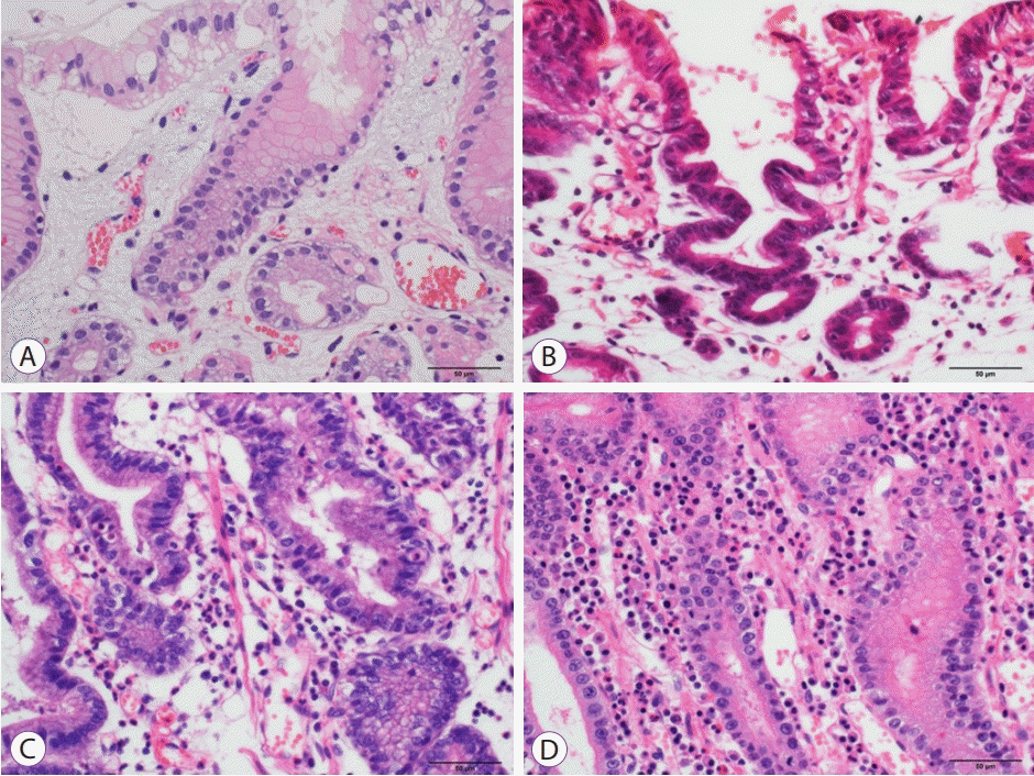
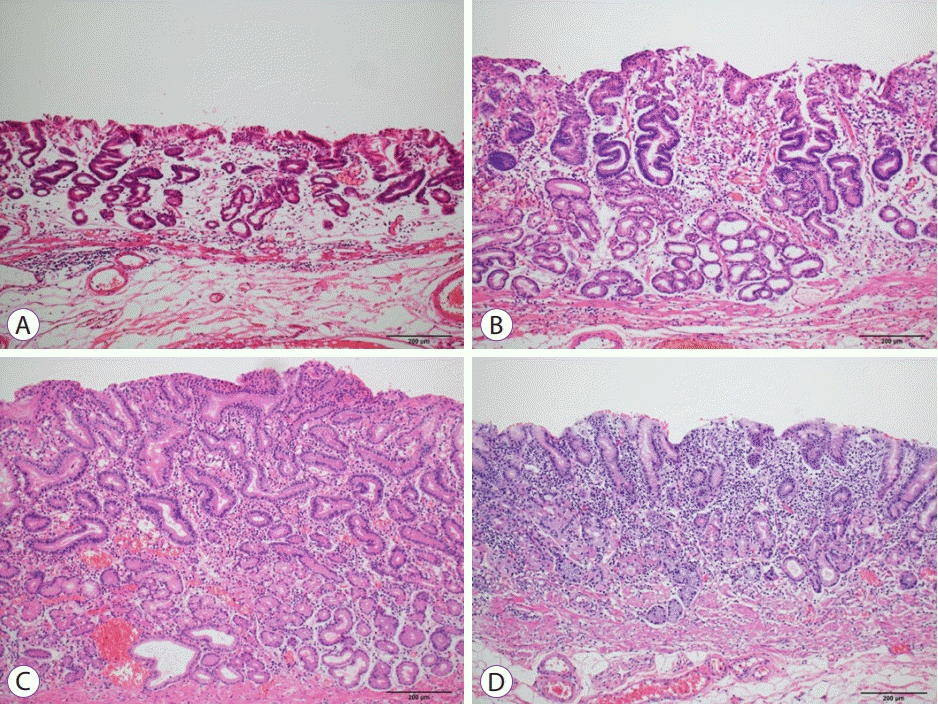
We classified the endoscopic grade of remnant gastritis based on previous reports [19]: grade 1, mild redness; grade 2, intermediate grade (between grades 1 and 3); grade 3, severe redness; and grade 4, apparent erosion (Fig. 1). Endoscopic evaluation was performed independently by two endoscopists (Kei Matsumoto and Nobuaki Ikezawa). In case of disagreement in their assessment, the disparity was discussed and resolved. Additionally, remnant gastritis was assessed pathologically based on the criteria of the updated Sydney System for neutrophils and mononuclear cells [20]. Infiltration of neutrophils and mononuclear cells were classified into four grades: 0, absent; 1, mild; 2, moderate; and 3, severe (Figs. 2, 3). The pathological evaluations were performed on non-cancerous ESD specimens by a single pathologist without using endoscopic images.

ESD procedure

A conventional endoscope with a single accessory channel (GIFQ240, Q260J; Olympus Medical Systems, Tokyo, Japan) was used. FlushKnife BT (DK-2618JN; FUJIFILM, Tokyo, Japan) and FlushKnife BTS (DK2620JBS; FUJIFILM, Tokyo, Japan), 2.5 mm in length, were used for the circumferential mucosal incision and submucosal dissection, respectively, of all cases. Moreover, a transparent hood (D-201-10704; Olympus, Tokyo, Japan, 16675; TOP, Tokyo, Japan) was attached to the tip of the endoscope to ensure a clear view. A short ST hood (DH-28GR, Fujifilm, Japan) and a FlushKnife (DK2618JN10, Fujifilm, Tokyo, Japan) of 1.0 mm length were partially used, especially in cases with severe fibrosis. VIO 300D (ERBE Elektromedizin, GmbH, Tübingen, Germany) was used as the electrosurgical unit. In cases where intraoperative perforation occurred, endoclips were used to close the perforation. To prevent delayed perforation in cases without intraoperative perforation, post-resection prophylaxis was performed using endoclips or polyglycolic acid (PGA) sheets with fibrin glue, at the operator’s discretion. The procedure time was defined as the time from injection into the submucosal layer to resection of the lesion. All procedures were reviewed and evaluated using recorded videos. When bleeding occurred during ESD, two or three attempts were made to achieve hemostasis using the tip of the endoknives. Then, hemostats were used if bleeding would not stop. The number of bleeding episodes that required hemostats were counted. A difficult case was defined as ESD lasting ≥120 minutes, involving piecemeal resection or perforation during the procedure, according to a previous report [21].

Adverse events

Post-ESD bleeding was defined as bleeding requiring endoscopic hemostasis or other procedures with a hemostatic effect, a >2 g/dL decrease in hemoglobin compared to the latest preoperative hemoglobin level, or other obvious bleeding or massive melena [22]. Perforation was diagnosed endoscopically during ESD or by the presence of free air on plain abdominal radiography or computed tomography scan.

Statistical analysis

Proportions of categorical variables were analyzed using the two-sided Fisher’s exact test and the Chi-square test. Continuous variables were compared using Student’s t-test, and non-continuous variables were assessed using the Wilcoxon rank-sum test. Statistical significance was set at P < 0.05. Statistical analyses were performed using JMP software version 10 (SAS Institute, Cary, NC, USA).

RESULTS

Clinicopathological features and clinical outcomes

During the study period, 34 patients (34 lesions) underwent ESD for RGCs at the anastomotic sites. The clinicopathological features of the patients and lesions are shown in Table 1. Of the 34 patients, there were 29 men and five women, and the median age is 74 years (range, 53–84). The types of reconstruction were B-I in 12 patients (35%), B-II in 19 patients (56%), and RY in three patients (9%). The median resected specimen size was 58.5 mm (range, 24–97). The median tumor size was 25 mm (range, 7–70). The depth of invasion was M or SM1 in 26 patients (77%) and SM2 or deeper in eight patients (24%).
The procedure-related parameters of ESD are shown in Table 2. The median operation time was 170 minutes (range, 39–639 minutes). The en bloc resection rate was 94% (32/34), while the en bloc with R0 resection rate was 74% (25/34). The median number of bleeding episodes during ESD was five (range, 0–17). Prophylaxis after resection was performed using endoclips in four patients (12%) and PGA sheets with fibrin glue in eight patients (24%). Seventy-four percent of the cases (25/34) were reported as difficult. In terms of adverse events, intraoperative perforation, delayed bleeding, and delayed perforation occurred in five (15%), four (12%), and one (3%) out of the 34 patients, respectively. Of the five patients with perforation, two patients did not require emergency surgery because the peritonitis was localized and improved with antibiotics. However, the remaining patients underwent emergency surgery for panperitonitis. Moreover, one case of delayed perforation required emergency surgery.

Comparison of clinicopathological features of patients and lesions between the B-II and non-B-II groups

A comparison of the clinical features of patients and lesions between the B-II and non-B-II groups is shown in Table 3. The interval from the previous surgery to the time of RGC resection was significantly longer in the B-II group than in the non-B-II group (35 vs. 7 years; p<0.001). Similarly, the B-II group’s median resected specimen size and tumor size were significantly larger than those in the non-B-II group (p<0.001 and p=0.048, respectively). Pathological evaluation of remnant gastritis tended to show more mononuclear cells in the B-II group than in the non-B-II group, although the difference was not significant (p=0.069). Endoscopic evaluation of remnant gastritis reported more severe gastritis in the B-II group than in the non-B-II group. Moreover, the color and location of the lesions were not significantly different between the two groups. The lesions in both groups were mostly discolored, and a considerable number of them were of the 0-IIa type (Fig. 4).

Comparison of procedure-related parameters between the B-II and non-B-II groups

A comparison of procedure-related parameters between the B-II and non-B-II groups is shown in Table 4. Operation time in the B-II group was significantly longer than that in the non-B-II group (p=0.004). The frequency of bleeding episodes during ESD was significantly higher in the B-II group than in the non-B-II group (p=0.014). Furthermore, there was no significant difference in the frequency of adverse events and the number of difficult cases between the two groups.

DISCUSSION

In this study, we investigated the clinicopathological characteristics of RGCs at the anastomotic sites after distal gastrectomy and the ESD outcomes between the B-II and non-B-II groups. Regarding clinicopathological characteristics, the B-II group had a longer interval from previous surgery and larger lesions than in the non-B-II group. Endoscopic evaluation of remnant gastritis revealed more severe gastritis in the B-II group than in the non-B-II group, and pathological evaluation tended to show more mononuclear cells in the B-II group. Concerning treatment outcome, the operation time was significantly longer, and the frequency of bleeding episodes was significantly higher in the B-II group.
According to the updated Sydney system, neutrophil infiltration is associated with acute inflammation and tissue damage, while mononuclear cell infiltration is indicative of chronic inflammation [20]. Compared to other reconstruction methods, B-II is presumed to result in more profound chronic inflammation due to prolonged and persistent reflux. This may be related to differences in clinicopathological characteristics and ESD outcomes. Larger lesions were observed in the B-II group in this study, and similarly, another study reported that RGCs in B-II were larger than those in B-I [23]. In the anastomosis of B-II, which is severely inflamed and has a narrow lumen, it may have been time-consuming and difficult to identify small-sized tumors due to the redness and bleeding from contact with the scope. Moreover, our data showed a high number of discolored type 0-IIa lesions at the anastomotic site. Thus, noting these findings before passing through the anastomosis may enable the identification of small lesions.
Furthermore, the B-II group had a longer operative time and more frequent bleeding episodes. This may have been due to differences in lesion size and background mucosa. In the submucosa of patients with severe residual gastritis, such as in the B-II group, neovascularization may be induced, causing frequent intraoperative bleeding. Therefore, this may prolong the operative time and predispose the patient to more frequent bleeding episodes. ESD, especially in post-B-II cases, may be more time-consuming than surgery. Nonetheless, since total gastrectomy is required in surgery, ESD is more desirable in terms of postoperative quality of life, even if it takes a longer time.
The duodenal muscular layer is thinner and more fragile than the stomach, which may result in intraoperative and postoperative perforations. In the duodenum, small perforations can progressively widen from exposure to bile and pancreatic juice, eventually leading to severe panperitonitis (Supplemental Fig. 1). To minimize the effect of bile and pancreatic juice, it is important to reduce the extent of duodenal resection and to close the mucosal defect on the duodenal side. Conversely, making an incision close to the lesion on the duodenal side results in an inadequate mucosal flap, and access to the submucosal layer would become more difficult. To overcome the mentioned concerns, we can perform the traction method on the duodenal side and facilitate access to the submucosal layer. The closure of the mucosal defect on the duodenal side after resection can prevent bile and pancreatic juice exposure. Various closure methods, such as methods that utilize endoclips [24,25], the Over-The-Scope Clip system [26], and PGA sheets [27,28] have been recently developed. The closure of at least the duodenal side using one of these methods can reduce the risk of delayed perforation (Supplemental Fig. 2), which occurred in one patient in the B-II group.
This study had some limitations. First, this was a retrospective, single-center study with a limited sample size. Second, we did not evaluate the long-term outcomes of ESD. Third, this study has a selection bias and may not fully characterize RGCs because it only included patients who underwent ESD and excluded surgical or observational cases. Finally, because postoperative examinations were not performed systematically, it was impossible to compare the interval from the previous surgery sufficiently. Despite these limitations, this study seems novel, and the results seem clinically meaningful because the study included the largest number of RGCs at anastomotic sites detected after distal gastrectomy. To the best of our knowledge, this is the first study to reveal the impact of different reconstruction methods on the clinicopathological characteristics of RGCs at anastomotic sites after distal gastrectomy and on ESD outcomes.
In conclusion, compared to non-B-II, the RGCs at the anastomotic sites in B-II featured larger lesions with a background of severe remnant gastritis. Compared to ESD in the non-B-II group, ESD in the B-II group was associated with a longer operative time and more frequent bleeding episodes.

Supplementary Material

Supplemental Fig. 1.

Perforation on the duodenal side. (A) Small perforation on the duodenal side (red arrow). (B) Perforation size progressively became larger.
ce-2021-084-suppl.pdf

Supplemental Fig. 2.

Technical tips for endoscopic submucosal dissection. (A) Make an incision close to the lesion on the duodenal side. (B-D) Perform the traction method on the duodenal side to facilitate access to the submucosal layer. (E, F) After resection, close the mucosal defect on the duodenal side.
ce-2021-084-suppl.pdf

Notes

Conflicts of Interest: The authors have no potential conflicts of interest.

Funding

None.

Author Contributions

Conceptualization: Shinwa Tanaka, Takashi Toyonaga

Data curation: Kei Matsumoto, Nobuaki Ikezawa, Mari Nishio, Masanao Uraoka, Tomoatsu Yoshihara

Formal analysis: KM, Hiroya Sakaguchi, Hirofumi Abe, Tetsuya Yoshizaki

Supervision: Hiroshi Yokozaki, Yuzo Kodama

Writing-original draft: KM, ST

Writing-review&editing: Madoka Takao, Toshitatsu Takao, Yoshinori Morita

REFERENCES

1. Toyonaga T, Man-i M, East JE, et al. 1,635 endoscopic submucosal dissection cases in the esophagus, stomach, and colorectum: complication rates and long-term outcomes. Surg Endosc. 2013; 27:1000–1008.
2. Ohashi M, Katai H, Fukagawa T, Gotoda T, Sano T, Sasako M. Cancer of the gastric stump following distal gastrectomy for cancer. Br J Surg. 2007; 94:92–95.
3. Mezhir JJ, Gonen M, Ammori JB, Strong VE, Brennan MF, Coit DG. Treatment and outcome of patients with gastric remnant cancer after resection for peptic ulcer disease. Ann Surg Oncol. 2011; 18:670–676.
4. Kaneko K, Kondo H, Saito D, et al. Early gastric stump cancer following distal gastrectomy. Gut. 1998; 43:342–344.
5. Sinning C, Schaefer N, Standop J, Hirner A, Wolff M. Gastric stump carcinoma - epidemiology and current concepts in pathogenesis and treatment. Eur J Surg Oncol. 2007; 33:133–139.
6. Ohira M, Toyokawa T, Sakurai K, et al. Current status in remnant gastric cancer after distal gastrectomy. World J Gastroenterol. 2016; 22:2424–2433.
7. Lee JY, Choi IJ, Cho SJ, et al. Endoscopic submucosal dissection for metachronous tumor in the remnant stomach after distal gastrectomy. Surg Endosc. 2010; 24:1360–1366.
8. Hoteya S, Iizuka T, Kikuchi D, Yahagi N. Clinical advantages of endoscopic submucosal dissection for gastric cancers in remnant stomach surpass conventional endoscopic mucosal resection. Dig Endosc. 2010; 22:17–20.
9. Hirasaki S, Kanzaki H, Matsubara M, Fujita K, Matsumura S, Suzuki S. Treatment of gastric remnant cancer post distal gastrectomy by endoscopic submucosal dissection using an insulation-tipped diathermic knife. World J Gastroenterol. 2008; 14:2550–2555.
10. Nishide N, Ono H, Kakushima N, et al. Clinical outcomes of endoscopic submucosal dissection for early gastric cancer in remnant stomach or gastric tube. Endoscopy. 2012; 44:577–583.
11. Osumi W, Fujita Y, Hiramatsu M, et al. Endoscopic submucosal dissection allows less-invasive curative resection for gastric tube cancer after esophagectomy - a case series. Endoscopy. 2009; 41:777–780.
12. Tanaka S, Toyonaga T, Morita Y, et al. Endoscopic submucosal dissection for early gastric cancer in anastomosis site after distal gastrectomy. Gastric Cancer. 2014; 17:371–376.
13. Katai H, Nunobe S, Saka M, Fukagawa T, Sano T. [Reconstruction after distal gastrectomy]. Nihon Geka Gakkai Zasshi. 2008; 109:264–268.
14. Kobori O, Shimizu T, Maeda M, et al. Enhancing effect of bile and bile acid on stomach tumorigenesis induced by N-methyl-N’-nitro-N-nitrosoguanidine in Wistar rats. J Natl Cancer Inst. 1984; 73:853–861.
15. Kim MC, Kim W, Kim HH, et al. Risk factors associated with complication following laparoscopy-assisted gastrectomy for gastric cancer: a large-scale korean multicenter study. Ann Surg Oncol. 2008; 15:2692–2700.
16. Zong L, Chen P. Billroth I vs. Billroth II vs. Roux-en-Y following distal gastrectomy: a meta-analysis based on 15 studies. Hepatogastroenterology. 2011; 58:1413–1424.
17. Lee MS, Ahn SH, Lee JH, et al. What is the best reconstruction method after distal gastrectomy for gastric cancer? Surg Endosc. 2012; 26:1539–1547.
18. Japanese Society of Gastric Cancer Eds. Gastric cancer treatment guidelines. Tokyo, Japan: Kanehara Shuppan;2018.
19. Kubo M, Sasako M, Gotoda T, et al. Endoscopic evaluation of the remnant stomach after gastrectomy: proposal for a new classification. Gastric Cancer. 2002; 5:83–89.
20. Dixon MF, Genta RM, Yardley JH, Correa P. Classification and grading of gastritis. The updated Sydney System. International Workshop on the Histopathology of Gastritis, Houston 1994. Am J Surg Pathol. 1996; 20:1161–1181.
21. Yano T, Hasuike N, Ono H, et al. Factors associated with technical difficulty of endoscopic submucosal dissection for early gastric cancer that met the expanded indication criteria: post hoc analysis of a multi-institutional prospective confirmatory trial (JCOG0607). Gastric Cancer. 2020; 23:168–174.
22. Tajiri H, Kitano S. Complications associated with endoscopic mucosal resection: definition of bleeding that can be viewed as accidental. Dig Endosc. 2004; 16:S134–S136.
23. Nomura K, Hoteya S, Kikuchi D, Inoshita N, Iizuka T. Utility of endoscopic submucosal dissection in the remnant stomach and clinical outcomes for different reconstruction methods. Digestion. 2019; 100:254–261.
24. Takamaru H, Saito Y, Yamada M, et al. Clinical impact of endoscopic clip closure of perforations during endoscopic submucosal dissection for colorectal tumors. Gastrointest Endosc. 2016; 84:494–502.e1.
25. Kato M, Takeuchi Y, Yamasaki Y, et al. Technical feasibility of line-assisted complete closure technique for large mucosal defects after colorectal endoscopic submucosal dissection. Endosc Int Open. 2017; 5:E11–E16.
26. Dohi O, Yoshida N, Naito Y, et al. Efficacy and safety of endoscopic submucosal dissection using a scissors-type knife with prophylactic over-the-scope clip closure for superficial non-ampullary duodenal epithelial tumors. Dig Endosc. 2020; 32:904–913.
27. Takimoto K, Hagiwara A. Filling and shielding for postoperative gastric perforations of endoscopic submucosal dissection using polyglycolic acid sheets and fibrin glue. Endosc Int Open. 2016; 4:E661–E664.
28. Sakaguchi H, Takao T, Takegawa Y, et al. Efficacy of the envelope method in applying polyglycolic acid sheets to post-endoscopic submucosal dissection ulcers in living pigs. Clin Endosc. 2021; 54:64–72.
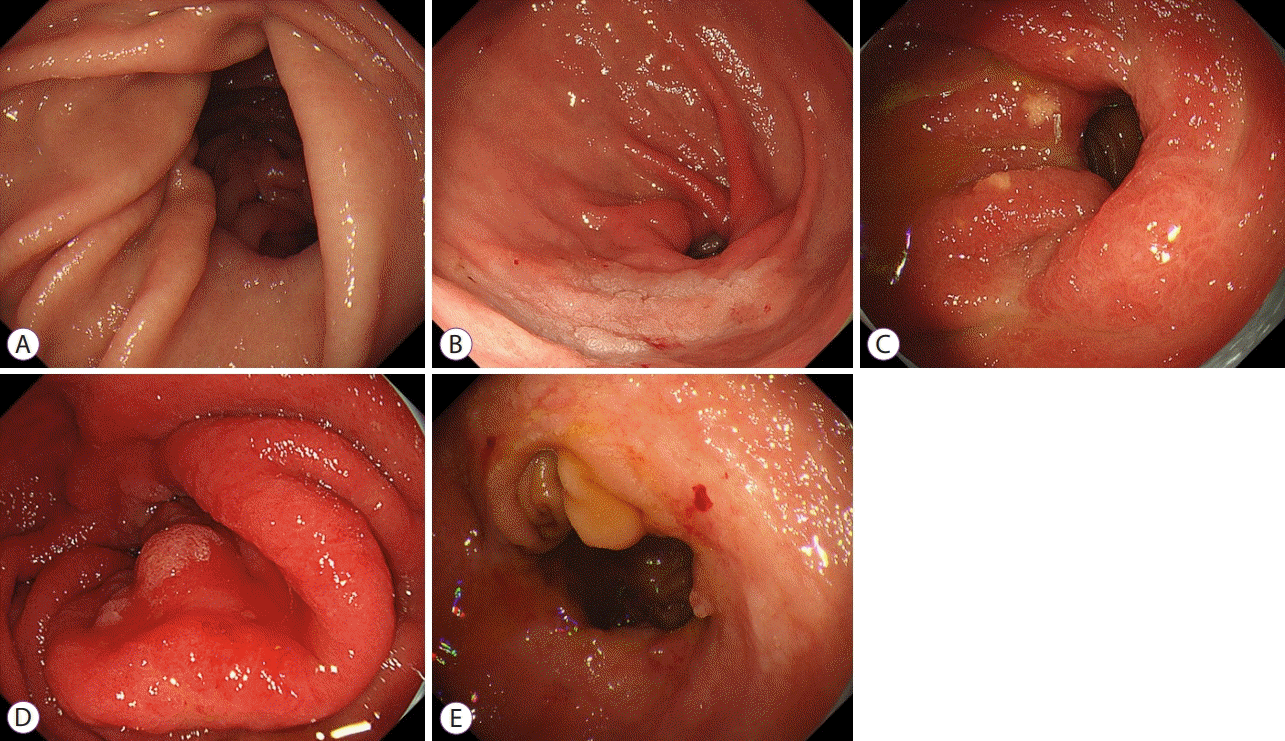
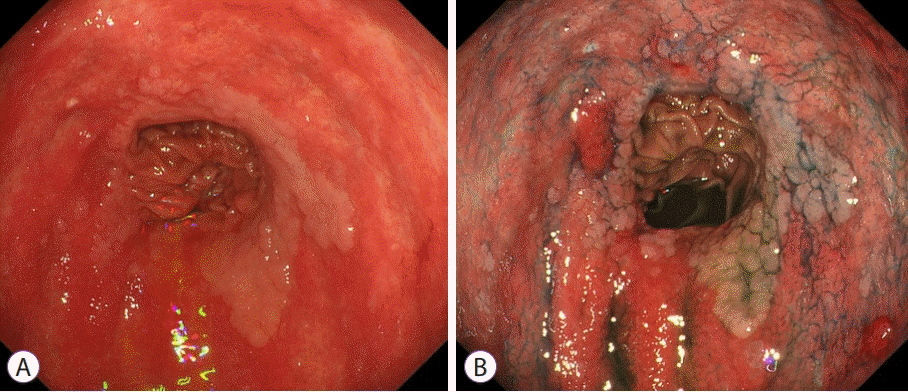
Fig. 1.
Endoscopic grading of remnant gastritis. (A) Grade 0, normal mucosa. (B) Grade 1, mild redness. (C) Grade 2, intermediate grade (between grades 1 and 3). (D) Grade 3, severe redness. (E) Grade 4, apparent erosion.
ce-2021-084f1.tif
Fig. 2.
Pathological grading of remnant gastritis (neutrophils). (A) 0, absent. (B) 1, mild. (C) 2, moderate. (D) 3, severe.
ce-2021-084f2.tif
Fig. 3.
Pathological grading of remnant gastritis (mononuclear cells). (A) 0, absent. (B) 1, mild. (C) 2, moderate. (D) 3, severe.
ce-2021-084f3.tif
Fig. 4.
Remnant gastric cancers at the anastomotic sites after Billroth II. (A) White light observation. (B) After spraying indigo carmine.
ce-2021-084f4.tif
Table 1.
The Clinicopathological Features of Patients and Lesions
n=34
Age (years) 74 (53–84)
Sex
 Male / Female 29 (85) / 5 (15)
Interval from previous surgery (years) 20 (1–58)
Type of reconstruction
 B-I / B-II / RY 12 (35) / 19 (56) / 3 (9)
Morphological type
 0-I / 0-IIa / 0-IIa+IIc / 0-IIb+IIc / 0-IIb 1 (3) / 16 (47) / 10 (29) / 5 (15) / 1 (3) / 1 (3)
Histologic type
 tub1 / tub2 / por / sig 18 (53) / 14 (41) / 1 (3) / 1 (3)
Color of lesion
 Discolored / No change in color / Red 21 (62) / 9 (26) / 4 (12)
Location of lesion (L / G / A / P) 7 (21) / 9 (26) / 4 (12) / 14 (41)
Resected specimen size (mm) 58 (24–97)
Tumor size (mm) 25 (7–70)
Depth of invasion
 M / SM1 / SM2 or deeper 22 (65) / 4 (12) / 8 (24)
Pathological grade of remnant gastritis
 Neutrophils cells 1.23 (1–3)
 Mononuclear cells 1.06 (1–2)
Endoscopic grade of remnant gastritis 1.7 (1–3)

Data are presented as the number (%) or median (range).

A, anterior wall; B-I, Billroth-I reconstruction; B-II, Billroth-II reconstruction; G, greater cuvature; L, lesser curvature; M, mucosa; P, posterior wall; RY, Roux-en-Y reconstruction.

Table 2.
Procedure-Related Parameters
n=34
Operation time (min) 170 (39–639)
En bloc resection 32 (94)
En bloc with R0 resection 25 (74)
Adverse events 9 (26)
 Intraoperative perforation 5 (15)
 Delayed bleeding 4 (12)
Emergency surgery 3 (9)
The number of bleeding episode during ESD 5 (0–17)
Difficult cases 25 (74)

Data are presented as the number (%) or median (range).

ESD, endoscopic submucosal dissection.

Table 3.
Comparison of Clinicopathological Features of Patients in B-II and Non-B-II Groups
B-II group (n=19) Non-B-II group (n=15) p value
Age (years) 74.0 (62–82) 71.6 (53–84) 0.31
Sex 0.38
Male / Female 17 (89) / 2 (11) 12 (80) / 3 (20)
Interval from previous surgery (years) 35 (3-58) 7 (1–20) 0.00002
Morphological type
0-I / 0-IIa / 0-IIc / 0-IIa+IIc / 0-IIb / 0-IIb+IIc 1 (5) / 8 (42) / 5 (26) / 4 (21) / 0 (0) / 1 (5) 0 (0) / 8 (53) / 5 (33) / 1(7) / 1 (7) / 0 (0)
Histologic type
tub1 / tub2 / por / sig 7 (37) / 11 (58) / 1 (5) / 0 (0) 11 (73) / 3 (20) / 0 (0) / 1 (7)
Resected specimen size (mm) 63 (24–97) 40.5 (25–74) 0.00077
Tumor size (mm) 28 (7–70) 19 (8–53) 0.048
Depth of invasion
M / SM1 / SM2 or deeper 11 (61) / 2 (11) / 5 (28) 10 (67) / 2 (13) / 3 (20)
Pathological grade of remnant gastritis
Neutrophils 1.33 (1–3) 1.08 (1–2) 0.12
Mononuclear cells 1.28 (0–2) 0.77 (0–2) 0.069
Endoscopic grade of remnant gastritis 2 (1–3) 1.33 (1–3) 0.0075
Color of lesion
Discolored / No change in color / Red 11 (58) / 5 (26) / 3 (16) 10 (67) / 4 (27) / 1 (7)
Location of lesion (L : G : A : P) 3 (16) / 4 (21) / 4 (21) / 8 (42) 4 (27) / 5 (33) / 0 (0) / 6 (40)

Data are presented as the number (%) or median (range).

A, anterior wall; B-II, Billroth-II reconstruction; G, greater cuvature; L, lesser curvature; M, mucosa; P, posterior wall.

Table 4.
Comparison of B-II and Non-B-II Groups in Terms of Procedure-Related Parameters
B-II group (n=19) Non-B-II group (n=15) p value
Operation time (min) 238 (48–639) 121 (39–225) 0.004
Adverse events 5 (27.8) 4 (26.7)
 Intaroperative perforation 4 (22.2) 1 (6.7) 0.25
 Delayed bleeding 1 (5.6) 3 (20) 0.22
Emergency surgery 2 (11.1) 1 (6.7) 0.59
Number of bleeding episodes during ESD 7 2 0.014
Difficult case 16 (84.2) 9 (60) 0.12

Data are presented as the number (%) or median (range).

ESD, endoscopic submucosal dissection.

TOOLS
Similar articles