Journal List > J Korean Med Sci > v.37(21) > 1162223

Choi, Lee, Park, Lee, Kim, Kim, Park, Kim, Kim, Jang, Bae, Yang, Park, and Cho: Impact of the COVID-19 Pandemic on Patient Delay and Clinical Outcomes for Patients With Acute Myocardial Infarction

Abstract

Background

It has been known that the fear of contagion during the coronavirus disease 2019 (COVID-19) creates time delays with subsequent impact on mortality in patients with acute myocardial infarction (AMI). However, difference of time delay and clinical outcome in patients with ST-segment elevation myocardial infarction (STEMI) or non-STEMI between the COVID-19 pandemic and pre-pandemic era has not been fully investigated yet in Korea. The aim of this study was to investigate the impact of COVID-19 pandemic on time delays and clinical outcome in patients with STEMI or non-STEMI compared to the same period years prior.

Methods

A total of 598 patients with STEMI (n = 195) or non-STEMI (n = 403) who underwent coronary angiography during the COVID-19 pandemic (February 1 to April 30, 2020) and pre-pandemic era (February 1 to April 30, 2017, 2018, and 2019) were analyzed in this study. Main outcomes were the incidence of time delay, cardiac arrest, and in-hospital death.

Results

There was 13.5% reduction in the number of patients hospitalized with AMI during the pandemic compared to pre-pandemic era. In patients with STEMI, door to balloon time tended to be longer during the pandemic compared to pre-pandemic era (55.7 ± 12.6 minutes vs. 60.8 ± 13.0 minutes, P = 0.08). There were no significant differences in cardiac arrest (15.6% vs. 10.4%, P = 0.397) and in-hospital mortality (15.6% vs. 10.4%, P = 0.397) between pre-pandemic and the pandemic era. In patients with non-STEMI, symptom to door time was significantly longer (310.0 ± 346.2 minutes vs. 511.5 ± 635.7 minutes, P = 0.038) and the incidence of cardiac arrest (0.9% vs. 3.5%, P = 0.017) and in-hospital mortality (0.3% vs. 2.3%, P = 0.045) was significantly greater during the pandemic compared to pre-pandemic era. Among medications, angiotensin converting enzyme inhibitors/angiotensin type 2 receptor blockers (ACE-I/ARBs) were underused in STEMI (64.6% vs. 45.8%, P = 0.021) and non-STEMI (67.8% vs. 57.0%, P = 0.061) during the pandemic.

Conclusion

During the COVID-19 pandemic, there has been a considerable reduction in hospital admissions for AMI, time delay, and underuse of ACE-I/ARBs for the management of AMI, and this might be closely associated with the excess death in Korea.

Graphical Abstract

jkms-37-e167-abf001.jpg

INTRODUCTION

As the coronavirus disease 2019 (COVID-19) infection spreads around the world, each country was reporting many excess deaths. Excess deaths were defined as any death that exceeds the expected number of deaths based on the number of deaths over the past several years.12 During the COVID-19 pandemic, there are several reports regarding excess death that does not directly associate with COVID-19 infection because of lock-down and stay-at-home campaign. It has been known that the fear of contagion during the COVID-19 pandemic creates time delays with subsequent impact on mortality in patients with acute myocardial infarction (AMI).3 However, difference of time delay and clinical outcome in patients with ST-segment elevation myocardial infarction (STEMI) or non-STEMI between pre-pandemic and pandemic era has not been fully investigated yet in Korea. Therefore, the aim of this study was to investigate the impact of COVID-19 pandemic on time delay and clinical outcome in patients with STEMI or non-STEMI.

METHODS

Study design and patient population

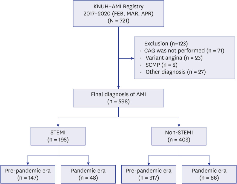
This observational study included 721 consecutive patients who were diagnosed with AMI at admission during pre-pandemic era (February, March, April 2017, 2018, and 2019) and COVID-19 pandemic era (February, March, April 2020). Patients who were registered in the Kyungpook National University Hospital—AMI registry within the study period were enrolled in this study. AMI was diagnosed based on the presence of acute myocardial injury detected by abnormal cardiac biomarkers in the setting of evidence of acute myocardial ischemia. STEMI and non-STEMI were defined according to fourth universal definition of myocardial infarction.4 Study flow diagram was shown in Fig. 1. Of these, patients who did not receive coronary angiography (n = 71) or were diagnosed with variant angina (n = 23), stress induced cardiomyopathy (n = 2), or other diagnoses (n = 27) were excluded. Baseline characteristics of the patients who did not receive coronary angiography are shown in Supplementary Table 1. Finally, 598 patients who underwent coronary angiography in the pre-pandemic and pandemic era with same diagnosis during the same period were analyzed in this study. Among them, 195 patients were diagnosed as STEMI at pre-pandemic (n = 147) and pandemic era (n = 48), whereas 403 patients were diagnosed as non-STEMI at pre-pandemic (n = 317) and pandemic era (n = 86), respectively.
Fig. 1

Study flow diagram.

AMI = acute myocardial infarction, FEB = February, MAR = March, APR = April, CAG = coronary angiography, SCMP = stress induced cardiomyopathy, STEMI = ST-segment elevation myocardial infarction.
jkms-37-e167-g001

Clinical assessment

Baseline demographic and clinical characteristics including age, sex, cardiovascular risk factors such as hypertension, diabetes mellitus, hyperlipidemia, and current smoking, and presenting characteristics were collected at the time of admission. Electrocardiogram was recorded and analyzed by attending cardiologists. Left ventricular ejection fraction was assessed by two-D echocardiography prior to hospital discharge. Venous blood samples were obtained at the time of admission. All patients’ data and procedural details were collected at the time of admission. All patients’ medications were collected during hospitalization.

Definition of time variables

Transfer time in hospital was defined as transfer time from emergency room (ER) to catheterization laboratory. Symptom-to-door time was defined as the time from symptom onset to ER arrival in our hospital. Door-to-balloon time was defined as the time of ER arrival to the first passage of an intracoronary device.567

Clinical outcomes

The primary outcomes were the incidence of time delay, cardiac arrest, and in-hospital mortality. During the follow-up period, clinical outcome data were obtained by reviewing medical records and interviewing patients by telephone.

Statistical analyses

Data were expressed as mean ± standard deviation for continuous variables and as percentages for categorical variables. Comparisons between baseline variables were assessed using Student’s t-test for continuous variables and Pearson’s χ2 test for categorical variables. Normality test was performed for all continuous variables. Analyses were conducted to compare eras (pre-pandemic versus pandemic) and were stratified based on presentation (non-STEMI or STEMI). The cumulative incidence rates of cardiac arrest and in-hospital death between pre-pandemic and pandemic eras were estimated by Kaplan-Meier curve using the log-rank test. For all analyses, a 2-sided P value < 0.05 was considered statistically significant. Statistical analysis was performed using the Statistical Package for the Social Sciences, version 20.0 (IBM Corp., Armonk, NY, USA).

Ethics statement

The protocol was approved by the ethics committee of Kyungpook National University Hospital (KNUH 2021-09-026) as minimal-risk research using data collected for routine clinical practice and waived the requirement for informed consent.

RESULTS

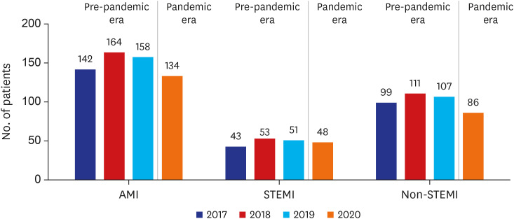
During the pandemic era, there was 13.5% reduction in the number of patients hospitalized with AMI compared with pre-pandemic era, which was mainly driven by 18.8% reduction in non-STEMI (Fig. 2).
Fig. 2

Frequency of hospital admission of overall AMI, STEMI, and non-STEMI from February to April in 2017, 2018, 2019, and 2020.

AMI = acute myocardial infarction, STEMI = ST-segment elevation myocardial infarction.
jkms-37-e167-g002
Baseline characteristics are presented in Table 1. There were no significant differences in demographic characteristic including age, sex, body mass index between pre-pandemic and pandemic era both STEMI and non-STEMI. In initial presentation, systolic (P = 0.002) and diastolic blood pressure (P = 0.003) was significantly lower in the pre-pandemic era compared with those of pandemic era in STEMI, but not in non-STEMI. Among cardiovascular risk factors, dyslipidemia (P = 0.008) was significantly higher during pandemic era compared with pre-pandemic era in non-STEMI. There were no significant differences of prevalence of other cardiovascular risk factors hypertension, diabetes mellitus, previous ischemic heart disease, chronic kidney disease, and current smoking between pre-pandemic and pandemic era both STEMI and non-STEMI.8 Left ventricular ejection fraction (P = 0.069) was lower during pandemic era in non-STEMI, but not in STEMI. Among laboratory findings, low-density lipoprotein cholesterol level was significantly lower during pandemic era compared with pre-pandemic era in STEMI. There were no significant differences in the serum levels of hemoglobin, glucose, hemoglobin A1c, lipid profiles, and creatinine between two eras in both STEMI and non-STEMI. Among medications, the use of P2Y12 inhibitors were lower in the pre-pandemic era, whereas the use of angiotensin converting enzyme inhibitors/angiotensin type 2 receptor blockers (ACE-I/ARBs) were lower during pandemic era in both STEMI and non-STEMI.
Table 1

Baseline characteristics of study subjects

jkms-37-e167-i001
Variables STEMI P value Non-STEMI P value
Pre-pandemic era (n = 147) Pandemic era (n = 48) Pre-pandemic era (n = 317) Pandemic era (n = 86)
Demographics
Age, yr 65.9 ± 12.8 61.9 ± 12.5 0.059 67.5 ± 10.9 68.8 ± 11.3 0.343
Male 109 (74.1) 38 (79.2) 0.484 223 (70.3) 59 (68.6) 0.755
Body mass index, kg/m2 23.5 ± 3.3 23.0 ± 3.2 0.439 23.8 ± 3.3 27.7 ± 32.5 0.291
Initial presentation
Killip class ≥ 2 31 (21.1) 6 (12.5) 0.382 9 (2.8) 3 (3.5) 0.898
Systolic blood pressure, mmHg 128.2 ± 32.7 145.9 ± 31.4 0.002 147.2 ± 26.4 146.3 ± 28.5 0.825
Diastolic blood pressure, mmHg 77.0 ± 20.2 87.7 ± 23.2 0.003 86.0 ± 18.1 86.5 ± 16.8 0.834
Heart rates, beats/min 82.5 ± 21.6 82.3 ± 20.3 0.961 82.3 ± 18.6 85.2 ± 17.4 0.258
Cardiovascular risk factors
Hypertension 69 (46.9) 25 (52.1) 0.536 181 (57.1) 56 (65.9) 0.144
Diabetes mellitus 42 (28.6) 10 (20.8) 0.293 124 (39.1) 29 (34.1) 0.399
Dyslipidemia 24 (16.3) 10 (20.8) 0.475 74 (23.3) 32 (37.6) 0.008
Previous ischemic heart disease 19 (12.9) 8 (16.7) 0.515 100 (31.5) 27 (31.8) 0.969
CKD or azotemia 7 (4.8) 1 (2.1) 0.417 33 (10.4) 8 (9.4) 0.787
Current smoking 44 (29.9) 17 (35.4) 0.477 76 (24.0) 19 (22.4) 0.755
LVEF 46.6 ± 10.1 48.6 ± 7.6 0.250 51.6 ± 11.4 49.0 ± 11.7 0.069
Laboratory findings
Hemoglobin, g/dL 13.2 ± 2.1 13.5 ± 2.0 0.328 13.2 ± 1.9 12.9 ± 2.0 0.261
Glucose, mg/dL 186.9 ± 101.5 163.9 ± 59.6 0.289 169.7 ± 83.9 157.3 ± 96.2 0.423
HbA1c, % 6.3 ± 1.2 6.2 ± 2.1 0.880 6.3 ± 1.31 6.1 ± 1.1 0.477
Total cholesterol, mg/dL 159.9 ± 42.1 155.1 ± 40.3 0.492 154.1 ± 42.4 158.2 ± 42.4 0.442
Triglyceride, mg/dL 108.2 ± 57.2 127.2 ± 78.6 0.074 109.2 ± 72.7 120.8 ± 91.7 0.228
LDL-C, mg/dL 106.3 ± 39.6 90.6 ± 34.4 0.015 99.7 ± 41.6 96.9 ± 39.0 0.581
HDL-C, mg/dL 44.9 ± 13.0 43.8 ± 11.1 0.596 45.8 ± 15.0 43.7 ± 11.2 0.244
Serum creatinine, mg/dL 1.13 ± 0.83 1.06 ± 1.60 0.692 1.25 ± 1.44 1.28 ± 1.48 0.894
Medication during hospitalization
Aspirin 142 (96.6) 48 (100.0) 0.196 305 (96.2) 83 (96.5) 0.897
P2Y12 inhibitors 137 (93.2) 48 (100.0) 0.064 285 (89.9) 83 (96.5) 0.054
Beta-blockers 119 (81.0) 47 (97.9) 0.004 269 (84.9) 76 (88.4) 0.410
ACE-I/ARB 95 (64.6) 22 (45.8) 0.021 215 (67.8) 49 (57.0) 0.061
Statin 112 (98.2) 48 (100.0) 0.356 230 (95.4) 86 (100.0) 0.044
Calcium-channel blockers 61 (41.5) 15 (31.2) 0.206 169 (53.3) 46 (53.5) 0.977
Oral anticoagulants 16 (10.9) 4 (8.3) 0.613 39 (12.3) 7 (8.1) 0.282
Data are expressed as mean ± standard deviation or number (percent).
STEMI = ST-segment elevation myocardial infarction, CKD = chronic kidney disease, LVEF = left ventricular ejection fraction, HbA1c = hemoglobin A1c, LDL-C = low-density lipoprotein cholesterol, HDL-C = high-density lipoprotein cholesterol, ACE-I = angiotensin converting enzyme inhibitor, ARB = angiotensinogen type 2 receptor blocker.
Angiographic and procedural characteristics are presented in Table 2. In patients with STEMI, there were no significant differences in diseases vessel, multivessel disease, treated vessel, pre thrombolysis in myocardial infarction flow, fluoroscopy time procedure time, frequency of angioplasty, and the number of stents between pre-pandemic and pandemic era. The amount of contrast was significantly higher during pandemic era compared to that of pre-pandemic era (P = 0.002). In patients with non-STEMI, left main coronary artery diseases was significantly higher during the pandemic era compared with pre-pandemic era (P = 0.037). Accordingly, percutaneous coronary intervention of left main coronary artery disease was performed more frequently during pandemic era compared with pre-pandemic era (P = 0.032).
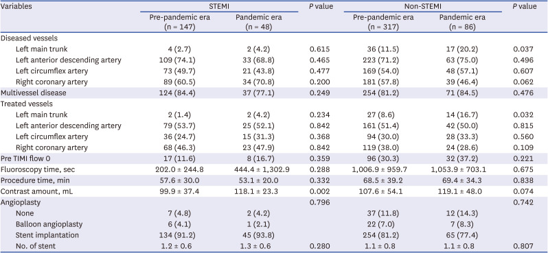
Table 2

Angiographic and procedural characteristics in study subjects

jkms-37-e167-i002
Variables STEMI P value Non-STEMI P value
Pre-pandemic era (n = 147) Pandemic era (n = 48) Pre-pandemic era (n = 317) Pandemic era (n = 86)
Diseased vessels
Left main trunk 4 (2.7) 2 (4.2) 0.615 36 (11.5) 17 (20.2) 0.037
Left anterior descending artery 109 (74.1) 33 (68.8) 0.465 223 (71.2) 63 (75.0) 0.496
Left circumflex artery 73 (49.7) 21 (43.8) 0.477 169 (54.0) 48 (57.1) 0.607
Right coronary artery 89 (60.5) 34 (70.8) 0.200 181 (57.8) 39 (46.4) 0.062
Multivessel disease 124 (84.4) 37 (77.1) 0.249 254 (81.2) 71 (84.5) 0.476
Treated vessels
Left main trunk 2 (1.4) 2 (4.2) 0.234 27 (8.6) 14 (16.7) 0.032
Left anterior descending artery 79 (53.7) 25 (52.1) 0.842 161 (51.4) 42 (50.0) 0.815
Left circumflex artery 36 (24.7) 15 (31.3) 0.368 94 (30.0) 28 (33.3) 0.560
Right coronary artery 68 (46.3) 23 (47.9) 0.842 119 (38.0) 24 (28.6) 0.109
Pre TIMI flow 0 17 (11.6) 8 (16.7) 0.359 96 (30.3) 32 (37.2) 0.221
Fluoroscopy time, sec 202.0 ± 244.8 444.4 ± 1,302.9 0.288 1,006.9 ± 959.7 1,053.9 ± 703.1 0.675
Procedure time, min 57.6 ± 30.0 53.1 ± 20.0 0.332 68.5 ± 39.2 69.4 ± 34.3 0.838
Contrast amount, mL 99.9 ± 37.4 118.1 ± 23.3 0.002 107.6 ± 54.1 119.1 ± 48.0 0.074
Angioplasty 0.796 0.742
None 7 (4.8) 2 (4.2) 37 (11.8) 12 (14.3)
Balloon angioplasty 6 (4.1) 1 (2.1) 22 (7.0) 7 (8.3)
Stent implantation 134 (91.2) 45 (93.8) 254 (81.2) 65 (77.4)
No. of stent 1.2 ± 0.6 1.3 ± 0.6 0.280 1.1 ± 0.8 1.1 ± 0.8 0.807
Data are expressed as mean ± standard deviation or number (percent).
STEMI = ST-segment elevation myocardial infarction, TIMI = thrombolysis in myocardial infarction.
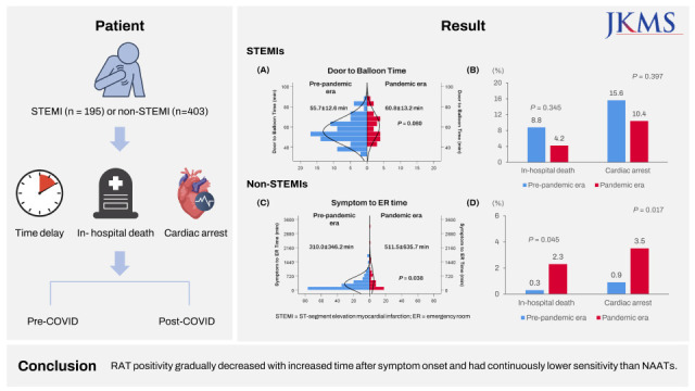
Time variables related to logistics of care of AMI are presented in Table 3. Time distribution of symptom to ER time, ER to catheterization lab arrival time, and door to balloon time between pre-pandemic and pandemic era in both STEMI and non-STEMI are shown in Supplementary Fig. 1. In patients with STEMI, there was no significant difference in symptom to ER time, symptom to catheterization lab arrival time, symptom to balloon time, and ER to catheterization lab time between pre-pandemic and pandemic era. Door to balloon time was shorter in the pre-pandemic era compared with pandemic era (P = 0.080). In patients with non-STEMI, symptom to ER time was significantly longer during the pandemic compared with pre-pandemic era (P = 0.038). Patients who arrived ER within 360 minutes (72.7% vs. 57.1%, P = 0.040) and within 720 minutes (87.6% vs. 75.5%, P = 0.040) were significantly higher in the pre-pandemic era compared with pandemic era in non-STEMI, but not in STEMI (Fig. 3).
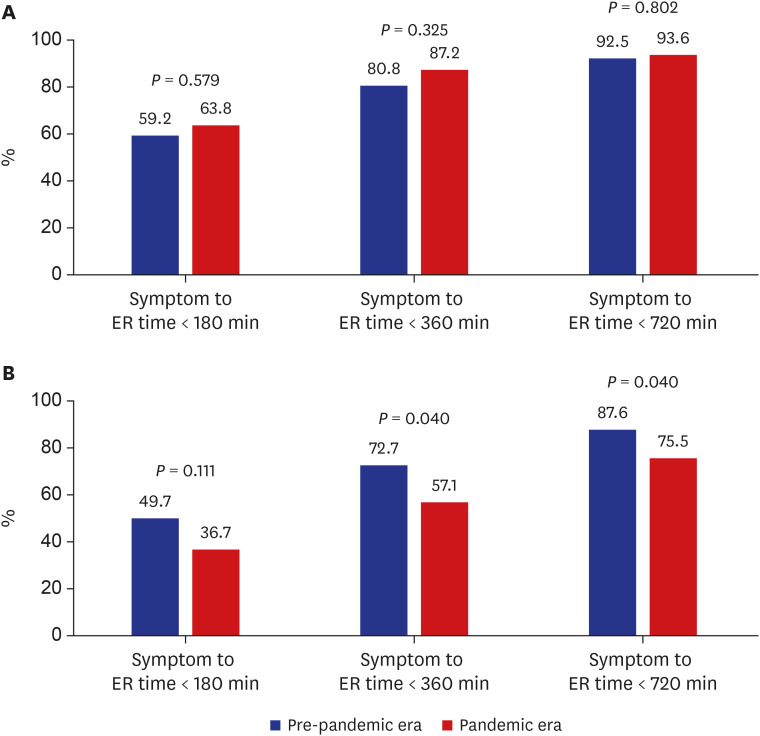
Table 3

Time variables in study subjects

jkms-37-e167-i003
Variables STEMI P value P valuea Non-STEMI P value P valuea
Pre-pandemic era (n = 147) Pandemic era (n = 48) Pre-pandemic era (n = 317) Pandemic era (n = 86)
Symptom to ER time, min 202.0 ± 244.8 444.4 ± 1,302.9 0.364 0.288 310.0 ± 346.2 511.5 ± 635.7 0.038 0.047
Symptom to cath lab time, min 241.3 ± 244.8 487.0 ± 1,304.2 0.119 0.215 2,773.3 ± 2,812.1 3,042.0 ± 2,855.6 0.561 0.475
Symptom to balloon time, min 258.2 ± 244.5 505.3 ± 1,304.6 0.356 0.356 2,614.8 ± 2,694.3 3,139.6 ± 3,047.1 0.294 0.335
ER to cath lab time, min 38.8 ± 12.5 42.6 ± 10.2 0.175 0.183 2,666.9 ± 2,999.5 2,635.8 ± 2,990.1 0.941 0.639
Door to balloon time, min 55.7 ± 12.6 60.8 ± 13.2 0.080 0.111 2,619.0 ± 3,014.7 2,665.2 ± 3,114.4 0.921 0.799
Length of stay, days 6.82 ± 4.39 5.20 ± 1.44 0.074 0.024 6.48 ± 5.82 6.07 ± 5.43 0.555 0.359
Data are expressed as mean ± standard deviation or number (percent).
STEMI = ST-segment elevation myocardial infarction, ER = emergency room, cath = catheterization, lab = laboratory.
aNon-parametric test.
Fig. 3

Comparison of patient delay in (A) ST-segment elevation myocardial infarction and (B) non-ST-segment elevation myocardial infarction between pre-pandemic and pandemic era.

ER = emergency room.
jkms-37-e167-g003
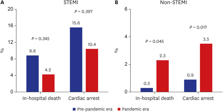
In patients with STEMI, there were no significant differences in in-hospital death (8.8% vs. 4.2%, P = 0.345) and cardiac arrest (15.6% vs. 10.4%, P = 0.397) between pre-pandemic and pandemic era (Fig. 4). In patients with non-STEMI, in-hospital death (2.3% vs. 0.3%, P = 0.045) and cardiac arrest (3.5% vs. 0.9%, P = 0.017) were significantly higher during pandemic era compared with pre-pandemic era.
Fig. 4

The incidence of in-hospital death and cardiac arrest in (A) STEMI and (B) non-STEMI between pre-pandemic and pandemic era.

STEMI = ST-segment elevation myocardial infarction.
jkms-37-e167-g004

DISCUSSION

The main findings of this observational study are as follows. First, the number of patients who were admitted with a suspicion of AMI were decreased during the pandemic. Second, there were considerable time delays including system delay in STEMI and patient delay in non-STEMI during the pandemic. Third, the incidence of in-hospital death and cardiac arrest was significantly higher during the pandemic in non-STEMI, but not in STEMI.
There are three intriguing findings in our study. First, there has been a reduction in hospital admissions for AMI,9 and this might be closely related to the excess death in Korea during the COVID-19 pandemic. Little is known about the associations between patient delay and clinical outcomes during the COVID-19 pandemic in Korea. The COVID-19 pandemic is spreading rapidly around the world, causing hundreds of thousands of excess deaths in many countries.1 However, some of excess death might not be directly related to COVID-19 infection. In the United States, mortality increased by 20% during the COVID-19 pandemic from March to July 2020.10 However, COVID-19 was a documented cause of only 67% of these excess deaths. In Daegu city, Korea, crude death rate increased during the COVID-19 pandemic from February to April 2020 (Supplementary Fig. 2). However, COVID-19 was a documented cause of about 40% of these excess deaths. Interestingly, Western countries reported a decrease in hospitalization for acute cardiovascular disease (CVD) such as AMI and stroke during the COVID-19 pandemic.1112 It might be explained by true population-level reduction of acute CVD because of shifts in dietary pattern (i.e., decreased consumption of high-sodium, fast food intake),131415 reduced exposure to ambient air pollution,16171819 telemedicine,2021 decline in ambulatory CVD clinic visits, outpatient testing, deferral of elective procedures, stay-at-home messaging from government and media,222324 and seasonal variation. However, one of the possible explanations is that many patients were reluctant to visit the hospital due to the fear of contacting COVID-19.2526 Recently, Google Trends meta-data showed that search volume for chest pain is strongly correlated with COVID-19 case numbers in the Unites States. This indicates that the fear of contacting COVID-19 may be leading patients to self-triage using internet searches instead of hospital admission.27 In our study, there has been a considerable decrease in hospital admission for AMI. In addition, AMI patients visited the hospital later than usual. Although our study excluded suspected AMI patients with cardiac enzyme elevation, cardiac arrest, cardiogenic shock, or ventricular tachycardia/fibrillation who did not receive coronary angiography, the number of these patients were numerically lower in the pandemic era (Supplementary Fig. 3). Therefore, it seems to be evident that acute care of CVD may be delayed, deferred, or abbreviated during the COVID-19 pandemic. Considering the high mortality of CVD, this late presentation may increase ‘collateral COVID-19 mortality’28 and contribute to these excess deaths during the COVID-19 pandemic.29 Therefore, patients suffering from non-COVID-19 related acute CVD continue to receive timely, evidence based and high-quality care.
Second, logistics of AMI care were delayed during the COVID-19 pandemic. However, the pattern of time delay and the effect on in-hospital death and cardiac arrest were different between STEMI and non-STEMI. In the present study, patients with STEMI have a trend for longer door to balloon time during the pandemic. This system delay might be because it took time to put on personal protective equipment and to transfer patients due to uncertainty about COVID-19 infection.303132 Interestingly, the incidence of in-hospital death and cardiac arrest was lower during the pandemic. Patients with STEMI may have died while staying at home without coming to the hospital despite symptoms.3334 In addition, symptom to ER time was significantly longer and the incidence of in-hospital death and cardiac arrest was significantly higher in non-STEMI during the pandemic. Patients delay in non-STEMI due to the fear of possibility contacting patients with COVID-19 at ER is thought to have led to greater in-hospital death and cardiac arrest during the pandemic.2526 Therefore, it seems that the system delay of STEMI and the patient delay of non-STEMI differently contributed to worsening the prognosis of patients with AMI during the pandemic.
Third, ACE-I/ARBs were significantly underused for the management of AMI during the COVID-19 pandemic. In Korea, this information is interesting and valuable because there have been few real-world data regarding usage of ACE-I/ARBs for AMI in the early period of COVID-19 pandemic. Previous studies showed that COVID-19 patients with diabetes mellitus and hypertension appear to have worse prognosis due to overexpression of ACE2 receptor in airway alveolar epithelial cells because SARS-CoV-2 uses the ACE2 receptor to enter the lungs in a mechanism.353637383940 Therefore, there has been some concerns regarding the negative effect of ACE-I/ARBs by upregulation of ACE2 receptors that patients receiving ACE-I/ARBs may be more susceptible to COVID-19 infection and have poorer outcomes. Major cardiology scientific associations have rejected these correlation hypotheses for now.414243 However, in the present study, it has been documented that ACE-Is/ARBs were significantly underused for the management of AMI in the early period of the pandemic although there is no significant evidence to support an association between COVID-19 and ACE-I/ARBs. The suboptimal use of ACE-I/ARBs during the pandemic may affect the clinical outcome in this study.
This study has certain limitations that should be noted. First, because this study was a single center and observational study, we could not completely exclude the possibility of residual confounding factors that were not available in our registry. Second, we collected symptom onset time based on the patient’s memory. Therefore, we could not completely exclude the possibility of recall bias. Third, the reason for late presentation was not collected in our registry. Therefore, our results should only be regarded as hypothesis generating. However, the limitations of the study should not undermine the strength of this study, namely that it includes patients encountered in day-to-day clinical practice before and during the pandemic. Despite these limitations, we believe that our data could provide the clinical insight necessary to understand contemporary management and prognosis for AMI during the pandemic.
In conclusion, during the COVID-19 pandemic, there has been a considerable reduction in hospital admissions for AMI, time delay, and underuse of ACE-I/ARBs for the management of AMI, and this might be closely associated with the excess death in Korea.

Notes

Disclosure: The authors have no potential conflicts of interest to disclose.

Author Contributions:

  • Conceptualization: Lee JH.

  • Data curation: Park HK, Lee E, Kim MS, Kim HJ, Park BE, Kim HN, Kim N, Jang SY, Bae MH, Yang DH, Park HS, Cho Y.

  • Formal analysis: Lee JH, Choi H.

  • Methodology: Lee JH, Choi H.

  • Software: Lee JH.

  • Validation: Lee JH.

  • Investigation: Choi H.

  • Writing - original draft: Lee JH, Choi H.

  • Writing - review & editing: Lee JH, Choi H.

References

1. Our World in Data. Excess mortality during the coronavirus pandemic (COVID-19). Updated 2021. Accessed October 4, 2021. https://ourworldindata.org/excess-mortality-covid .
2. Korean Statistical Information Service (KOSIS). COVID-19-time excess death analysis. Updated 2021. Accessed October 4, 2021. https://kosis.kr/covid/statistics_excessdeath.do .
3. Helal A, Shahin L, Abdelsalam M, Ibrahim M. Global effect of COVID-19 pandemic on the rate of acute coronary syndrome admissions: a comprehensive review of published literature. Open Heart. 2021; 8(1):e001645. PMID: 34083389.
4. Thygesen K, Alpert JS, Jaffe AS, Chaitman BR, Bax JJ, Morrow DA, et al. Fourth universal definition of myocardial infarction (2018). J Am Coll Cardiol. 2018; 72(18):2231–2264. PMID: 30153967.
5. Nathan AS, Raman S, Yang N, Painter I, Khatana SA, Dayoub EJ, et al. Association between 90-minute door-to-balloon time, selective exclusion of myocardial infarction cases, and access site choice: insights from the cardiac care outcomes assessment program (COAP) in Washington State. Circ Cardiovasc Interv. 2020; 13(9):e009179. PMID: 32883103.
6. Chandrasekhar J, Marley P, Allada C, McGill D, O’Connor S, Rahman M, et al. Symptom-to-balloon time is a strong predictor of adverse events following primary percutaneous coronary intervention: results from the Australian Capital Territory PCI Registry. Heart Lung Circ. 2017; 26(1):41–48. PMID: 27451348.
7. Park J, Choi KH, Lee JM, Kim HK, Hwang D, Rhee TM, et al. Prognostic implications of door-to-balloon time and onset-to-door time on mortality in patients with ST -segment-elevation myocardial infarction treated with primary percutaneous coronary intervention. J Am Heart Assoc. 2019; 8(9):e012188. PMID: 31041869.
8. Visseren FL, Mach F, Smulders YM, Carballo D, Koskinas KC, Bäck M, et al. 2021 ESC guidelines on cardiovascular disease prevention in clinical practice. Eur Heart J. 2021; 42(34):3227–3337. PMID: 34458905.
9. Aldujeli A, Hamadeh A, Briedis K, Tecson KM, Rutland J, Krivickas Z, et al. Delays in presentation in patients with acute myocardial infarction during the COVID-19 pandemic. Cardiol Rev. 2020; 11(6):386–391.
10. Woolf SH, Chapman DA, Sabo RT, Weinberger DM, Hill L, Taylor DD. Excess deaths from COVID-19 and other causes, March-July 2020. JAMA. 2020; 324(15):1562–1564. PMID: 33044483.
11. Morelli N, Rota E, Terracciano C, Immovilli P, Spallazzi M, Colombi D, et al. The baffling case of ischemic stroke disappearance from the casualty department in the COVID-19 era. Eur Neurol. 2020; 83(2):213–215. PMID: 32289789.
12. Aguiar de Sousa D, Sandset EC, Elkind MS. The curious case of the missing strokes during the COVID-19 pandemic. Stroke. 2020; 51(7):1921–1923. PMID: 32466737.
13. Ruiz-Roso MB, Knott-Torcal C, Matilla-Escalante DC, Garcimartín A, Sampedro-Nuñez MA, Dávalos A, et al. COVID-19 lockdown and changes of the dietary pattern and physical activity habits in a cohort of patients with type 2 diabetes mellitus. Nutrients. 2020; 12(8):2327.
14. Navarro-Cruz AR, Kammar-García A, Mancilla-Galindo J, Quezada-Figueroa G, Tlalpa-Prisco M, Vera-López O, et al. Association of differences in dietary behaviours and lifestyle with self-reported weight gain during the COVID-19 lockdown in a university community from Chile: a cross-sectional study. Nutrients. 2021; 13(9):3213. PMID: 34579090.
15. Sorić T, Brodić I, Mertens E, Sagastume D, Dolanc I, Jonjić A, et al. Evaluation of the food choice motives before and during the COVID-19 pandemic: a cross-sectional study of 1232 adults from Croatia. Nutrients. 2021; 13(9):3165. PMID: 34579041.
16. Venter ZS, Aunan K, Chowdhury S, Lelieveld J. COVID-19 lockdowns cause global air pollution declines. Proc Natl Acad Sci U S A. 2020; 117(32):18984–18990. PMID: 32723816.
17. Tanzer-Gruener R, Li J, Eilenberg SR, Robinson AL, Presto AA. Impacts of modifiable factors on ambient air pollution: a case study of COVID-19 shutdowns. Environ Sci Technol Lett. 2020; 7(8):554–559.
18. Azuma K, Kagi N, Kim H, Hayashi M. Impact of climate and ambient air pollution on the epidemic growth during COVID-19 outbreak in Japan. Environ Res. 2020; 190:110042. PMID: 32800895.
19. Son JY, Fong KC, Heo S, Kim H, Lim CC, Bell ML. Reductions in mortality resulting from reduced air pollution levels due to COVID-19 mitigation measures. Sci Total Environ. 2020; 744:141012. PMID: 32693269.
20. Hollander JE, Carr BG. Virtually perfect? Telemedicine for COVID-19. N Engl J Med. 2020; 382(18):1679–1681. PMID: 32160451.
21. Portnoy J, Waller M, Elliott T. Telemedicine in the era of COVID-19. J Allergy Clin Immunol Pract. 2020; 8(5):1489–1491. PMID: 32220575.
22. Okuhara T, Okada H, Kiuchi T. Examining persuasive message type to encourage staying at home during the COVID-19 pandemic and social lockdown: a randomized controlled study in Japan. Patient Educ Couns. 2020; 103(12):2588–2593.
23. Knell G, Robertson MC, Dooley EE, Burford K, Mendez KS. Health behavior changes during COVID-19 pandemic and subsequent “Stay-at-Home” orders. Int J Environ Res Public Health. 2020; 17(17):6268.
24. Limbers CA, McCollum C, Greenwood E. Physical activity moderates the association between parenting stress and quality of life in working mothers during the COVID-19 pandemic. Ment Health Phys Act. 2020; 19:100358. PMID: 33072187.
25. Gluckman TJ, Wilson MA, Chiu ST, Penny BW, Chepuri VB, Waggoner JW, et al. Case rates, treatment approaches, and outcomes in acute myocardial infarction during the coronavirus disease 2019 pandemic. JAMA Cardiol. 2020; 5(12):1419–1424. PMID: 32766756.
26. Marotta M, Gorini F, Parlanti A, Chatzianagnostou K, Mazzone A, Berti S, et al. Fear of COVID-19 in patients with acute myocardial infarction. Int J Environ Res Public Health. 2021; 18(18):9847. PMID: 34574770.
27. Ciofani JL, Han D, Allahwala UK, Asrress KN, Bhindi R. Internet search volume for chest pain during the COVID-19 pandemic. Am Heart J. 2021; 231:157–159. PMID: 33010246.
28. Baldi E, Sechi GM, Mare C, Canevari F, Brancaglione A, Primi R, et al. Out-of-hospital cardiac arrest during the COVID-19 outbreak in Italy. N Engl J Med. 2020; 383(5):496–498. PMID: 32348640.
29. Bhatt AS, Moscone A, McElrath EE, Varshney AS, Claggett BL, Bhatt DL, et al. Fewer hospitalizations for acute cardiovascular conditions during the COVID-19 pandemic. J Am Coll Cardiol. 2020; 76(3):280–288. PMID: 32470516.
30. Mahmud E, Dauerman HL, Welt FG, Messenger JC, Rao SV, Grines C, et al. Management of acute myocardial infarction during the COVID-19 pandemic: a position statement from the Society for Cardiovascular Angiography and Interventions (SCAI), the American College of Cardiology (ACC), and the American College of Emergency Physicians (ACEP). J Am Coll Cardiol. 2020; 76(11):1375–1384. PMID: 32330544.
31. Primessnig U, Pieske BM, Sherif M. Increased mortality and worse cardiac outcome of acute myocardial infarction during the early COVID-19 pandemic. ESC Heart Fail. 2021; 8(1):333–343. PMID: 33283476.
32. Park DW, Yang Y. Delay, death, and heterogeneity of primary PCI during the COVID-19 pandemic: an international perspective. J Am Coll Cardiol. 2020; 76(20):2331–2333. PMID: 33183507.
33. Kitahara S, Fujino M, Honda S, Asaumi Y, Kataoka Y, Otsuka F, et al. COVID-19 pandemic is associated with mechanical complications in patients with ST-elevation myocardial infarction. Open Heart. 2021; 8(1):e001497. PMID: 33547221.
34. Metzler B, Siostrzonek P, Binder RK, Bauer A, Reinstadler SJ. Decline of acute coronary syndrome admissions in Austria since the outbreak of COVID-19: the pandemic response causes cardiac collateral damage. Eur Heart J. 2020; 41(19):1852–1853. PMID: 32297932.
35. Sparks MA, Crowley SD, Gurley SB, Mirotsou M, Coffman TM. Classical Renin-Angiotensin system in kidney physiology. Compr Physiol. 2014; 4(3):1201–1228. PMID: 24944035.
36. Li G, He X, Zhang L, Ran Q, Wang J, Xiong A, et al. Assessing ACE2 expression patterns in lung tissues in the pathogenesis of COVID-19. J Autoimmun. 2020; 112:102463. PMID: 32303424.
37. Rico-Mesa JS, White A, Anderson AS. Outcomes in patients with COVID-19 infection taking ACEI/ARB. Curr Cardiol Rep. 2020; 22(5):31. PMID: 32291526.
38. Yang X, Yu Y, Xu J, Shu H, Xia J, Liu H, et al. Clinical course and outcomes of critically ill patients with SARS-CoV-2 pneumonia in Wuhan, China: a single-centered, retrospective, observational study. Lancet Respir Med. 2020; 8(5):475–481. PMID: 32105632.
39. Guan WJ, Ni ZY, Hu Y, Liang WH, Ou CQ, He JX, et al. Clinical characteristics of coronavirus disease 2019 in China. N Engl J Med. 2020; 382(18):1708–1720. PMID: 32109013.
40. Zhang JJ, Dong X, Cao YY, Yuan YD, Yang YB, Yan YQ, et al. Clinical characteristics of 140 patients infected with SARS-CoV-2 in Wuhan, China. Allergy. 2020; 75(7):1730–1741. PMID: 32077115.
41. Hippisley-Cox J, Young D, Coupland C, Channon KM, Tan PS, Harrison DA, et al. Risk of severe COVID-19 disease with ACE inhibitors and angiotensin receptor blockers: cohort study including 8.3 million people. Heart. 2020; 106(19):1503–1511. PMID: 32737124.
42. Flacco ME, Acuti Martellucci C, Bravi F, Parruti G, Cappadona R, Mascitelli A, et al. Treatment with ACE inhibitors or ARBs and risk of severe/lethal COVID-19: a meta-analysis. Heart. 2020; 106(19):1519–1524. PMID: 32611676.
43. Rossi GP, Sanga V, Barton M. Potential harmful effects of discontinuing ACE-inhibitors and ARBs in COVID-19 patients. Elife. 2020; 9:e57278. PMID: 32250244.

SUPPLEMENTARY MATERIALS

Supplementary Table 1

Baseline characteristics of patients who did not receive coronary angiography
jkms-37-e167-s001.doc

Supplementary Fig. 1

Time distribution of study subjects. (A) Symptom to ER time, (B) ER to cath lab time, and (C) door to balloon time in STEMI. (D) Symptom to ER time, (E) ER to cath lab time, and (F) door to balloon time in non-STEMI.
jkms-37-e167-s002.doc

Supplementary Fig. 2

The crude death rates (A) and the number of death and excess death (B) in Daegu city, Korea from 2017 to 2020. The 2020 Daegu non-COVID-19 indicates the number of people who died from non-COVID-19 related illness. The 2020 Daegu COVID-19 indicates the number of people who died from COVID-19 infection.
jkms-37-e167-s003.doc

Supplementary Fig. 3

Frequency of hospital admission of overall suspected acute MI patients with cardiac enzyme elevation, cardiac arrest, cardiogenic shock, or ventricular tachycardia/fibrillation who were not perform coronary angiography from February to April in 2017, 2018, 2019, and 2020.
jkms-37-e167-s004.doc
TOOLS
Similar articles