

 PDF
PDF Citation
 Print
Print


Introduction
Inflammatory bowel disease (IBD) represents a group of inflammatory disorder conditions of the gastrointestinal tract. There are a large number of patients suffering from IBD worldwide, which leads to a diminished quality of life and sometimes even death. Over the last decade, mesenchymal stromal cells (MSCs) have reported as an attractive therapy strategy in the treatment of IBD because of their immunosuppressive effect (1). Accumulating evidence suggests that the immunosuppressive effect of MSCs is, at least in part, mediated by exosomes (2, 3). Exosomes are extracellular membrane-enclosed microvesicles (40~100 nm) that many important components including proteins, microRNA, and mRNA (4). Recent studies have shown encouraging therapeutic activities of MSCs-derived exosomes (MSC-Exo) in various inflammatory-related animal models (5–7), although MSC-Exo are not as effective as their in anti-inflammatory ability in vitro (8–11). Compared with their cellular counterpart, exosomes may be safer and affordable, and could be easily stored without loss of function (12). Thus, MSC-Exo have led to the development of cell-free therapeutic approach as a safe and more advantageous alternative to MSCs-based therapy strategy (13–15). Recently, some studies have investigated that the therapeutic efficacy of secretome, extracts or conditioned medium from MSCs in animal model of induced colitis (16–19).
The result of preclinical research is encouraging, but the major barriers to clinical studies include standardized characterization and safety issue of MSC-Exo. Exosomes-based therapy needs clinically significant numbers of well-defined, safe, and efficient MSCs. Currently, the exosomes used in these studies are prepared from MSCs supplemented with classical fetal bovine serum (FBS). FBS is an animal-derived product, and its usage has raised safety concern because it might be a source of zoonotic infections and xenogenic antigens. It has been reported that commercial FBS (20~50%) is virus-positive (20). A study has reported that FBS proteins (7~30 mg) are linked to a standard preparation of 1×10⁸ MSCs (21). If so, it’s very possible that preparation of exosomes from MSCs grown in FBS carries a certain amount of FBS proteins, which can lead to immunological reactions when transplanted into the body (22–24). Moreover, FBS has unknown exact composition, and the batch-to-batch variability can ultimately lead to variations in cells and exosomes performance (20). For these reasons, the regulatory authorities encourage the usage of chemically-defined media as an alternative to FBS, especially for clinical-grade production of MSCs and MSC-Exo (25). Evaluation of the therapeutic potential and application of MSC-Exo in future clinical trials demands production under serum-free and xeno-free, and defined medium conditions (26, 27). The usage of defined medium will be very advantageous for reproducibility of exosomes production.
We previously developed for the first time, in the literature to date, a serum and xeno-free, chemically defined and no plate-coating-based culture system for the isolation and expansion of MSCs (28). Umbilical cord-derived MSCs cultured in this chemically defined medium fulfilled the biological characteristics of MSCs, and retained the similar immunosuppressive capability with those in classical FBS-containing media. Thus, we speculate that MSC-Exo in defined medium may also mimic the beneficial effects of MSCs in the immunosuppressive capability. In this study, we investigated the immunosuppressive function in vitro and the therapeutic potential of MSC-Exo in defined medium in a mice model of colitis.
Materials and Methods
Ethics statement
This study was approved by Ethics Committee of the Beijing Friendship Hospital affiliated to Capital Medical University. All animal experiments were conducted in accordance with the relevant guidelines and regulations of Animal Ethics Committee of the Beijing Friendship Hospital affiliated to Capital Medical University.
Production of umbilical cord-derived MSCs
The umbilical cord samples were obtained from healthy pregnant women. All women provided written informed consent. UCMSCs were prepared as described previously (28). The cord was rinsed with phosphate-buffered saline (PBS). The vessels were removed. Wharton’s jelly tissues were cut into 1~2 mm3 pieces and digested for 60 min in an enzyme cocktail (hyaluronidase 5 U/mL, collagenase 125 U/mL and dispase 50 U/mL; Sigma, St. Louis, MO, USA) at 37°C in a shaking incubator. The total nucleated cells were filtered through a 70 μM mesh and plated in xeno-free defined medium at 37°C, 5% CO2. The defined medium was prepared as described previously (28). After removing non-adherent cells on 5 days, the adherent cells were passaged when reached 90% confluences. UCMSCs at passages 5 were used for experiments.
Preparation of MSC-Exo
UCMSCs were conditioned in PBS for 48 h at 90% confluence. Exosomes were prepared from the conditioned PBS according to the method previously described (29). Briefly, PBS was collected and centrifuged at 500 ×g for 10 min, 3000 ×g for 20 min and 10000 ×g for 30 min to remove cells and debris. The supernatant was ultra-centrifuged at 100000 ×g for 100 min to collect the exosomes. The MSC-Exo were resuspended in PBS and filtered with a 0.22-μm microcentrifuge filter. The protein concentration was determined by BCA kit.
Characterization of MSC-Exo
The morphology of MSC-Exo was examined using a transmission electron microscope, and the size distribution was determined by a Zetasizer Nano ZS (Malvern Instruments, UK) according to the manufacturer’s instructions. The exosome-associated proteins including CD9, CD63 and CD81 were analyzed using western blotting.
Co-culture of PBMCs with MSC-Exo
PBMCs were obtained via Ficoll separation from healthy donors, who provided written informed consent. PBMCs (5×10⁵ cells/well in 96-well culture plate) were plated in RPMI 1640 medium containing 10% FBS, 10 μg/ml phytohemagglutinin (PHA, Sigma-Aldrich) and 0, 5 or 20 μg MSC-Exo. After 72 h, cultures were pulsed with cell counting kit-8 reagent (Dojindo, Kumamoto, Japan) and measured with a Model 450 microplate reader. The inflammatory cytokines was analyzed in supernatant by ELISA (BD, Biosciences, San Jose, CA, USA).
Colitis induction and treatment procedure
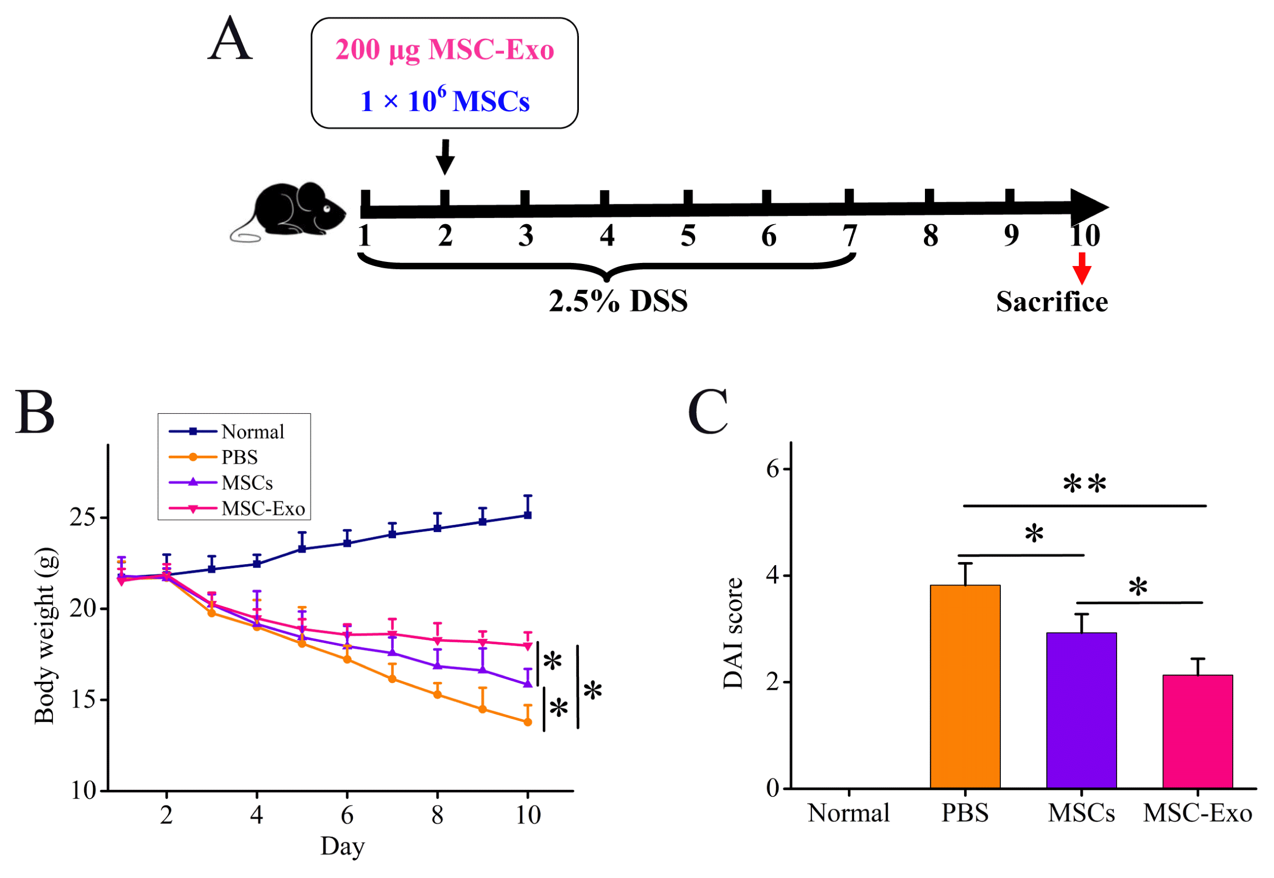
C57BL/6 mice (6~8 weeks) were administered with 2.5% DSS in the drinking water from day 1 to day 7. The mice were administered with MSC-Exo (200 μg/mouse) or MSCs (1×10⁶ cells/mouse) in 200 μL PBS by intraperitoneal injection on day 2, and sacrificed on day 10. 200 μL PBS was injected into DSS-treated mice as control group, while the mice that received drinking DSS-free water were used as normal group.
Clinical symptoms evaluation
Body weight was monitored daily, stool consistency and bleeding severity were recorded on day 10, and DAI was assessed (Supplementary Table S1).
Histological evaluation
Colon tissue samples were fixed in 4% paraformaldehyde, prepared and stained with hematoxylin and eosin (H&E staining). The intensity of inflammation was evaluated by histological score (Supplementary Table S2).
Cytokines assays
The colon was homogenized in lysis buffer containing 1% Triton X-100 and protease inhibitor cocktail and centrifuged at 13000 ×g for 20 min to collect the supernatant. The total protein concentration was determined by BCA Protein Assay. The levels of inflammatory cytokines such as IFN-γ, TNF-α, IL-6, IL-17, IL-10, and TGF-β1 were quantitatively measured by ELISA and corrected for the amount of total protein.
Results
Characterization of MSC-Exo
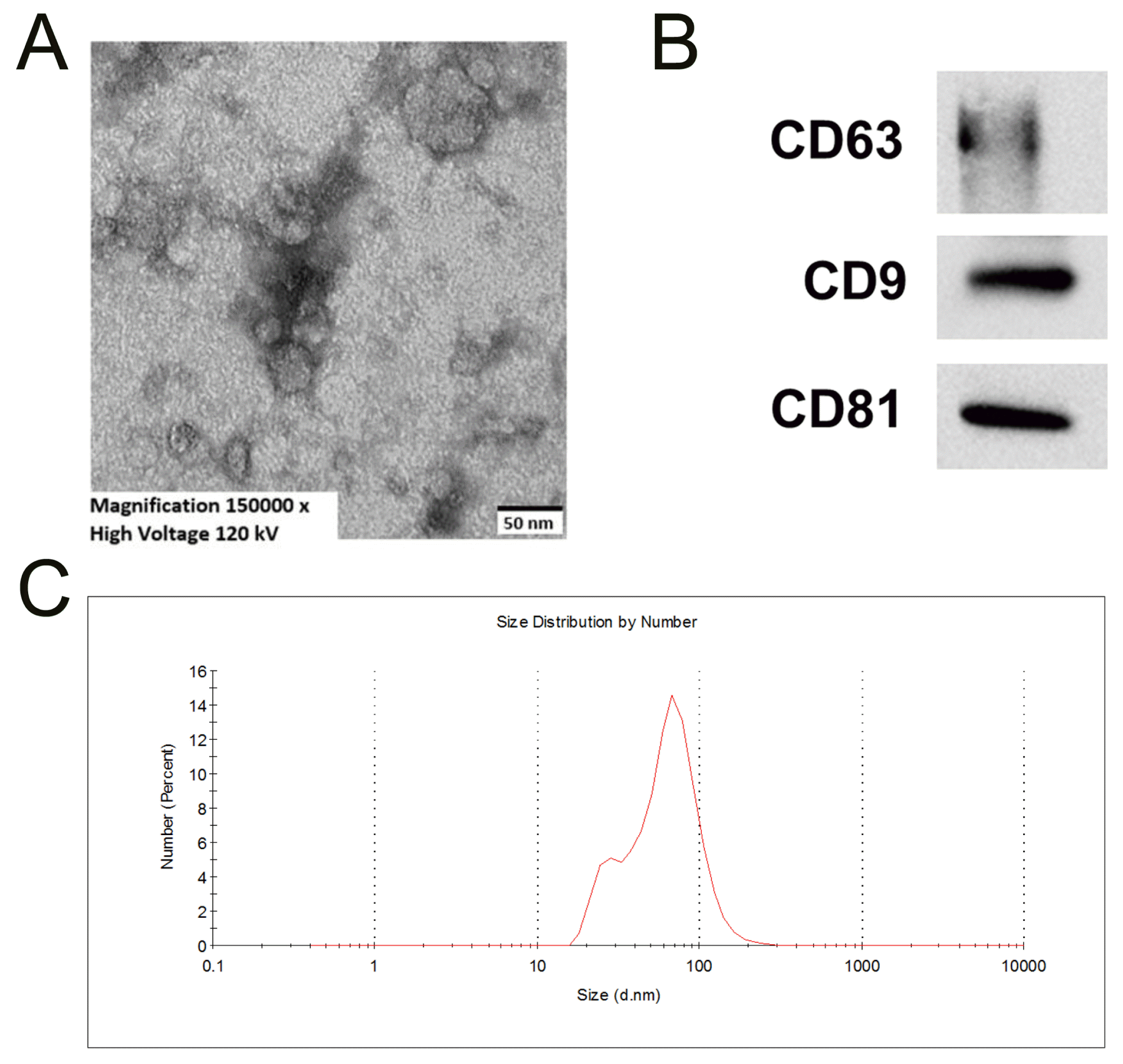
To validate whether exosomes were successfully isolated from UCMSCs cultured in defined medium, the characterization tests were performed. Transmission electron microscopy analysis showed spheroid shape of MSC-Exo, with a diameter between 40 and 100 nm (Fig. 1A). The particle size distribution of MSC-Exo was recorded (Fig. 1B). The MSC-Exo expressed CD9, CD63, and CD81 (Fig. 1C). These results demonstrate that we have successfully isolated and identified exosomes from UCMSCs cultured in defined medium.
MSC-Exo possessed a certain degree of immunosuppressive capability in vitro
Different levels of MSC-Exo were used to treat PBMCs. When in high concentration (20 μg) of MSC-Exo, the concentration of IFN-γ (Fig. 2A), TNF-α (Fig. 2B) and IL-1β (Fig. 2C) was significantly decreased, while TGF-β1 (Fig. 2D) and IL-10 (Fig. 2E) significantly increased compared with no MSC-Exo group. In consistence with the results in high concentration, the significant decrease of IFN-γ (Fig. 2A) and TNF-α (Fig. 2B) and increase of TGF-β1 (Fig. 2D) were also showed in low concentration (5 μg) of MSC-Exo. However, no significant differences in the level of IL-1β and IL-10 were detected (Fig. 2C and 2E). Moreover, MSC-Exo at these two concentrations had no effect on inhibiting the proliferation of peripheral blood mononuclear cells (PBMCs) (Fig. 2F). These data suggest that MSC-Exo possess a certain degree of immunosuppressive capability in vitro.
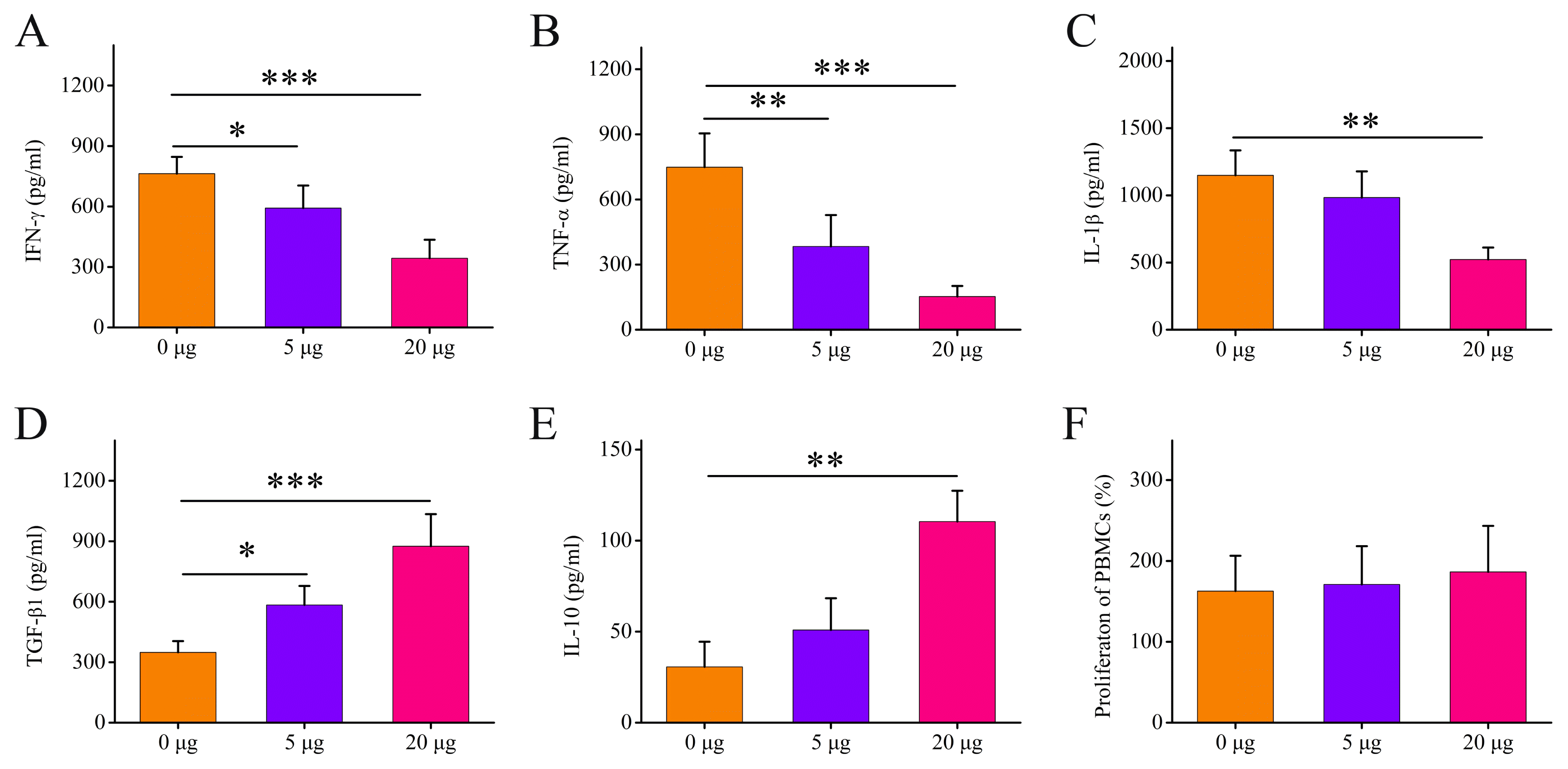
Fig. 2
MSC-Exo possessed a certain degree of immunosuppressive capability in vitro. The concentrations of the pro-inflammatory cytokines (A) IFN-γ, (B) TNF-α and (C) IL-1β and anti-inflammatory cytokines (D) TGF-β1 and (E) IL-10 were measured in the supernatant of PBMCs treated with different levels of MSC-Exo for 72 h. (F) The proliferation of PBMCs was evaluated after culture with different levels of MSC-Exo. Bars indicate means±SD. n=5; *p<0.05, **p<0.01, and ***p<0.001.
![]()
MSC-Exo or MSCs alleviated clinical symptom in DSS-induced colitis mice
To assess whether administering MSC-Exo ameliorated colitis, we used 2.5% DSS for 7 days to establish experimental models of DSS-induced colitis, and administered 200 μg MSC-Exo on days 2. The MSCs (1×10⁶) cultivated in defined medium were used as counterpart, and the same procedures performed. All the mice were sacrificed on day 10 (Fig. 3A). Our result showed that the administration of MSCs or MSC-Exo significantly improved clinical parameters including body weight (Fig. 3B) and disease activity index (DAI, Fig. 3C) compared with PBS-treated mice, suggesting that MSC-Exo significantly alleviated clinical symptom in DSS-induced colitis. In addition, 200 μg MSC-Exo significantly ameliorated the parameters compared with 1×10⁶ MSCs-treated mice (Fig. 3B and 3C).
MSC-Exo or MSCs alleviated colonic damage in DSS-induced colitis mice
To further investigate whether administering MSC-Exo alleviated colonic damage, the quantitative evaluation of colon length and histopathological examination was performed. Our result showed that the administration of MSCs or MSC-Exo significantly alleviated colonic damages such as colon length (Fig. 4A and 4B) and histological severity (Fig. 4C and 4D), suggesting that MSC-Exo or MSCs significantly alleviated colonic damage in DSS-induced colitis. In addition, the histopathological scoring in the MSC-Exo group was significantly lower than the MSCs group (Fig. 4D), although no significant difference in colon length was shown between two groups (Fig. 4B).
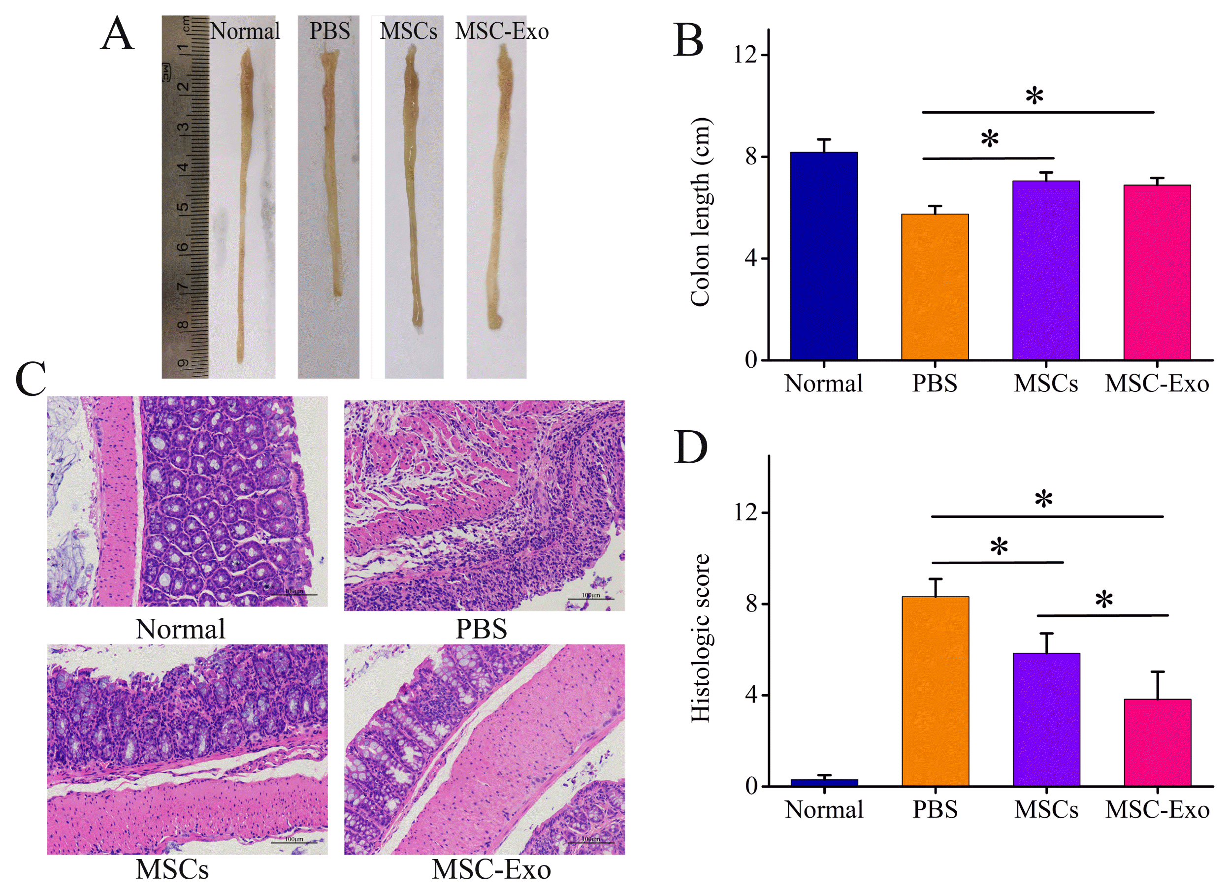
Fig. 4
MSC-Exo or MSCs alleviated colonic damage in DSS-induced colitis mice. (A) Representative images of colon length. (B) Colon length was quantitatively analyzed. (C) Representative H&E staining, bar=100 μm. (D) Corresponding severity score was determined. Bars indicate means± SD. n=5~8; *p<0.05.
![]()
MSC-Exo or MSCs reduced the inflammatory state in DSS-induced colitis mice
To characterize this therapeutic mechanism, we measured the inflammatory cytokines in colon tissue. The pro-inflammatory cytokines including IFN-γ (Fig. 5A), TNF-α (Fig. 5B), IL-6 (Fig. 5C), and IL-17 (Fig. 5D) were significantly down-regulated, while anti-inflammatory cytokines including IL-10 (Fig. 5E) and TGF-β1 (Fig. 5F) were significantly up-regulated in the MSCs or MSC-Exo-treated mice compared with the PBS-treated mice. These data suggest that MSC-Exo or MSCs significantly reduced the inflammatory state in DSS-induced colitis mice. Moreover, the levels of TNF-α (Fig. 5B), IL-6 (Fig. 5C), and IL-17 (Fig. 5D) were decreased, while IL-10 (Fig. 5E) and TGF-β1 (Fig. 5F) increased in the colon after treatment with MSC-Exo at a dose of 200 μg compared with 1×10⁶ MSCs. In addition, IFN-γ was not significant change after MSC-Exo treatment compared with MSCs (Fig. 5A).
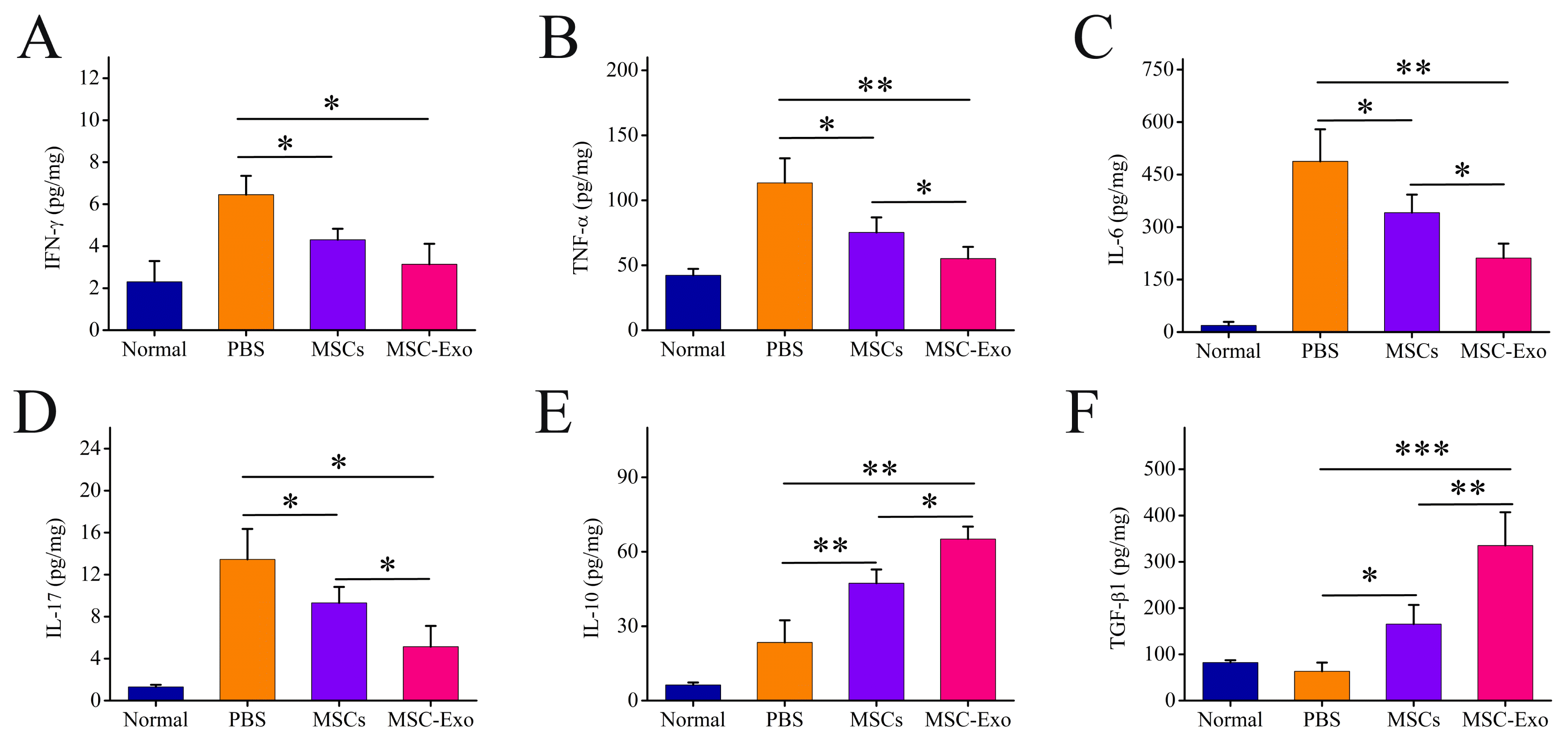
Fig. 5
MSC-Exo or MSCs reduced the infammatory state in DSS-induced colitis mice. The concentrations of the pro-inflammatory cytokines (A) IFN-γ, (B) TNF-α, (C) IL-6 and (D) IL-17 and anti-inflammatory cytokines (E) IL-10 and (F) TGF-β1 in colonic protein extracts were measured by ELISA. Bars indicate mean±SD, n=5~8; *p<0.05, **p<0.01 and ***p<0.001.
![]()
Discussion
The current study demonstrates that the exosomes from MSCs in defined medium possess a certain degree of immunosuppressive effect in vitro and exhibit a therapeutic capability in a mouse model of DSS-induced colitis through suppressing inflammation mechanism. To the best of our knowledge, this is the first report of defined medium-derived MSC-Exo. The translation of therapeutically valuable MSC-Exo into a therapeutic agent presents several major considerations including standardized characterization and safety issue of MSC-Exo. The usage of defined medium will be very advantageous for these considerations. Exosomes, one of several groups of extracellular vesicles released by MSCs, may exert different functions via the release of different kinds of molecules, depending on the cell culture environment (30). Further study is needed to answer whether the immunosuppressive effect of exosomes from MSCs cultured in defined medium is different from those in classical FBS-supplemented medium.
The increasing evidence has shown that the inhibitory effect of MSC-Exo on lymphocyte proliferation is minor when compared to their parental cells (8–10). Here, MSC-Exo failed to suppress PBMCs proliferation at dose levels up to 20 μg/mL, while MSCs in defined medium exhibit this response (28). This result is consistent with previous reports on MSC-Exo from various sources in FBS or platelet lysate-based medium (9–11), suggesting that this phenomenon is independent of culture conditions and sources. Further research is needed to answer whether MSC-Exo have an immunosuppressive role on the different immune cell subsets. Furthermore, our result showed that MSC-Exo reduced the concentration of pro-inflammatory cytokines and increased the secretion of anti-inflammatory cytokines during in vitro culture, suggesting that MSC-Exo display a certain degree of immunosuppressive activity. Similar fashion has been reported in FBS-based study (8, 11). The immunosuppressive potential of MSC-Exo has been actively observed and in vivo, although not as effective as their cellular counterpart (2, 6–8, 31, 32).
The second key finding in this study is that the single intraperitoneal injection of MSC-Exo in defined medium was able to significantly alleviate the clinical symptom and colonic damage in DSS-induced colitis. This result is consistent with previous studies based on FBS (16, 18, 19, 33). Intraperitoneal injection is selected as the most common administration route for MSC-Exo delivery in DSS-induced colitis, based on previous studies (16, 34). In line with the clinical and histological evaluation, the MSC-Exo treatment significantly downregulated the expression level of pro-inflammatory cytokines such as IFN-γ, TNF-α, IL-6, and IL-17, while upregulated anti-inflammatory cytokines such as IL-10 and TGF-β1. The performance of some cytokines is consistent in the previous study based on FBS (16, 18, 35), but some are different (19). The reason of differences remains unclear, but it may hint to the fact that culture condition may reflect the content of MSC-Exo and may therefore affect their potency. On the other hand, it cannot be excluded the differences of tissue source and method of isolation. In fact, multiple studies have confirmed the difference of exosomes caused by different sources and methods (13). Taken together, we suggest that MSC-Exo in defined medium exert therapeutic effects in a murine model of colitis through suppressing inflammation mechanism. Further functional in vivo and in vitro studies are needed to unveil the exact mechanism.
In addition, our results show that the use of MSC-Exo (200 μg) leads to an improved clinical symptom and histological severity in vivo when compared with MSCs at a dose of 1×10⁶ cells. One possible explanation is that exosomes act faster than cells. However, we did not detect differences in the colon length between MSCs and MSC-Exo treated mice. The reason is not known but we hypothesize that the in vivo environment is much more complex, or that the endpoints of evaluation was not appropriate. This is related to up-regulation anti-inflammatory responses and down-regulation of inflammatory responses. An increasing number of FBS-based studies have reported that MSC-Exo are equal or superior effectiveness as MSCs in various disease models (36–39), most studies are absence of comparable dosing, so it is difficult to draw conclusions about comparable therapeutic efficacy. The summary of the literature has shown some dose variations and a dose ranging (0.1~250 μg/animal) in various animal models (13). Some previous reports showed therapeutic effect of MSC-Exo at a dose of 200 μg or MSCs at a dose of 1×10⁶ cells in mice models of various diseases (33, 35, 40), and so defined as our dose in this study.
In conclusion, our results suggest the feasibility of MSC-Exo in defined medium for cell-free applications in the treatment of IBD. Additional studies are required to compare the composition and therapeutic benefits of exosomes from MSCs in defined medium and FBS-containing media.
 XML Download