

 PDF
PDF Citation
 Print
Print


Introduction
Recently, high-precision and high-dose radiation therapy techniques, such as intensity-modulated radiation therapy (IMRT), stereotactic body radiation therapy (SBRT), and stereotactic radiosurgery (SRS), have been widely used in the field of radiation therapy [1-4]. Such therapies have the advantage of increasing the therapeutic effect by irradiating sufficient radiation to the lesion to be treated while maximizing protection by minimizing the radiation dose to normal tissues; however, they require high precision and accuracy. Patient-specific dosimetric quality assurance (DQA) measurements are always performed to verify that radiation is accurately irradiated based on the patient treatment plan to guarantee the accuracy of such treatment. Here, patient-specific DQA is conducted using a solid detector such as a slit-type PMMA phantom, ionization chamber, film, or glass dosimeter or using an array-type detector and phantom composed of a single material into which the detector can be inserted [5-8]. A patient treatment plan follows a three-dimensional (3D) dose distribution because it is conducted in three dimensions, whereas the commonly used method is limited to evaluating a fragmentary dose distribution by measuring a one- (1D) or two-dimensional (2D) dose distribution. In this current research, we performed 3D dose evaluations by arranging several liquid scintillators and radiochromic films. However, an accurate evaluation remains restricted because liquid scintillators (besides those using gamma rays) have disadvantages such as low efficiency, long decay time, and reduced energy resolution, and films have limitations due to the air gaps and errors introduced when stacking multiple sheets [9,10]. The dose of each patient is evaluated using a single-material phantom fabricated with a material that is equivalent to water since the phantom cannot consider the density and shape of the tissue of each patient. There are only a limited number of studies that have focused on accurately verifying a high-precision radiation treatment plan because patient-specific DQA is limited to converting a 3D dose distribution into a 1D and 2D dose distribution and drawing a comparison. Thus far, various studies have been conducted to overcome these challenges and accurately validate the patient treatment plan. Silveira et al. [11] confirmed the actual patient treatment dose and applicability of the polymer gel. However, they could not perform dose verification as that using a human body because they employed a cylindrical phantom composed of a single material. Lee et al. [12] performed a 3D dosimetry with the polymer-gel located inside the insert of a liver cancer model made with a 3D printer during SBRT treatment; they were unable to conduct a dose evaluation in the environment that resembles the insides of the human body since only the liver was printed in 3D and inserted into a single-material phantom. Matrosic et al. [13] evaluated a 3D dose distribution by irradiating a therapeutic dose to a phantom coated with a polymer gel; this was conducted using a commercially available plastic phantom and not a material that mimics the human body.
For a more accurate dose evaluation, a phantom should be manufactured with the same shape as the human body instead of using a uniform shape. A phantom can be created to represent the same shape of the different organs and bone densities of the patient using 3D printing technology. Then, the dose distribution can be evaluated using 3D printed phantom. A patient-specific phantom can be manufactured using precise images, such as magnetic resonance imaging (MRI), computed tomography (CT), and positron emission tomography/computed tomography (PET/CT) images, with materials that have a density similar to that of the human body for considering the characteristics of each patient. Furthermore, the effectiveness of the precision treatment can be verified through the treatment planning system (TPS) for actual patient treatment and for performing a 3D gel dosimetry using a customized phantom [14,15].
In this study, a patient-customized phantom was manufactured using polymer gel and CT images to measure the 3D dose distribution. The usefulness of the 3D-printed gel-based patient-customized phantom was verified through a comparative evaluation with the actual 3D dose of the treated patient.
Go to : 
Materials and Methods
1. Producing the gel phantom with 3D printing
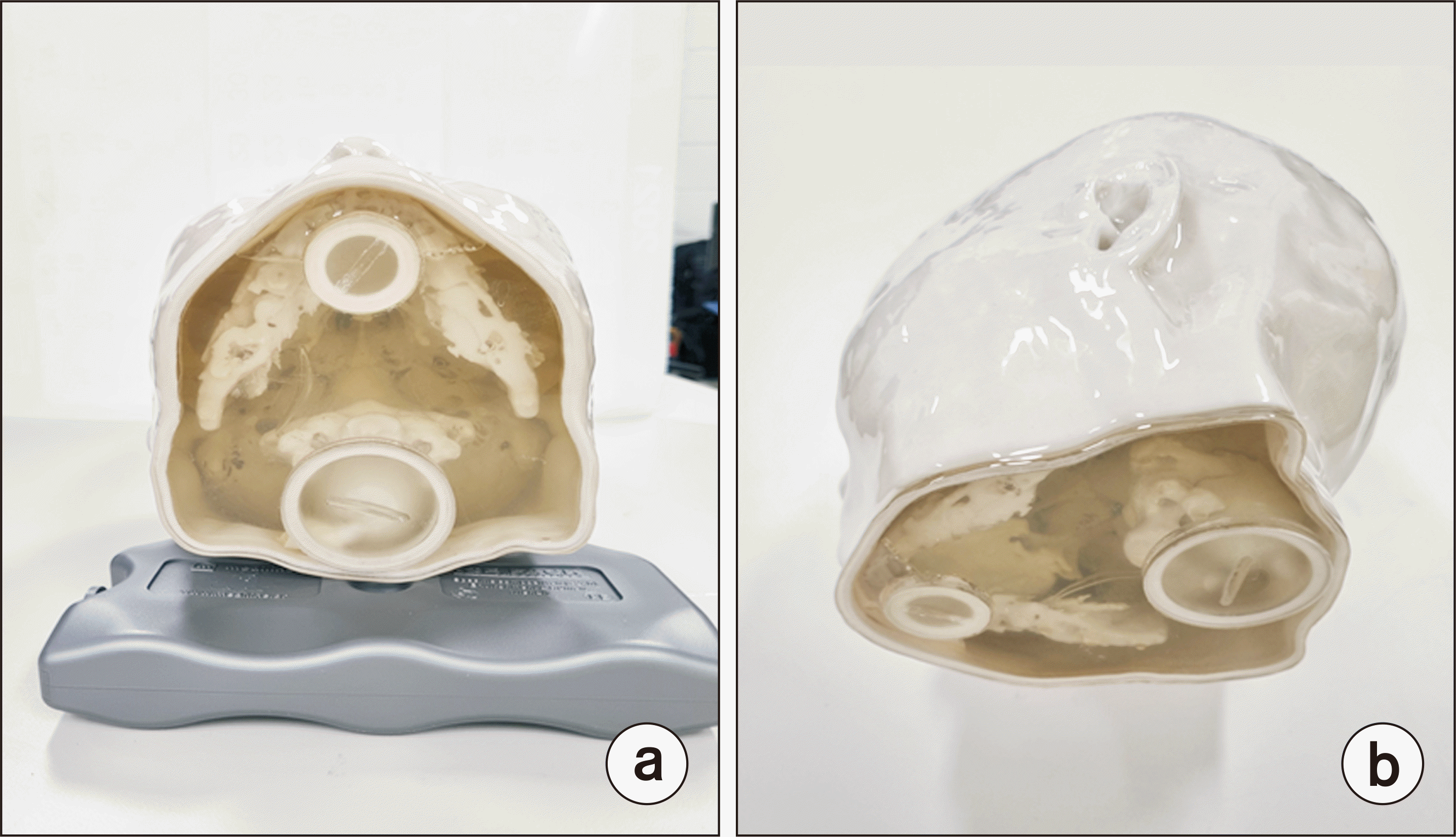
For this study, a head phantom was manufactured by RTsafe (RTsafe, Athens, Greece) using the CT images of the patient to produce a patient-customized phantom [13]. The phantom produced by RTsafe uses a ColorJet printing technique from Project 360 3D Printer (3D Systems, Morrisville, NC, USA). After spraying the liquefied material layer by layer, the material is solidified using a high-performance ultraviolet (UV) light to create the shape of the phantom. Each phantom layer printed is 0.1 mm thick, and the phantom can be manufactured based on the patient’s anatomy using the CT images. The 3D-printed head phantom comprises skin and bone structures, and the soft tissue is filled with a polymer gel, which is a water-equivalent material that enables a 3D dose evaluation (Fig. 1).
2. Patient-specific dosimetric quality assurance
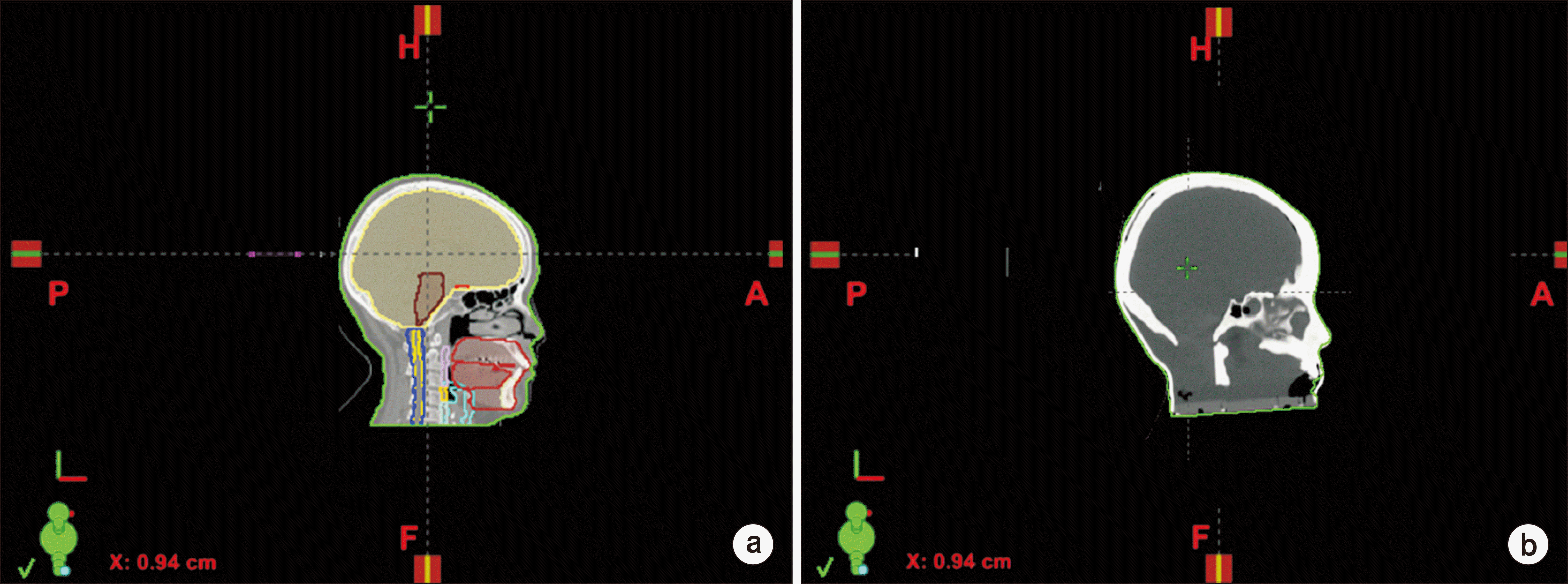
The CT images are captured to apply the patient-specific phantom to the patient-specific DQA. The tube voltage and current are considered under the same conditions as the CT image of the patient. Here, the slice thickness is set to 2.5 mm. The acquired CT image is then sent to Eclipse, which is a TPS. The rigid body image registration is conducted with the actual patient image. Then, this image and the Hounsfield unit (HU) are compared to evaluate the similarity. The treatment technology and dose irradiated to the patient are applied like in an actual setting to evaluate the usefulness of the manufactured phantom and gel dose evaluation in an actual treatment environment. To this end, the produced phantom was laid on the treatment table of Clinic iX (Varian Medical Systems, Palo Alto, CA, USA), the cone beam computed tomography (CBCT) was taken, and the setup was conducted using actual patient images. Then, IMRT was applied, and the energy was calculated as 6 MV. The CT image of the patient and the one from the 3D-printed phantom are shown in Fig. 2.
3. Polymer gel dosimetry
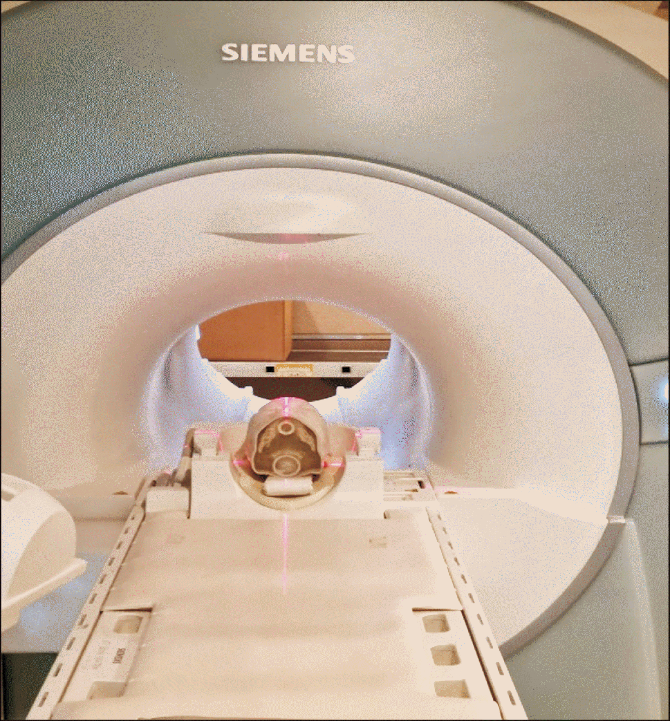
The irradiated phantom is analyzed through MRI scans, and at least 5 hours of post-irradiation is required. Therefore, MRI scans are performed after 24 hours of post-irradiation. Fig. 3 shows the shape of the phantom scanned via MRI. For the optimal conditions of the phantom, the room temperature was maintained at 20°C to 24°C, and the MRI image was captured using Siemens MRI (Siemens Medical Solutions USA, Inc., Knoxville, TN, USA) with a magnetic field of 3 Tesla (3T). The total scan time of the phantom was 35 minutes 58 seconds, and the voxel size was 1.4×1.4×2.0 mm3. 2D T2-weighted (T2-w) imaging was used, and images with 77 slices (slice thickness=2 mm, flip angle=180°, field of view (FOV)=350 mm, four constant times to echo (TEs) of 4.54 ms, and receiver bandwidth=781 Hz/Px) were acquired. The four TE/TRs measured over 2,000 ms of time repetition (TR) were TE[1]/TR 36/2,000 ms, TE[2]/TR 436/2,000 ms, TE[3]/TR 835/2,000 ms, and TE[4]/TR 1,230/2,000 ms. After scanning, the dose irradiated to the 3D polymer gel was analyzed using the MRI data as a 3D distribution. The detailed conditions are presented in Table 1.
Go to :
Results
1. Head phantom
The electron density value of the patient’s skull was 996±167 HU, and that of the head phantom bone material was 986±25 HU. This result covers the shortcomings of the resin powder that expresses high density, and it has been identified as the most challenging part of the existing ColorJet printing technology. Furthermore, the results indicate that resin powder with the added calcium has a density similar to that of human bones.
The electron density of the patient’s brain was 58±15 HU, and that of the phantom was 45±17 HU. A 2 Gy radiation dose was irradiated through the treatment plan using the CT image of the patient. Then, it was scanned with MRI to compare and analyze the 3D distribution of the radiation dose prescribed in the treatment plan. The results are presented in Fig. 4, which shows the dose values calculated by the post-irradiation MRI images and TPS. The dose calculated through the image of the phantom and TPS is shown by adjusting the brightness by 100%, 50%, and 0%. Fig. 4a shows that only high dose areas are depicted with the brightness and contrast adjusted, and it indicates how this high-dose area—the dose calculated by the TPS—is delivered to the phantom. Fig. 4b shows that also low dose areas are depicted with the brightness and contrast adjusted, and it indicates the extent to which the dose calculated by TPS is delivered outside the central high-dose area. These two images suggest that the dose calculated by the treatment plan for the target to be treated is designed adequately to match the phantom manufactured.

2. Profile comparison
Fig. 5 shows the comparison results of the 3D distribution of the gel investigated by the treatment plan through the MRI scan and analysis results of the dose profile calculated through TPS. Fig. 5a and b present the analysis results of the dose profile in the x-axis and y-axis directions in the coronal plane of the phantom, and Fig. 5c and d present the analysis results of the dose profile in the x-axis and y-axis directions in the transverse plane of the phantom.
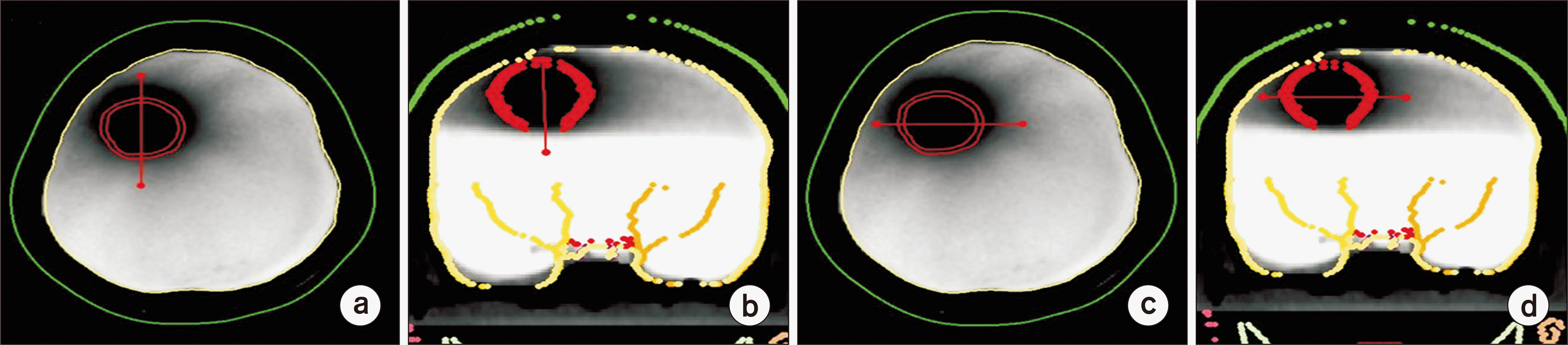 | Fig. 5(a) Dose profile in the x-axis direction of the coronal plane (T2 maps of the investigated phantom). The high-dose area is the dark area. (b) Dose profile in the y-direction of the coronal plane. (c) Dose profile in the x-direction of the transverse plane. (d) Dose profile in the y-axis direction of the transverse plane.
|
When comparing the 1D gamma index calculations for evaluating the agreement between the two data sets quantitatively, the passing criteria indicated a 2 mm distance to the agreement and a 5% dose difference. The dose profile results of the above evaluation are illustrated in Fig. 6. Based on the profile comparison, we confirmed that there was a shift between the dose distribution calculated from the TPS and the dose distribution irradiated to the phantom. Although the phantom was set up in the same position as the patient image in the treatment room using CBCT, only the error suggested after the first CBCT scan was applied, and a new scan was not conducted. This may have caused the setup to be inaccurate.
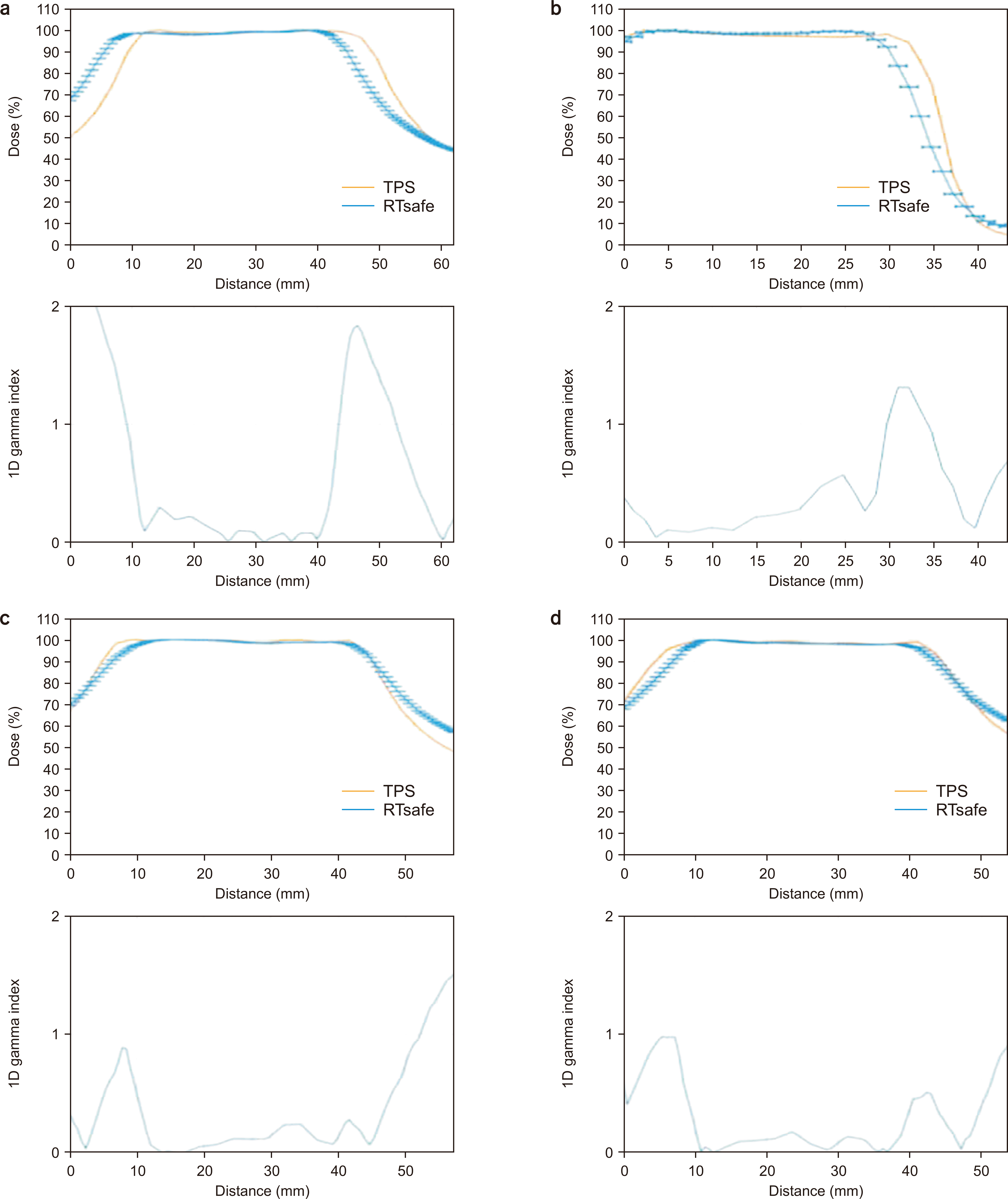 | Fig. 6Comparisons between one-dimensional (1D) profile and 1D gamma index values of the calculated (TPS) and measured doses: (a) x-axis direction of the coronal plane; (b) y-axis direction of the coronal plane; (c) x-axis direction of the transverse plane; (d) y-axis direction of the transverse plane. TPS, treatment planning system.
|
The 1D gamma index of the calculated dose and the absorbed dose matched the gamma passing criteria within 2 mm/2% when the graph was plotted for the absorbed dose from irradiating the gel phantom by 4.5 mm to the right. After correcting the error according to the position for the remaining directions, it was confirmed that the gamma passing rate was more than 95% in the gamma criteria of 2%/2 mm. Given the phantom setup and the low-dose exposure, it can be considered that the gel dosimetry conducted in this study will adequately match the dose from the TPS.
Go to :
Discussion
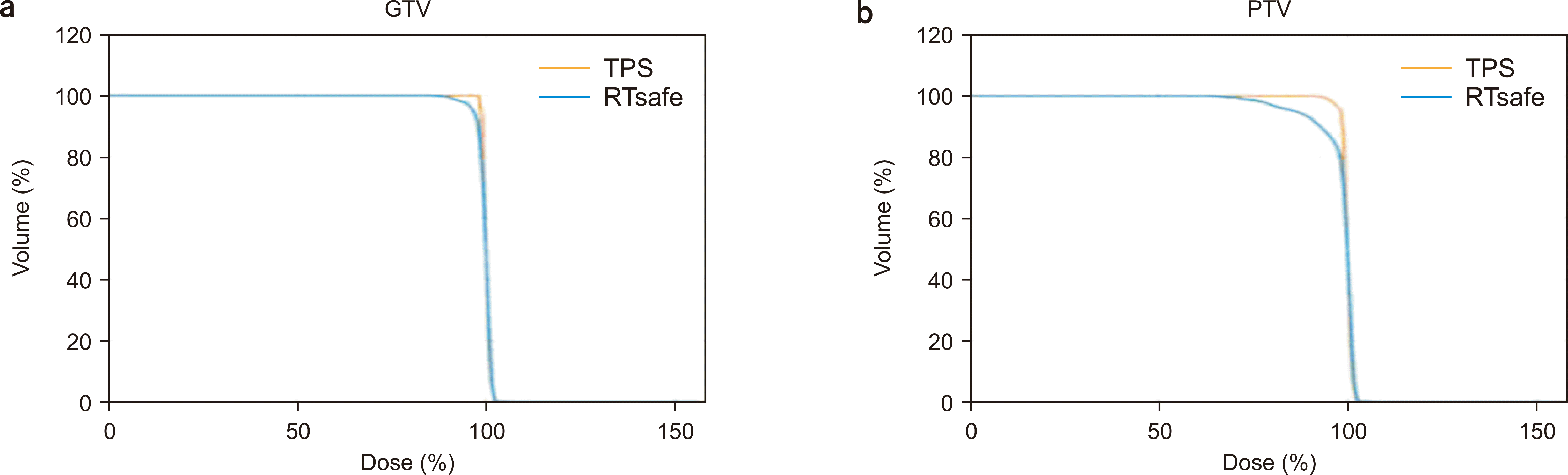
Based on the CT images of brain cancer patients, we used 3D printing technology to fabricate a patient-customized 3D phantom suitable for the density of each region, and performed 3D dose evaluation using a polymer gel inserted into the phantom. As suggested by Adam et al. [16], it was confirmed that the HU values of brain and bone were similar to those of the phantom produced in this study. Comparison of the HU values of the CT image of the patient and those of the phantom revealed that they were almost similar. These values were compared with the average HU values and their standard deviation obtained by designating three ROIs each to identify the homogeneity of the printed material. To analyze the three-dimensional dose distribution using the two-dimensional dose distribution, the dose volume histogram of PTV was compared. The results for the cumulative dose volume histogram of all PTVs are illustrated in Fig. 7 as a comparison between the planned relative dose distribution and the measured relative dose distribution. The difference between the calculated dose (TPS) and absorbed dose (RT-safe) in a gross tumor volume (GTV) can be observed from the results of the dose volume. As shown in Fig. 7, the high dose area of GTV matched well with the TPS results despite differences in the location of the scanned gel phantom. The difference between TPS and gel dosimetry for PTV showed more difference in the high dose area than the difference in GTV. The reason was analyzed as a difference due to the HU in the PTV area and the dose calculation algorithm. For comparing these results, the errors of the dose rate for the volumes of GTV and PTV are reduced when position correction is achieved for TPS and the measured gel.
Go to :
Conclusions
In this study, a comparative analysis of a patient-customized phantom fabricated using 3D printing technology and a 3D dosimetry and TPS using a gel was performed. The 3D-printed head phantom has advantages such as dose comparison among patient’s prescriptions, personalized quality assurance (QA), precision of phantom production, and various application possibilities. The accuracy of the patient treatment plan was verified through the DQA measurements conducted using pretreatment images and the results of the polymer gel dosimetry of an anthropomorphic phantom. The comparative analysis produced better results by validating the difficult-to-treat areas through QA using a customized 3D printing phantom. These results indicate that a customized phantom can be fabricated not only for the head but also for other body parts. It is expected that this study can be applied to the accurate patient specific dosimetry in high energy photon radiation therapy as well as particle radiation therapy.
Go to :
 XML Download