

 PDF
PDF Citation
 Print
Print


Introduction
Radiochromic films (RCFs) have been introduced for two-dimensional (2D) measurement in the field of radiation therapy [1,2], providing advantageous features such as a near water-equivalent material, high spatial resolution, and energy independence in the therapeutic energy range (i.e., megavoltage range) [3].
The principle of RCF dosimetry lies in measuring the color change of film, which, when irradiated, becomes dark due to the polymerization of diacetylene molecules. The variation in light intensity is measured by a scanner or densitometer [4], and the intensity is calibrated using the optical density (OD) [5]. The RCF for our study consists of an active layer and two matte-polyester substrates. The polyester laminates the side of the active layer for protection. However, attaching the film to a curved body for in vivo dosimetry is difficult because of the polyester’s rigidity [6].
Moreover, the sensitivity of the film depends on the amount of diacetylene molecules [7]. The active lithium salt of pentacosa-10,12-diynoic acid (LiPCDA) interacts with radiation, forming a free radial through polymerization. For EBT3, the darkness of the film is difficult to observe in the low-dose range owing to its thin active layer (28 µm) [8]. If the amount of diacetylene molecules increases, the scale of the color change increases for the same dose [9]. In other words, the sensitivity also increases as the film thickness increases.
Reflection scanning methods have been introduced to increase the sensitivity of films in the low-dose range [10]. Generally, transmission scanning is performed to measure the change in light intensity when the transmitted light intensity decreases in darkened areas [11]. In the case of reflection scanning, the reflected light intensity from the reflector (white surface) was measured [11].
This study employed films with active layers of various thicknesses fabricated for flexible dosimeters. Each active layer’s sensitivity was examined through transmission and reflection scanning. The optimal thickness of the active layer and the scanning option were determined to manufacture flexible dosimeters.
Go to : 
Materials and Methods
1. Active layer
Commercially available RCFs were supplied exclusively by a single company (Ashland, Bridgewater, NJ, USA). The active layer consists of gelatin, LiPCDA, and a 0.5% aqueous solution of tartrazine. The compositions of the active layer and LiPCDA are listed in Table 1 and 2, respectively.
The LiPCDA production process was as follows. Part A was produced by mixing gelatin (5 g) and water (45 g) and stirring at 70°C. Part B was manufactured by mixing an aqueous solution of tetraethylammonium hydroxide (9.85 g), 5.00 g of PCDA, and water (91.49 g) and stirring at 70°C. Part B was then mixed with an aqueous solution of lithium acetate (7.04 g). Part B was then mixed with an aqueous solution of lithium acetate (7.04 g), stirred, and then added to Part A at 35°C. The LiPCDA was ripened at 4°C in a freezer for 25 hours. An aqueous solution of 5 g of 0.5% wt tartrazine was added to the mixture. LiPCDA was dissolved at 40°C, ripened for 6 hours at 60°C, and sonicated for 108 seconds. These steps were repeated 5 times for the active layer material, which was then applied on a 75-µm-thick positron emission tomography film using an auto bar coater. The film’s total thickness depended on the active layer’s thickness. We fabricated films with thicknesses of 80, 90, 120, 140, and 170 µm, measuring the total thickness with vernier calipers.
2. Irradiation and film processing
Each set of the 3 film dosimeters was sampled from the same batch with the same film thickness. The size of each film was 3×3 cm2. Dosimetric leaves of 0.2, 0.5, 1, 2, and 3 Gy in the therapeutic dose range were exposed to the set of films. A 6-MV photon beam was generated using a Trilogy radiation therapy system (Varian Medical Systems, Houten, The Netherlands). The film set was placed at a depth of 10 cm in a solid water phantom with reference conditions (i.e., 100 cm source-to-surface distance, 10×10 cm2 field size). The machine was calibrated according to the American Association of Physicists in Medicine Task Group 51 protocol. The monitoring unit was set to irradiate a predetermined dose. Before irradiation, all films were scanned in the transmission and reflection mode using an Epson Expression 11000XL document scanner (Epson Seiko Corporation, Nagano, Japan) for an unexposed measurement [10]. The scanned images were saved in a tag image file format (TIFF) with 48-bit color and a spatial resolution of 300 dpi. The images were denoised using a median filter with a pixel size of 5×5.
After irradiation, the films were kept in a black envelope for self-development for 24 hours. Film scanning was performed to measure the exposure in a similar manner as with the unexposed measurement. The black surface was scanned for the background measurement in the reflection mode.
The scanned images were analyzed using RIT113 Classic software version 6.71 (Radiological Imaging Technology, Inc., Colorado Springs, CO, USA). The RGB channels were extracted from the TIFF image. The red channel was used only to calculate the netOD for the measurement value [12].
For the film calibration, the netOD was calculated as follows [7]:
where Munexp is the unexposed measurement, Mexp is the exposed measurement, and Mbkg is the background measurement. The relationship between the dose and netOD was expressed using the regression curve of the second-order polynomial equation. The response sensitivity of the film was determined as the slope of the tangent at each dose in the regression curve.
Lastly, the intra-batch difference was examined to determine the film’s uniformity. Three pieces of film were randomly obtained in one batch. The films were scanned to evaluate the netOD following the necessary protocol.
Go to :
Results|Discussion
Fig. 1a and b shows the calibration curve of the film within the 0 to 3-Gy range for the reflection and transmission modes, respectively. For the transmission and reflection modes, the sensitivity increased as the film’s thickness increased, and the netOD had similar values at 3 Gy. For the transmission mode, the dose response showed more linearity than for the reflection mode.
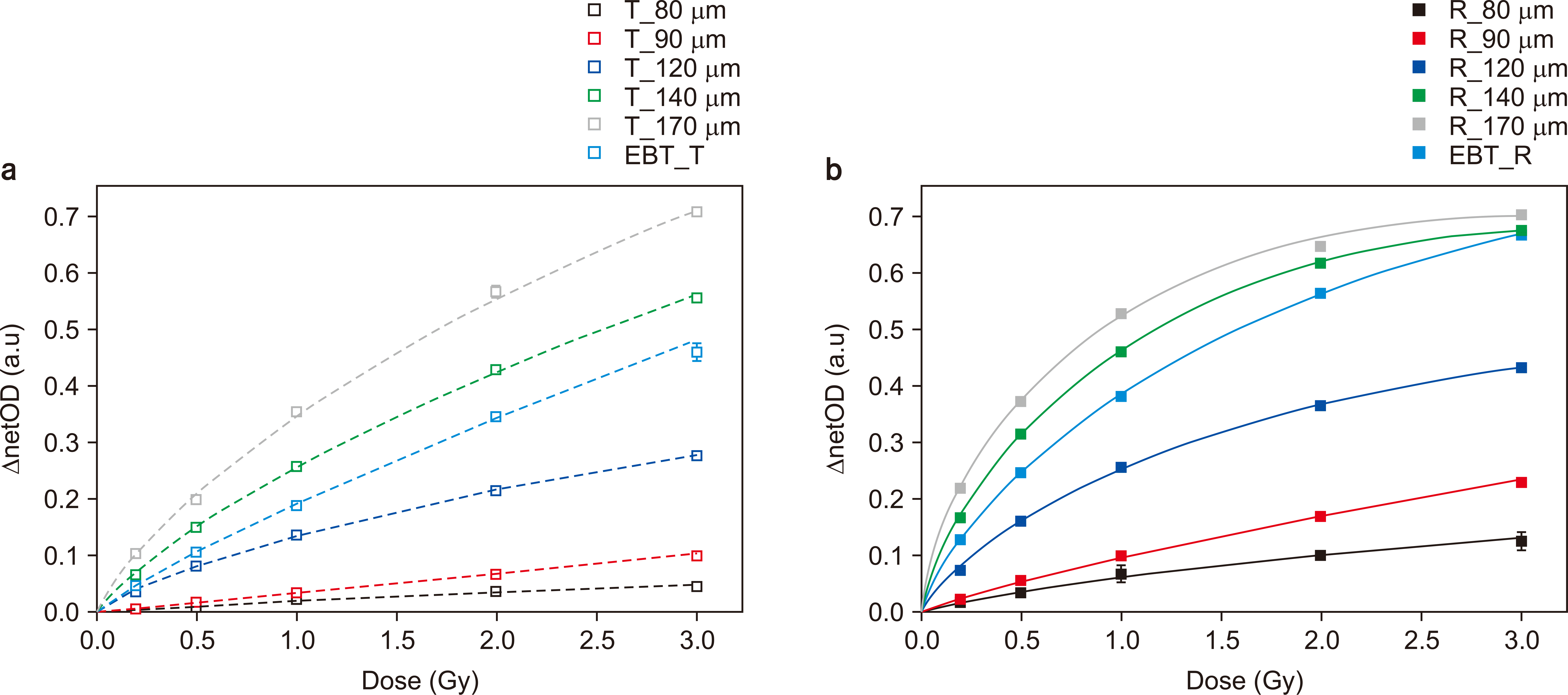
In the case of the reflection mode, the dose response increased dramatically in the low-dose range (<1 Gy), except for the films with thicknesses of 80 and 90 µm. The value of the netOD varied rapidly as the film’s thickness increased. However, the Hurter and Driffield curve showed a supralinear-curve relationship at doses greater than 2 Gy.
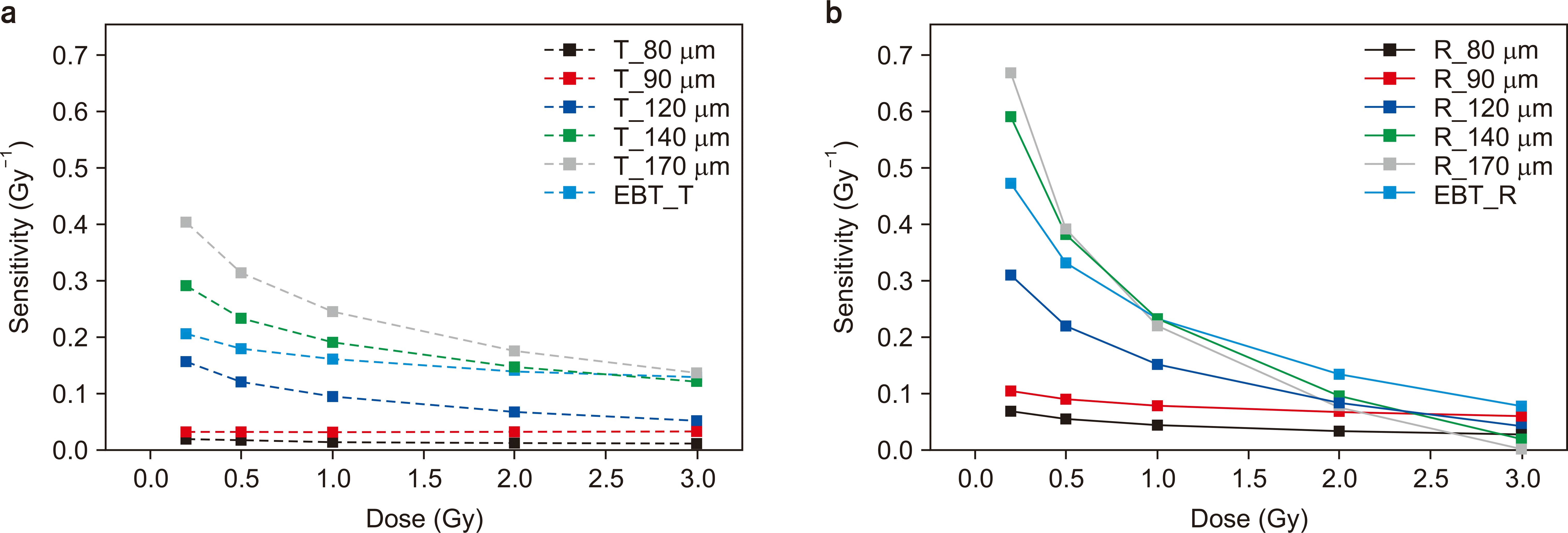
The sensitivity of the transmission and reflection scans was compared as a function of the dose (Fig. 2). The sensitivity of the reflection scan was higher than that of the transmission scan within 0 to 1 Gy, and the sensitivity steadily decreased with increasing doses. The sensitivity of the two modes presented comparable values (0.1–0.2) at 2 Gy and was saturated beyond that. The sensitivity of the reflection scan gradually decreased to lower than 0.1, which is the value of the transmission scan at approximately 3 Gy.
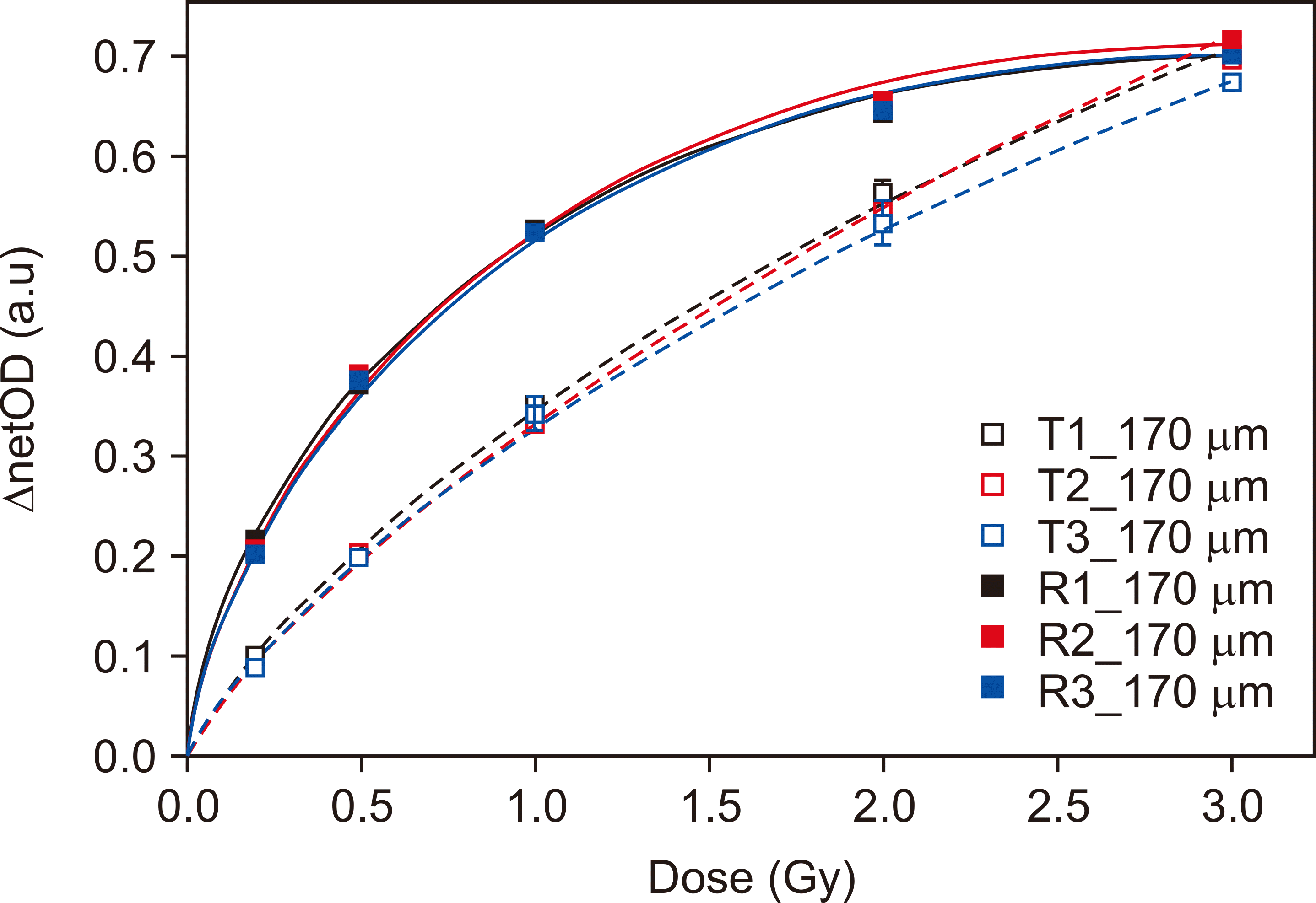
The results of the intra-batch test are shown in Fig. 3. At range doses lower than 1 Gy, the netOD difference of the intra-batch was larger for the reflection scan. In contrast, the netOD of the high dose showed a major difference for the transmission scan. The maximum netOD difference of the intra-batch was 5.5% at 2 Gy and 7.4% at 0.2 Gy in the transmission and reflection scans, respectively.
In this study, we manufactured an LiPCDA-based film evaluated its dosimetric characteristics. The film’s sensitivity increased as the thickness of the active layer increased in response to an increase in the number of diacetylene molecules. Even when the film was irradiated with the same dose, the polymerization reaction was much higher for the thicker films. Moreover, the light is more attenuated because the transmission length of the thick film is long. In conclusion, the intensity of the transmitted light is lower. However, the flexibility of the film decreased with increasing film thickness. Moreover, the cost of production is also higher owing to rising material costs. To evaluate film thickness, a far more accurate measuring instrument is needed. Viable alternatives include a ultrasonic water depth gauge.
The film thickness was the dominant factor for determining film sensitivity. In this study, we used micro-vernier calipers to measure the thickness. However, the accuracy and reproducibility of the device depends on operator skill.
In the case of the reflection scan, the film sensitivity was higher than that for the transmission scan below 1 Gy. For the reflection scan, the light underwent attenuation twice, which has the same effect as with an increase in transmission length. Therefore, the value of the netOD varies significantly. However, the netOD of the reflection scan was saturated beyond 2 Gy, although the sensitivity of the thick film was closer to 0 in the reflection mode. In a relatively higher dose range, the sensitivity of the transmission mode was higher. When the transmission mode was applied, the dynamic range was wide.
The intra-batch variation was checked for uniformity and was slightly higher than that of the commercial RCF. Film uniformity is an important quality factor, and therefore the manufacturing process needs to be improved to ensure uniformity.
Go to :
Conclusions
We performed a dosimetric test for an LiPCDA-based film and confirmed the sensitivity and uniformity of the transmission and reflection modes. In the low-dose range, the thicker film was appropriate for the reflection mode. In contrast, the transmission mode showed a better result. Therefore, a proper scan mode should be selected according to the dose range.
Go to :
 XML Download