

 PDF
PDF Citation
 Print
Print


Introduction
With the development of radiation therapy technology, intensity-modulated radiation therapy (IMRT) and volumetric modulated radiation therapy (VMAT) are being widely used in various cancer treatments [1-4]. They can modulate fluence maps by using the multi-leaf collimator (MLC) to deliver high-dose radiation to treatment targets while delivering minimal dose of radiation to surrounding normal organs [5]. To deliver optimized dose distribution to patients, fluence complexity must be improved, and many MLC movements are required for its implementation. As such, the MLC is a critical factor in radiation therapy.
The head scatter factor (Sc) is the relative ratio of all scattered radiation amounts from the head of the linear accelerator (linac). According to the American Association of Physicists in Medicine (AAPM) Task Group (TG)-74 report, the main causes of scattered radiation include the primary collimator, flattening filter, jaw, and MLCs [6]. The AAPM TG-74 report has mentioned that scattered radiation by the MLC is no longer negligible as IMRT and VMAT use several small and irregular MLC fields [6]. This led to the increased importance of scattered radiation from the MLC, and research was needed accordingly. Nevertheless, previous studies have developed models that did not consider scattered radiation from MLCs; these models were dual- and three-source models in which Sc is calculated by measuring the area of the model using detector’s eye view (DEV) [7-9]. When the MLC is located in a specific area as shown in Fig. 1a, it is exposed to the radiation field and creates scattered radiation; however, it is not considered in DEV; thus, it does not affect the Sc calculation. Only when the MLCs enter the DEV area (Fig. 1b), it can affect the Sc calculation. The MLC cannot be considered as present in region “a” of Fig. 1c (the region where the MLC-defined square field size is from 8×8 to 15×15 cm2 after the jaw-defined field size is fixed to 15×15 cm2); thus, the Sc value in the field where the scattered radiation from MLCs is not considered is calculated similarly, resulting in a difference in the measured Sc value. In the “b” region (the region where the MLC-defined square field size is 4×4 to 8×8 cm2 after the jaw-defined field size is fixed to 15×15 cm2), the position of the MLC affects the DEV; thus, the calculated Sc value decreases as the field size decreases. In terms of area “a,” it is a very small difference between the calculated and measured Sc values, but in the case of IMRT or VMAT, which use hundreds of segments and control points, the combined difference can be large. Previously, we have developed an additional model that can consider scattered radiation from the MLCs [10]. A model for considering scattered radiation from MLCs was located at the MLC location in the linac. The model was developed including line and area sources with scatter interface which was used for determining affected and unaffected areas. Details for scatter interface have been mentioned in the previous study [10]. This model has been verified using a 6 MV photon beam. The results showed that the model was more accurate than the existing model, and particularly, the accuracy increased for very small and irregular fields. For this developed model, the accuracy of the independent verification program could be improved, and a more accurate pretreatment quality management evaluation was enabled.
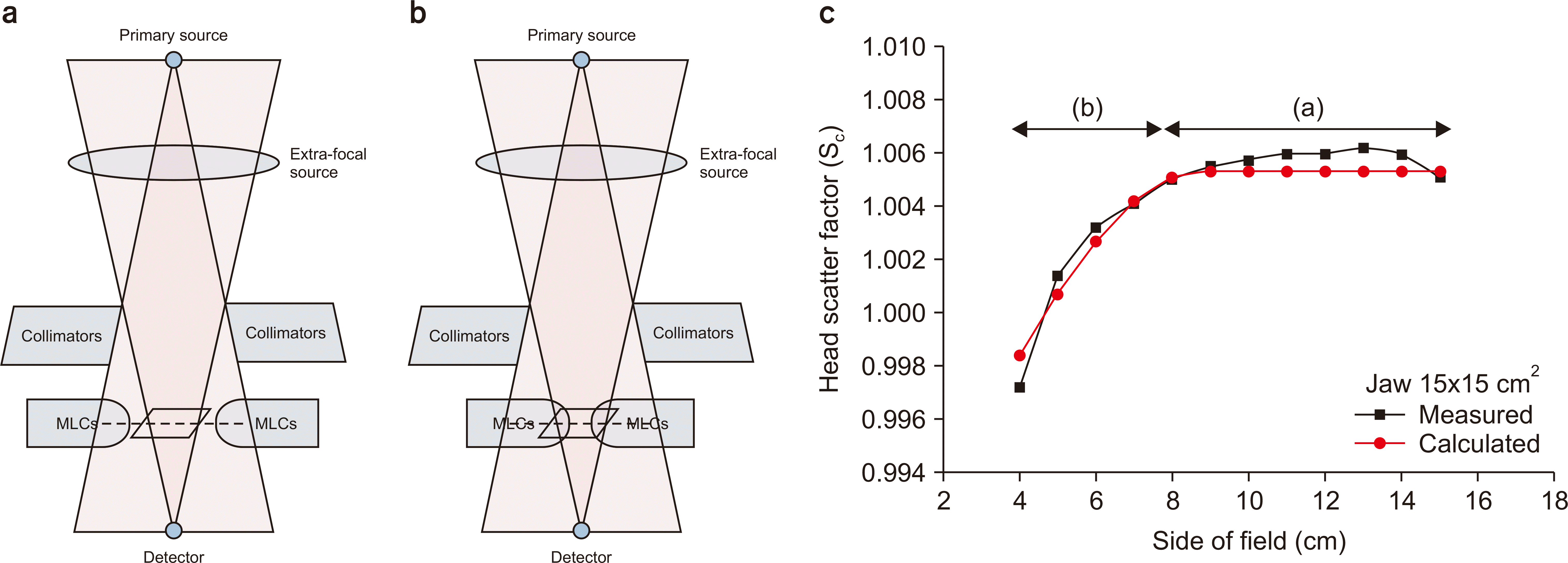 | Fig. 1Schematics of the geometrical relationship between the jaws and multi-leaf collimators (MLCs) in terms of the beam’s eye view, detector’s eye view, and scatter interface for (a) unaffected area, (b) affected area, and (c) head scatter factor (Sc) as a function of the MLC-defined square field size ranging from 4×4 to 15×15 cm2 at a fixed jaw setting of 15×15 cm2. The calculated Sc values are derived from the dual-source model.
|
The unflattened filter (FFF) photon beam is a photon beam from which the flattened filter has been removed in the linac. For small fields, it has radiation quality and profiles similar to those of the flattened filter (FF) but has the advantage of rapidly ending the radiation treatment when dose rates increase by 2–4 times. As the demand for stereotactic ablative radiotherapy (SABR) increased, the use of unflattened filter photon beams also increased. The MLC models developed in the previous studies have not yet been verified for unflattened filter photon beams. When one of the most important factors of scattered radiation, the flattening filter, is removed, it is expected to have a significant influence on the model. Therefore, this study aims to verify the developed model for the unflattened filter photon beam.
Go to : 
Materials and Methods
1. Measurement for head scatter factors
Various Sc sets were measured for the development of the dual-source model (DSM) and the MLC scatter model. A 6 MV unflattened photon beam from a Varian Clinac® TrueBeam STx linear accelerator that is equipped with 60 pairs of millennium MLCs (Varian Medical Systems, Palo Alto, CA, USA) has been used. Measurements have been performed using a 0.125 cm3 cylindrical ionization chamber (Model 31010; PTW Freiburg, Freiburg, Germany) and a mini phantom (Model 670; CIRS Inc., Norfolk, VA, USA). The source-to-chamber distance was 100 cm, and the depth was 10 cm.
For the development of the DSM, the Sc of various jaw-defined square and rectangular fields has been measured. Here, all MLCs were in a retracted state. For the development of the MLC scatter model, the jaw was fixed, and then the Sc of various MLC-defined square fields was measured. The field setup for this measurement is illustrated in Table 1. To reduce the uncertainty in Sc measurement, the measurement was repeated at least 5 times, and the standard deviation was within 0.3%. The reference field was a jaw-defined size of 10×10 cm2.
Table 1
Measurement cases for modeling and evaluating the dual-source and MLC scatter source
![]()
2. Development of source models
The DSM proposed by Jiang et al. [11], as explained in previous studies, is a model that considers all the scattered radiation from the head of the linac and the backscattered radiation reentering the monitor chamber, with an extrafocal source located at the location of the existing FF. Sc is obtained by measuring the area radiated from the DEV to the extrafocal source. As mentioned earlier, for all scattered radiation from the head, the scattered radiation from the MLC was not considered [10,11]. It was subjected to a process of optimizing the DSM parameters using the Sc values for various jaw-defined fields. The jaw-defined fields used during this process include a total of 3 sets, 4×4 to 40×40 cm2 square fields alongside the rectangular field, i.e., 10×4 to 10×40 cm2 and 4×10 to 40×10 cm2 with both the X and Y jaws fixed at 10 cm.
In this study, the DSM parameters were iteratively optimized using the trust-region-reflective algorithm for nonlinear least squares (MathWorks, Inc., Natick, MA, USA) to match the calculated Sc with the measured one for the jaw-defined square and rectangular fields (Table 1). The objective function was the chi-square difference between the measured and calculated Sc values for the jaw-defined fields with the fully retracted MLCs. Optimization was performed within the maximum number of iterations (400) until the objective function reached below a termination tolerance (10−6) [10].
The MLC scatter model developed in previous studies consists of line and area source, that is, scattered radiation from the end of the MLC tip and scattered radiation from the MLC area, respectively [10]. If the jaw-defined field is fixed and the MLC-defined field increases, the scattered radiation from the tip of the MLC increases, but the scattered radiation from the MLC area decreases. By formulating each of these, Sc,MLC can be defined as follows:
The calculated Sc,MLC can be used to obtain the Sc for an MLC-defined field. The fields used for modeling included a set of Sc values obtained when the MLC-defined field is 4×4 to 10×10 cm2 with a jaw fixed at 10×10 cm2 and a set of Sc values obtained when the MLC-defined field is 4×4 to 20×20 cm2 with a jaw fixed at 20×20 cm2, i.e., a total of two sets. Details are available in previous studies [10].
Similarly, the parameters were optimized using the same optimization methods as in DSM to attain the best fit between the measured and calculated Sc,MLC values.
3. Evaluation of source models
For the verification of the DSM, the calculated Sc values were compared to the three Sc set measurement values of the jaw-defined fields used in the existing modeling. For further verification, the calculated values were also compared with the Sc set measurement values that were not used in the existing modeling. These are Sc measurement values for a total of 4 sets of 4×4 to 4×40 cm2 and 4×4 to 40×4 cm2 with X and Y jaws fixed at 4 cm along with 40×4 to 40×40 cm2 and 4×40 to 40×40 cm2 with X and Y jaws fixed at 40 cm, respectively, which are the rectangular fields.
To verify the MLC scatter source model, two Sc set measurement values of MLC-defined fields used in the existing modeling and calculated values have been compared. For further verification, the calculated values have also been compared to the Sc set measurement values that were not used in the existing modeling. This is a set of Sc values when an MLC-defined field is 4×4 to 15×15 cm2 in a state where the jaw is fixed at 15×15 cm2. The equation for evaluating the accuracy using percent error is as follows:
Go to :
Results
1. Evaluation of jaw-defined square fields
In this study, the DSM has been evaluated using jaw-defined square fields ranging from 4×4 to 40×40 cm2 and rectangular fields with one pair of jaws fixed at 4, 10, or 40 cm while the other pair varied from 4 to 40 cm. Fig. 2 shows the Sc values calculated by DSM and measured Sc values for the jaw-defined field size. Table 2 quantifies and shows the difference between the measured Sc value and the calculated Sc value for each measurement set. Overall, Sc calculated using the DSM and the measured Sc showed a good agreement. It can be identified that all difference values are within 0.212%. This suggests that the DSM has been properly optimized and modeled for the measured values.
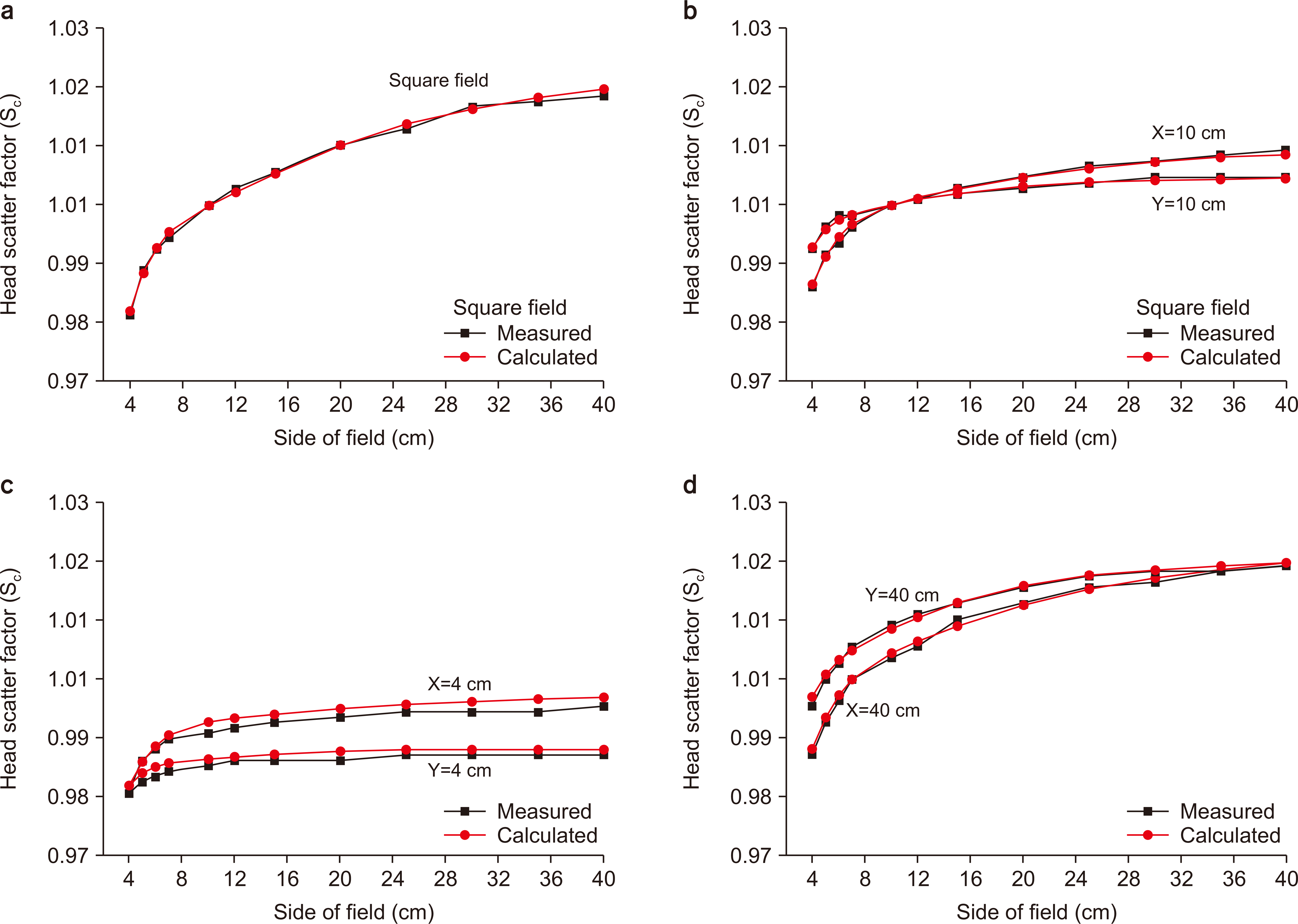 | Fig. 2Comparison between the calculated and measured head scatter factor (Sc) values for (a) square fields ranging from 4×4 to 40×40 cm2; (b) rectangular fields with one pair of jaws fixed at 10 cm, while the other pair varied from 4 to 40 cm; (c) rectangular fields with one pair of jaws fixed at 4 cm while the other pair varied from 4 to 40 cm; and (d) rectangular fields with one pair of jaws fixed at 40 cm, while the other pair varied from 4 to 40 cm. The calculated Sc values are derived from the dual-source model.
|
Table 2
Differences between the measured and calculated Sc values for jaw-defined field sizes
![]()
2. Evaluation of multi-leaf collimator-defined square fields
The optimization parameters for the MLC scatter source model have been determined using nonlinear least squares. Table 3 demonstrates the optimization parameters for line and area source of MLC scatter sources. There was no clear trend for each parameter. These optimization parameters were used to model the MLC scatter source, the calculated Sc values were compared with the measured Sc values, and Sc values were calculated using only DSM (Fig. 3). For all jaw-defined fields, if the MLC-defined field size becomes greater than or equal to the specific MLC-defined field size, the Sc value calculated using the DSM remained flat, which causes a difference from the measured Sc value. In the case of the calculated Sc value with the addition of the MLC scatter source, its accuracy with the measured Sc value appeared to be high. Table 4 demonstrates the quantified degree of accuracy. Overall, a small difference has been observed between the calculated value and the measured value, but the additional use of the MLC scatter source model resulted in a significantly smaller difference in probability. Particularly, if the jaw-defined field was 15×15 cm2, a difference of 0.055% has been observed when an additional MLC scatter source was used.
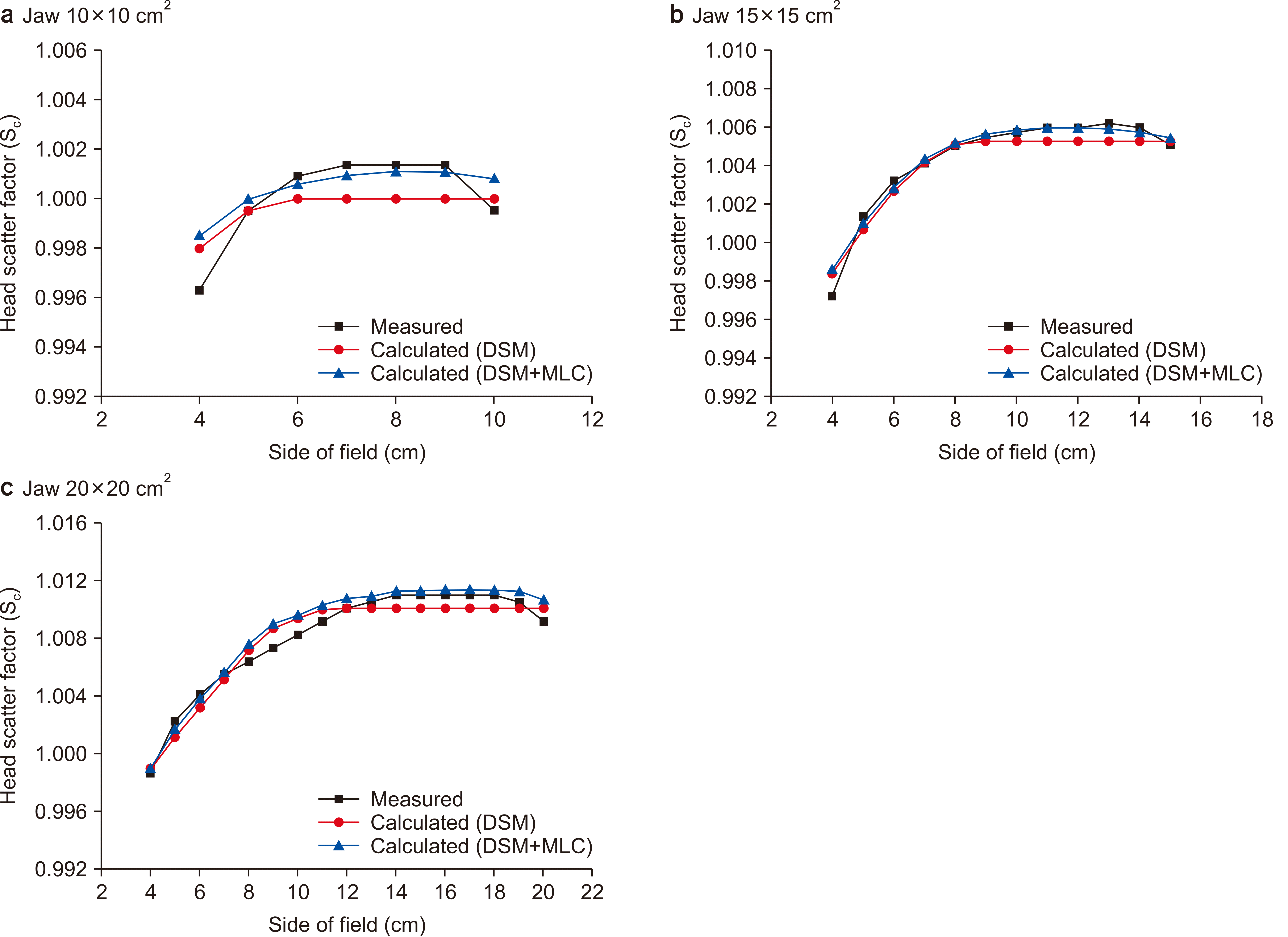 | Fig. 3Comparison between the measured and calculated head scatter factor (Sc) values for fixed jaw defined fields of (a) 10×10 cm2, (b) 15×15 cm2, and (c) 20×20 cm2. Sc is calculated based on the dual-source model (DSM) and DSM in conjunction with the multi-leaf collimator (MLC) scatter source (DSM+MLC).
|
Table 3
Optimal parameter values of the multi-leaf collimator scatter source for an unflattened 6 MV beam
![]()
Table 4
Differences between the measured and calculated Sc values for MLC-defined square field sizes ranging from 4×4 cm2 to the fixed jaw opening size
![]()
Go to :
Discussion
Previous studies have developed sources that are capable of modeling scattered radiation from the MLCs for flattened photon beams and have evaluated their performance [10]. When only DSM was used, the jaw-defined field area showed a high accuracy with the measured value, but when the MLC-defined field was used, it showed a low accuracy. When considering the scattered radiation from the MLCs by adding the MLC scatter source model developed in the previous study, the accuracy was higher than the Sc calculated using the DSM. However, previous studies have considered only the flattened photon beam, while this study has evaluated the performance when an unflattened photon beam was applied to the same MLC scatter source model.
Since the unflattened photon beam does not pass through FF, the Sc value is relatively less than that of the flattened beam [12,13]. Other studies have also examined these characteristics. However, the characteristic or tendency of Sc depending on the field size or depending on X or Y is the same as in the flattened beam [12,13]. Therefore, the DSM and MLC models could also be applied to the unflattened beam. The optimization parameter values for the MLC model are smaller than the optimization parameters obtained in previous research, which indicates that the amount of scattered radiation decreased compared with the flattened beam, resulting in a decrease in the associated weight value.
This study has several limitations. Measurements for small field sizes (less than 3×3 cm2) could not be performed, which are mainly used in SABR. The difficulty in applying the modeling can be attributed to the physical limitation of the detector size and high measurement uncertainty for small fields [14,15]. It is assumed that more accurate modeling is possible if a small field size Sc is measured using a detector optimized for small fields, such as a diamond detector. Moreover, the amount of scattered radiation varies for high photon energy, and this should be considered. In a future study, we plan to apply scattered radiation modeling from the MLCs for high photon energy and evaluate its performance. Furthermore, since this study is empirical, Monte Carlo simulations are required to more accurately model scattered radiation from the MLC. Fig. 3c demonstrates that between MLC-defined field sizes of 8×8 and 12×12 cm2, the Sc values calculated using the two models (DSM and DSM + MLC) are overestimated compared to the measured values. This suggests that scattered radiation from the MLC cannot be accurately divided using DEV. A more accurate MLC scatter source can be developed if scattered radiation from the MLC obtained in various MLC-defined field regions has been analyzed using a simulation.
Go to :
Conclusions
This study aimed to evaluate the accuracy of Sc calculation by applying the previously developed MLC scatter source model to an unflattened photon beam. When considering scattered radiation from the MLCs by adding an MLC scatter source model, it showed a higher degree of agreement with the actual measured Sc value than when using only DSM in the same way as in previous studies. This MLC scatter source model is expected to be applied to the independent verification program in the future to enable more accurate dose calculation.
Go to :
 XML Download