Introduction
Bezoars are foreign particles from the accumulation of indigestible materials in the gastrointestinal system and are a rare cause of mechanical intestinal obstruction. They have various names according to the material they are composed of, including phytobezoars, trichobezoars, and lactobezoars. Among the several types of bezoars, the most encountered form is phytobezoar, which is associated with the consumption of fibrous foods [1,2].
Phytobezoars are common in certain regions or cultures with high consumption of fiber-rich foods. Gaya et al. [3] mentioned that this type of bezoar is the most commonly reported etiological factor of intestinal obstruction in certain geographical areas where very high fiber-containing foods (e.g., persimmons) are grown and consumed. Apart from fiber-rich food consumption, many risk factors that facilitate the development of bezoars have also been described, including diabetes mellitus, history of ulcer surgery, hypothyroidism, and dental problems in the elderly as a result of the inability to chew food properly [4].
Bezoars can be asymptomatic or present with a variety of gastrointestinal symptoms, including pain, abdominal fullness, discomfort, difficulty swallowing, nausea, and anorexia. In contrast, bezoars can be coincidentally found in asymptomatic patients by esophagogastroduodenoscopy or CT scans performed during a health check-up or follow-up of other diseases. Symptoms related to gastrointestinal bleeding, such as hematemesis, bloody or tarry stool, anemia, and fainting, are the result of the development of ulceration in the gastric mucosa due to pressure necrosis induced by the bezoar [5].
Endoscopic examination plays the most important role in the detection of gastric bezoars, as well as in the treatment of this disease. Contrast-enhanced CT has been shown to be 90% sensitive but only 57% specific in the identification of bezoars [6].
This case is a report on whether it is useful to remove bezoars by methods other than surgery in patients who are difficult to treat surgically due to their old age or poor general condition.
Go to : 
Case
An 89-year-old female presented with throbbing epigastric pain, general weakness, and intermittent melena for 1 month. She had diabetes, hyperlipidemia, and a history of hospitalization with a hip fracture 2 months previously. There were episodic attacks of non-bilious vomiting that involved blood.
Generally, the patient was thin, with no pallor or jaundice (body weight 156 cm, weight 48 kg). Her vital signs were normal. The abdomen was soft, and no tenderness or rebound tenderness on physical examination. The bowel sounds were normal, and there was no abnormal bruit. The initial laboratory findings were as follows: white blood cell count, 7,440 cells/μL; hemoglobin, 5.5 g/dL; platelet count, 192×103 cells/μL. Additionally, plain abdominal radiography showed no abnormal bowel gas pattern.
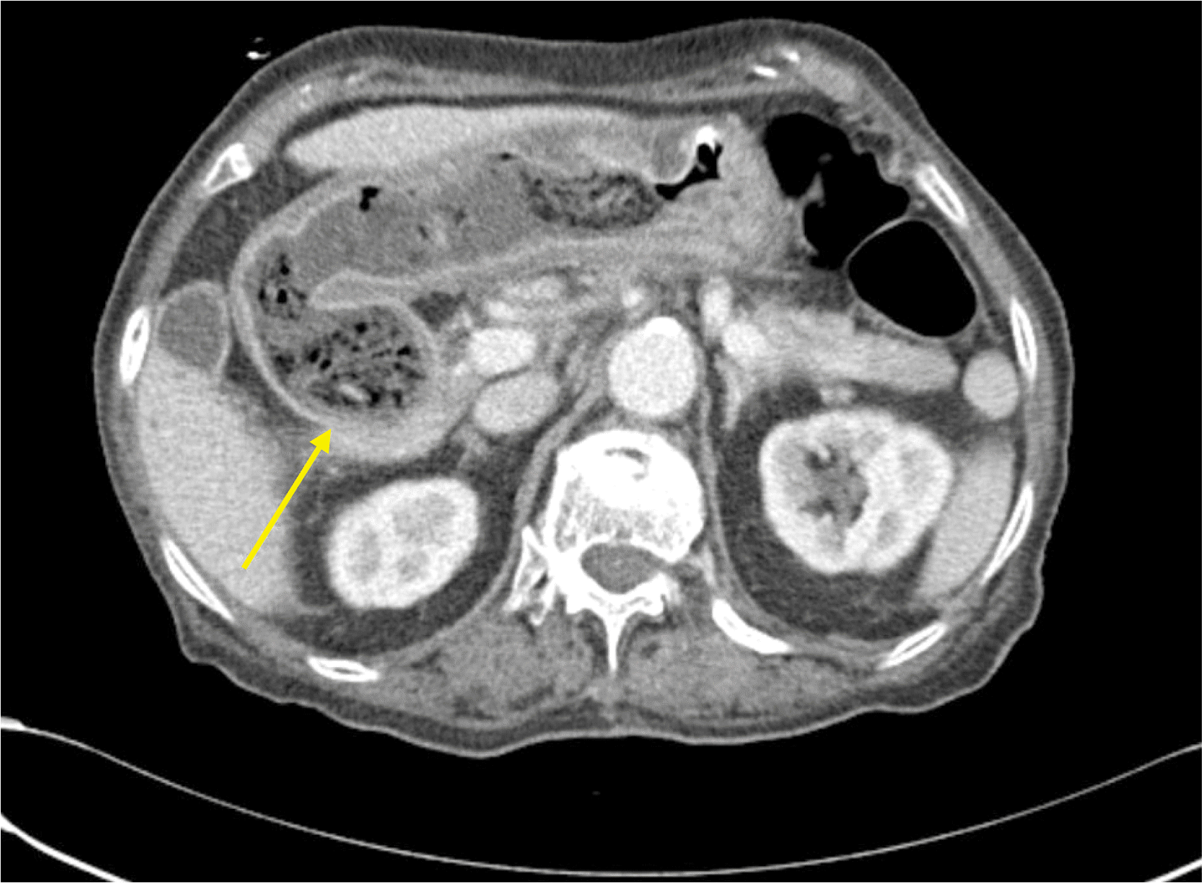
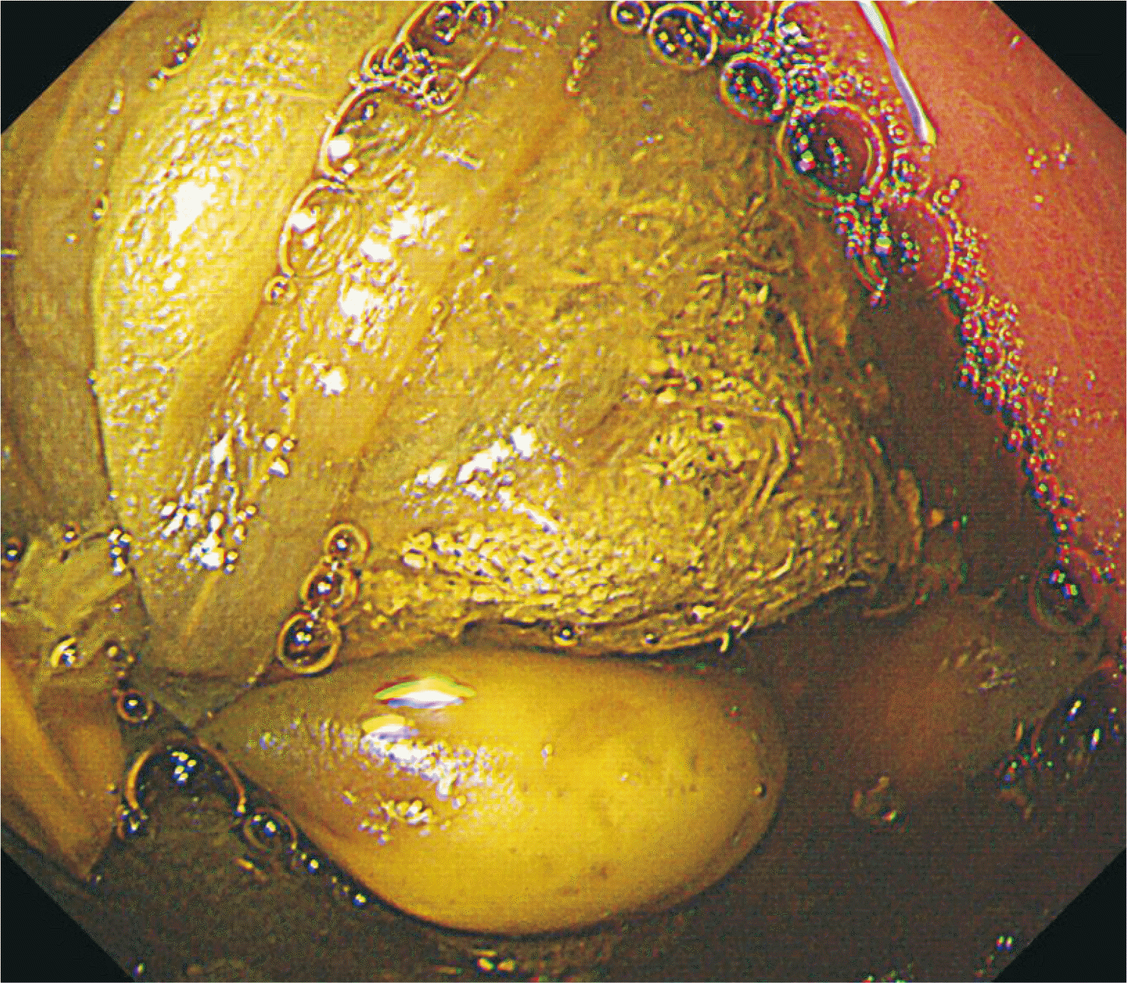
A CT scan of the abdomen it showed a 5-×4-cm, firm, atypically shaped mass at the stomach body and duodenal bulb with interspersed gas (Fig. 1). Esophagogastroduodenoscopy showed a mass of fiber impacting the antrum pylorus. Multiple ulcerations with blood clots were observed in the antrum and angle (biopsy was performed, and pathologic results showed a chronic ulcer, active bleeding, and healing with intestinal metaplasia) (Fig. 2).
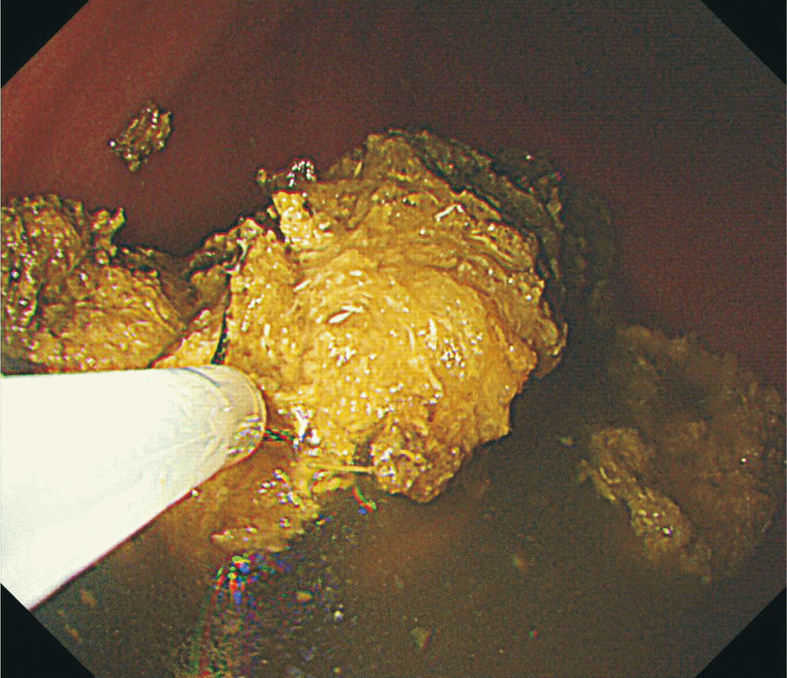
We assessed preoperative risks with open surgery owing to her old age and poor general condition. Besides, neither she nor her family wanted to have the surgery. The endoscopist attempted to remove the bezoar with forceps and a basket but failed at first. Thus, we planned to steep the bezoar in oil, use a snare to crumble it, and continue pouring oil to keep it from sticking. For 10 days, 30 mL of olive oil was injected three times a day through a Levin tube into the stomach, and the bezoar was soaked in olive oil. Cellulosic materials like Kimchi pieces were gradually separated without any undesirable side effects, such as diarrhea and nausea. We repeated the esophagogastroduodenoscopy three times at very short intervals to determine the state of the bezoar. In order to fragment the bezoar, we used an endoscopic snare three times, and at the end of the fourth endoscopy, we found that it had completely disappeared in the stomach. We confirmed that it broke into small pieces and descended into the small intestine, and abdominal radiography and CT were performed to follow broken bezoar fragments (Fig. 3). After the bezoar removal, the patient started food intake and was discharged without any complications.
Go to : 
Discussion
Various operative and non-operative or endoscopic techniques have been mentioned in the literature for phytobezoar treatment. Lots of studies have reported on lavage and aspiration with large esophagogastric tubes, chemical dissolution with Coca-Cola lavage or hydrolytic solutions, and mechanical disintegration with lithotripsy or an electrosurgical knife. Furthermore, endoscopic fragmentation has been achieved with biopsy forceps or polypectomy snares [7].
A number of articles have emphasized the usefulness of Coca-Cola administration for the dissolution of phytobezoars. However, persimmon phytobezoars may be resistant to such dissolution treatment because of their harder consistency compared with other types of phytobezoars [8].
A successful outcome of bezoar treatment with tablet-form gastroenterase (containing pepsin, pancreatic enzyme concentrate, cellulase, and dehydrocholic acid) was described in the 1970s. However, these tablets have been discontinued because of reported adverse reactions, including gastric ulceration, esophageal perforation, and hypernatremia. Cellulase, an enzyme that cleaves the leucoanthocyanidin-hemicellulose-cellulose bonds resulting in dissolution of the phytobezoar, has been widely used for phytobezoar treatment, since vegetables and fruits contain large amounts of cellulose. However, in many countries, cellulase is not readily available for ingestion as a commercial product or even as a medication under prescription [9]. In Korea, cellulase is not produced alone, but it is partly contained as a component in the digestive agent Bearse Tab (Daewoong Pharmaceutical, Seoul, Korea).
Surgical removal is inevitable for cases presenting with ileus or those with refractory bezoars. Bezoars were traditionally managed by open surgical retrieval (laparotomy). Recent papers have emphasized the importance of a minimally invasive surgical approach by laparoscopy in the management of gastrointestinal bezoars. Intraoperative endoscopic removal has also been reported [10-12].
There are several advantages to the use of olive oil for removing a phytobezoar. First, unlike enzymes, such as gastroenterase and cellulase, olive oil has no side effects for the gastrointestinal tract. Second, olive oil is easy to obtain and apply. We purchased olive oil from a market at an affordable price. Moreover, unlike powder or solid ingredients, olive oil is convenient to put in the Levin tube because it is liquid. Third, olive oil may act as a lubricant to form an oil film on the surface of the split phytobezoar to facilitate passage through the gastrointestinal tract. Of course, there is a concern that pieces of split phytobezoar will show difficulty in passing the ileocecal valve in older patients. A laboratory study by de Brito et al. [13] using rats reported that the function of the ileocecal junction appears to change with aging, which is associated with changes in the patterns of distribution of collagen and elastic fibers, resulting in increased tensile strength and firmness, but decreased elasticity. However, in humans, there is no clear evidence that aging is related to ileocecal valve incompetence.
If the elderly can be treated without surgery, conservative management will be more helpful in prolonging survival. From that point, the idea of dissolving a bezoar in olive oil is a new approach in patients who desire to avoid surgery. We could not find any papers stating that olive oil or other oils were used to dissolve bezoar, so this report will be the first case report.
Go to : 


 PDF
PDF Citation
 Print
Print


 XML Download