

 PDF
PDF Citation
 Print
Print


Introduction
Umbilical anomalies are common in infancy, and can result from failure of an embryologic process, thus understanding the basic anatomy and embryology is mandatory to identify and treat umbilical disorders [1]. Newborns are more vulnerable to the anomalies since they have moist umbilicus, and especially, umbilical discharge in infancy is often attributed to infection [2]. When serous, fecal or bilious drainage is observed in the umbilicus, it suggests patent omphalomesenteric duct (POMD) with fistula, as in this case. Newborns who present with drainage from the umbilicus can be quite frightening to parents and are in need of rapid diagnosis and treatment. Also, it is important to investigate if there is an underlying abnormality and causes the discharge since corrective surgical management could be required.
The omphalomesenteric duct (OMD) is a long narrow tube that joins the yolk sac to the midgut lumen of the developing fetus and provides nutrition until the placenta is established [1]. Usually, the duct fully disappears at the 5th to 7th week of gestational age. However, if the duct fails to be closed at appropriate times, it results in several residual structures called OMD remnants. Partial persistence of the OMD defects may lead to Meckel’s diverticulum, umbilical cyst, umbilical sinus, umbilical band, and POMD [2]. Also, complete persistence of the OMDs may lead to omphalomesenteric fistula. When a POMD is symptomatic, it presents before the age of 4 [2]. The symptoms include abdominal pain, rectal bleeding, umbilical drainage and umbilical hernia, and these may lead to gastrointestinal bleeding, intestinal obstruction, Meckel’s diverticulitis, and intussusception [3]. Eighty-five percent of the newborns less than 1 month old, and 77% of the infants between 12 and 24 months old with OMD were symptomatic [4,5].
Meckel’s diverticulum is known to be the most common (65%–80%) type of OMD remnants, and a POMD is the rarest type of OMD remnants [2]. Large number of parents visit outpatient clinic, thinking their child’s disease is mild. Usually, the disease starts with discharge or infection of the umbilicus, leading to the idea that antibiotics will be the enough treatment. However, in reality, the disease is more severe than expected, and may lead to surgery such as in this case. We report a case of POMD with review of literature.
Case
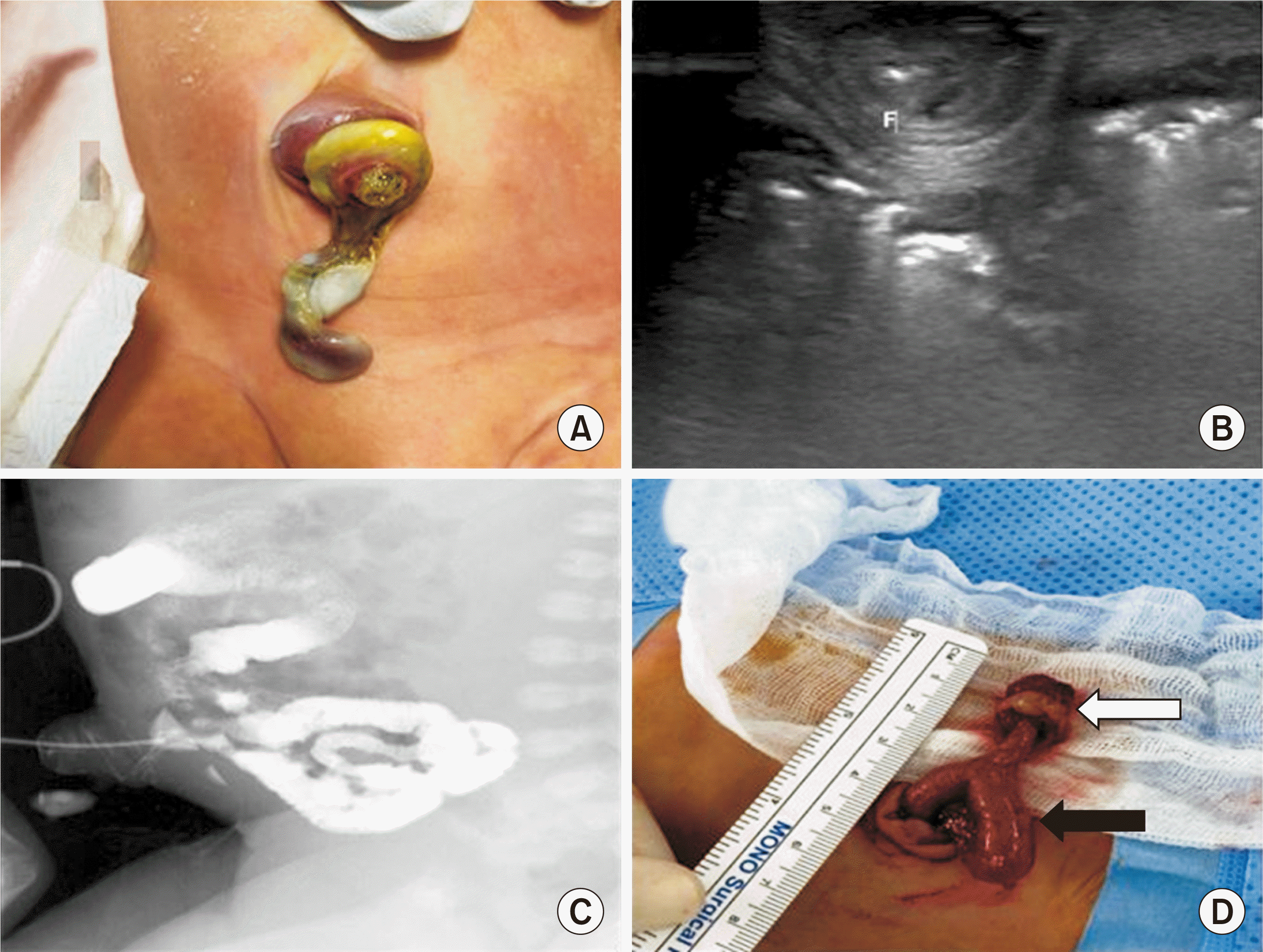
The male patient was born through vaginal delivery at a local clinic at the gestational age of 37 weeks and 6 days, with a birth weight of 3,220 g (50–75 percentile), and the patient was the first child of a 29-year-old mother with no significant past medical history. The prenatal screening test showed no abnormal results. An immediate postnatal examination was conducted after delivery, and no abnormalities were found except for a mild umbilical hernia. Swelling and discharge from the umbilicus started after the baby was born, and became aggravated. The patient was brought to the outpatient clinic of Ewha Womans University Mokdong Hospital on the 5th day of life for the evaluation of omphalitis. A yellowish discharge, found to be meconium was leaking from the opening in the umbilical stump, and an umbilical protrusion was noticed (Fig. 1A). There were no abdominal wall related abnormalities, and remainder of the physical exam showed no remarkable results other than the yellow discharge with umbilical hernia. The patient was hospitalized in the neonatal intensive care unit for further evaluation and treatment. Differential diagnosis were umbilical granuloma, intestinal perforation, meconium peritonitis and omphalomesenteric duct fistula [6].
Abdominal ultrasonography showed a mild umbilical hernia with an underlying OMD fistula connecting the terminal ileum to the umbilicus, and there was no definite evidence of associated peritonitis in the ultrasonography (Fig. 1B). Fistulography also indicated the patient had an umbilical hernia with a POMD connecting to the terminal ileum (Fig. 1C). Bladder wall thickening was noted. No urachal remnants were observed in the voiding cystourethrogram. Brain sonography and echocardiogram showed no abnormalities besides bilateral germinal matrix hemorrhage grade I and small secondum atrial septal defect with stretched patent foramen ovale 3.5 mm respectively.
Surgery was performed on the 3rd day of hospitalization (Fig. 1D). A circumferential skin incision was made at the umbilucus, followed by adhesiolysis around OMD remnant. Meckel’s diverticulum was also noticed. Small bowel was externalized and mesenteric vessels were ligated. Segmental resection of the small bowel and end to end anastomosis were carried out. On the postoperative 4th day, milk feeding was started and gradually increased to full feeding on the postoperative 6th day. The final pathologic report indicated a fibrous cord connecting the small intestine to gastric tissue showing ulceration, hemorrhage and fibrosis. In addition, a few benign ductal structures in the smooth muscle layer which were suggestive of OMD remnants were seen. The patient was discharged on the 8th postoperative day without any complications. At the last outpatient clinic follow-up, 5 months after surgery, no abnormalities were observed.
Discussion
The OMD usually disappears completely during the 5th to 7th weeks of gestational age, and the failure to close is termed an OMD fistula or vitelline fistula [2]. Many umbilical abnormalities such as fistulas, sinus tracts, cysts, mucosal remnants, and congenital bands require surgical correction, and are usually caused by OMD remnants [6]. When OMD is present from the ileum to the umbilicus, it results in the discharge of meconium from the umbilicus [6].
OMD abnormalities occur in about 2% of newborns, and 6% of the ducts remain patent. Exact etiology of incomplete obliteration is still unknown. In 1959, Soderlund [7] categorized these abnormalities into six sections, which are umbilical cyst, umbilical sinus with a band, umbilical polyp covered with intestinal mucosa, fibrous band containing a cyst, Meckel’s diverticulum, and POMD. Our case applies to both Meckel’s diverticulum and POMD. This categorization shows typical variations in OMD remnants. Out of these six sections, Meckel’s diverticulum is the most common type of OMD remnants, and a POMD is the rarest type of OMD remnants [8]. If the duct partially disappears leaving a blind pouch on the antimesenteric surface of the ileum, then Meckel’s diverticulum develops. If the distal segment of the duct remains unclosed, an omphalomesenteric sinus develops and if both ends of the duct remain, an open fistula with fecal material discharge develops [5]. With regard to epidemiology, although symptomatic forms are more common in males, there is an equal frequency between the sexes. The clinical presentation of the omphalomesenteric fistula is changeable and depends on the age, and can be asymptomatic [4,5]. When there are symptoms, they appear before the age of 4. For those with the fistula, 85% become symptomatic at less than 1 month of age, and 77% of the infants between at the age 1 and 2 [4,5]. The symptoms include abdominal pain, rectal bleeding, umbilical drainage and umbilical hernia, and these may lead to gastrointestinal bleeding, intestinal obstruction, Meckel’s diverticulitis, and intussusception [9]. The associated anomalies are intestinal atresia, cardiac malformation, cleft lip & palate and exomphalos [9]. Also, there is a report that trisomy 13 and Down syndrome are related to the anomalies.
According to Kling [10], intestinal obstruction is the most lethal OMD associated disorder, in which intestinal obstruction and vascular lesions occur rapidly and simultaneously, often requiring immediate surgical procedures regardless of the presence of hernia. The study reported that a simple umbilical hernia was easily detected, and so was an umbilical cyst. However, it was harder to detect OMD disorders, and most cases were diagnosed by ultrasonography or fistulography after birth [11]. POMD remnant, as in our case should not be confused with simple omphalitis, umbilical granuloma, umbilical hernia and patent urachus. Fecal discharge, as in our case mostly indicates patent OMD remnant and could be in need of surgical management. Ultrasonography and fistulography take major role in diagnosing OMD abnormalities. Because of the predominance of prevalence in congenital gastrointestinal malformation, ultrasonography is important in patients with OMD remnants. Also, fistulography, a contrast study through the umbilical stoma is another important diagnostic tool since it can figure out whether there is intraperitoneal communication with the bowel or extraperitoneal association with the urinary tract [12].
A review of the literature (Table 1) shows that both ultrasonography and fistulography were used as effective diagnostic tools of OMD fistula and other related remnants of the OMD [2-4,8,11,12]. Age at the presentation of the fistula varied from birth to 6 weeks of age and the range of the symptoms varied from umbilical drainage to mass protrusion. Also, the vast majority of literature suggests conducing early surgical resection in order to prevent further complication from the malformation. Segmental resection with end-to-end anastomosis was usually conducted, although other techniques such as transumbilical exploration and intraumbilical incision were used in some cases.
In summary, we report a case of POMD presenting with meconium discharge into the umbilical stump on the 5th day of Life. This disorder was not diagnosed in antenatal ultrasonography, and through the fistula, there was a meconium discharge. After having diagnostic examinations by using ultrasonography and fistulography, surgical management were immediately conducted. Careful inspection about umbilicus is needed when it comes to conducting physical examinations of neonate or infants. If fecal drainage is observed, POMD with fistula should be considered. Differential diagnosis and appropriate treatment, especially making decision whether to conduct surgical resection should be made, thus increasing the possibility of rapid recovery. The prognosis is usually good with early treatment in the absence of associated malformation.
 XML Download