

 PDF
PDF Citation
 Print
Print


Introduction
Kummell’s disease is an avascular necrosis disorder of the vertebral body after osteoporotic vertebral compression fracture. This disease involves delayed healing of a fractured vertebral body which can be seen in the intravertebral vacuum cleft with fluid or air inside it and needs more aggressive treatment [1,2]. The term was originally coined by Hermann Kummell for a condition wherein a minor asymptomatic spinal trauma leads to symptomatic, progressive painful back pain and functional limitations [2]. This condition is common in the elderly, who are susceptible to vertebral fractures owing to osteoporosis or osteonecrosis which are initially asymptomatic but progress and become symptomatic with change of vertebral body image [2].
Most vertebral fractures do not need aggressive treatment and can be treated by bed rest using braces and pain medications. However, some patients diagnosed with Kummell’s disease complain of severe back pain, deformity, and inability to walk, all of these lowering their quality of life [3,4]. Percutaneous vertebroplasty (PVP) and percutaneous kyphoplasty (PKP) are effective minimally invasive treatments. Bone cement injected during these treatments appropriately agglutinates fractures and resolves the pseudoarthrosis-caused instability by filling the vacuum cleft; this is thus expected to provide remarkable pain relief [3-5]. Among these, PVP is an option including shorter operating time, decreased cost that still provides comparable pain relief and vertebral height restoration as PKP for treating Kummell’s disease [6,7].
Several studies have reported on ways to perform PVP more effectively: spontaneous re-expansion of the vertebral body was reported when inserting the needle into the fractured vertebral body in Kummell’s disease [5,8]. In addition, manual compression after inserting cement into the vertebral body may help maintain the extended spine position and increase hyperlordosis [9-12].
Since no studies have implemented PVP in combination with the methods presented in previous studies, we performed PVP in our patient who had an osteoporotic compression fracture with osteonecrosis using the abovementioned combination therapy.
Go to : 
Case
An 84-year-old female (height 155 cm, weight 59 kg) visited our pain clinic with complaints of low back pain and severe radiating pain in the right lower extremity during walking (numeric rating scale [NRS], 7–8). Our patient had osteoporosis, hypertension, and stroke history and had taken anticoagulant medication.
Physical examination revealed no specific sign except for the tenderness of the spinous process at L3 level. Lumbar spine plain radiographs readings were compression fracture at L3 and L4, degenerative spondylosis, decrease in height of L3 vertebral body and vacuum change, all of which suggest Kummell’s disease (Fig. 1A, B). MRI showed subacute compression fracture of L3 with mild central stenosis and old compression fracture of L4 and L5 (Fig. 1C). From these findings, we concluded that her condition corresponded to Kummell’s disease with instability of L3.
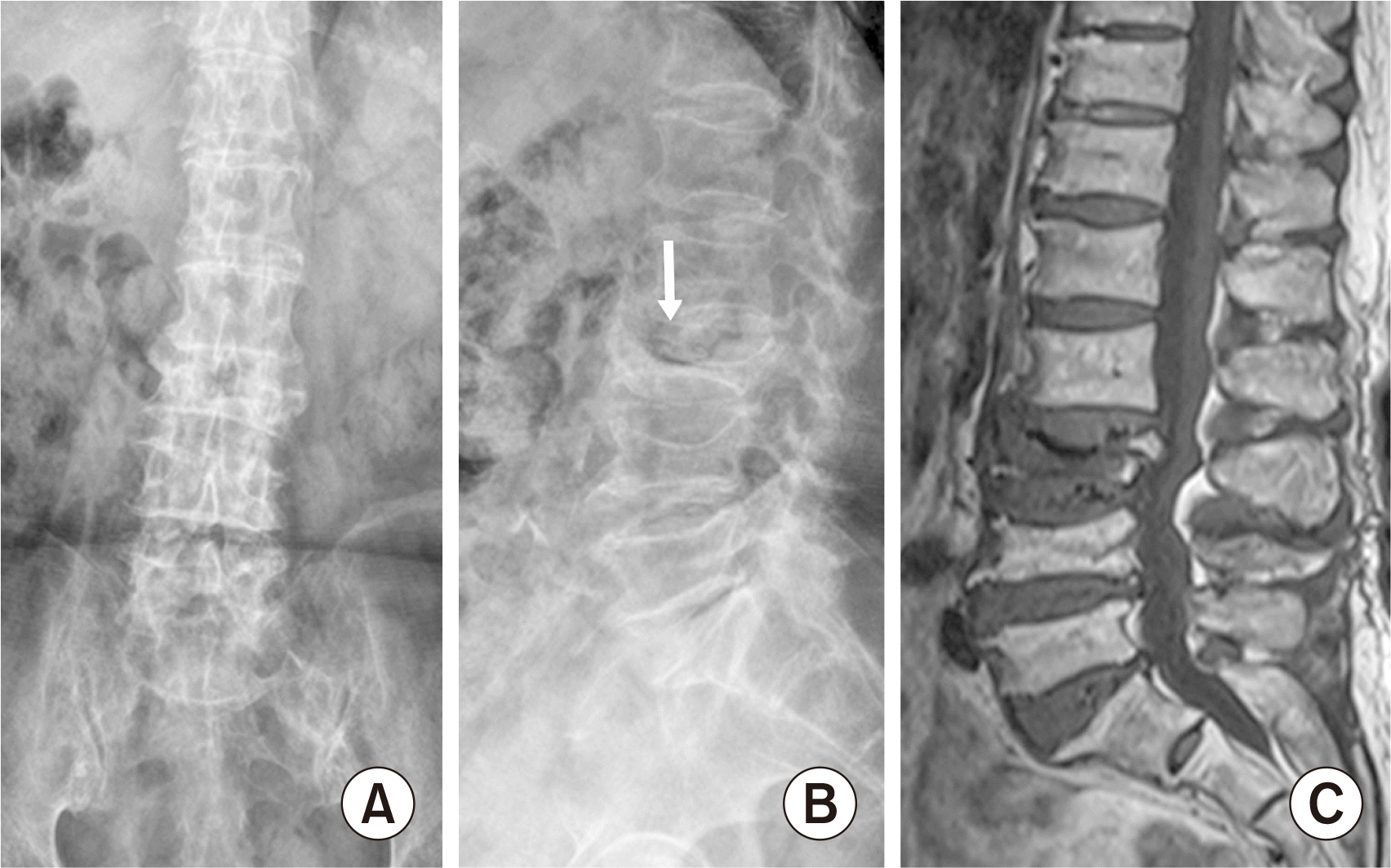 | Fig. 1Antero-posterior (A) and lateral (B) images of L spine plain radiography. They show severe vertebral collapse with vertebral vacuum cleft (arrow) at L3. T1-weighted lumbar MR image (C) shows low signal area in collapsed L3 vertebra. Informed consent for publication of the clinical images was obtained from the patient.
|
Despite our conservative treatments including right L3/4 transforaminal epidural steroid injections and bilateral facet joint blocks with medications such as NSAIDs, afloqualone, and gabapentin, she had little back pain relief (NRS 6–7). Therefore, we planned a PVP considering her age, life expectancy, and general conditions.
On the day of the procedure, she was given a prophylactic antibiotic (1 g cefazoline) and a durogesic patch was attached for analgesia. She was laid in prone position on the operating table (Spinal Table HM 604; Handok Medical, Seoul, Korea). We monitored her blood pressure, heart rate, and oxygen saturation, and the area was prepared and draped in an aseptic method with betadine. After disinfecting the skin, skin infiltration with 1% lidocaine was performed, and a vertebroplasty needle (HMN-NVP1103; Hyun Medics, Bucheon, Korea) was inserted targeting the right L3 vertebra pedicle. We removed the inlet cannula carefully and intentionally waited for about 5 minutes expecting the spontaneous reexpansion of the height of the vertebral column. Indeed, we could find the phenomenon of spontaneous recovery of the collapsed vertebral body (Fig. 2). Total 3 mL of polymethylmethacrylate (Spinefix; Teknimed, Vic-en-Bigorre, France) was injected into the vertebral body by split up into 1 mL, respectively. Shortly after polymethylmethacrylate injection, manual compression was done at the injection site for 15 minutes by loading the operator’s weight not only to control bleeding but also to make the L3 body firm and facilitate its expansion.
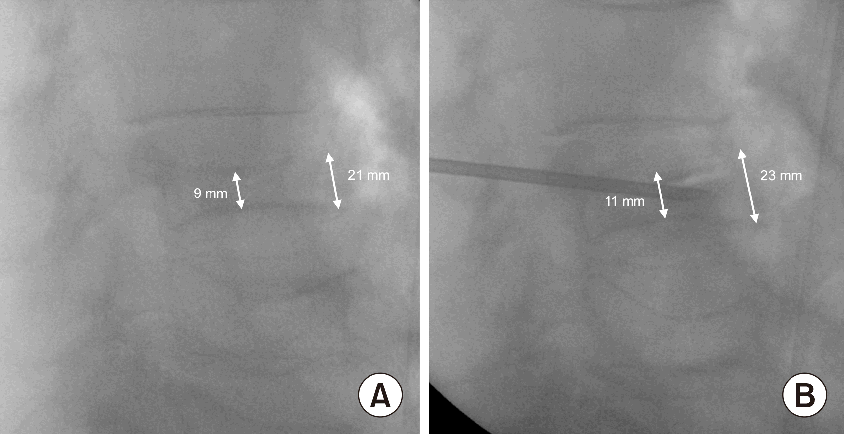 | Fig. 2The lateral image of C-arm fluoroscopy. There is the phenomenon of spontaneous recovery of the collapsed vertebral body. The height of the vertebral column increases from 9 to 11 mm centrally and from 21 to 23 mm anteriorly (A, B). Informed consent for publication of the clinical images was obtained from the patient.
|
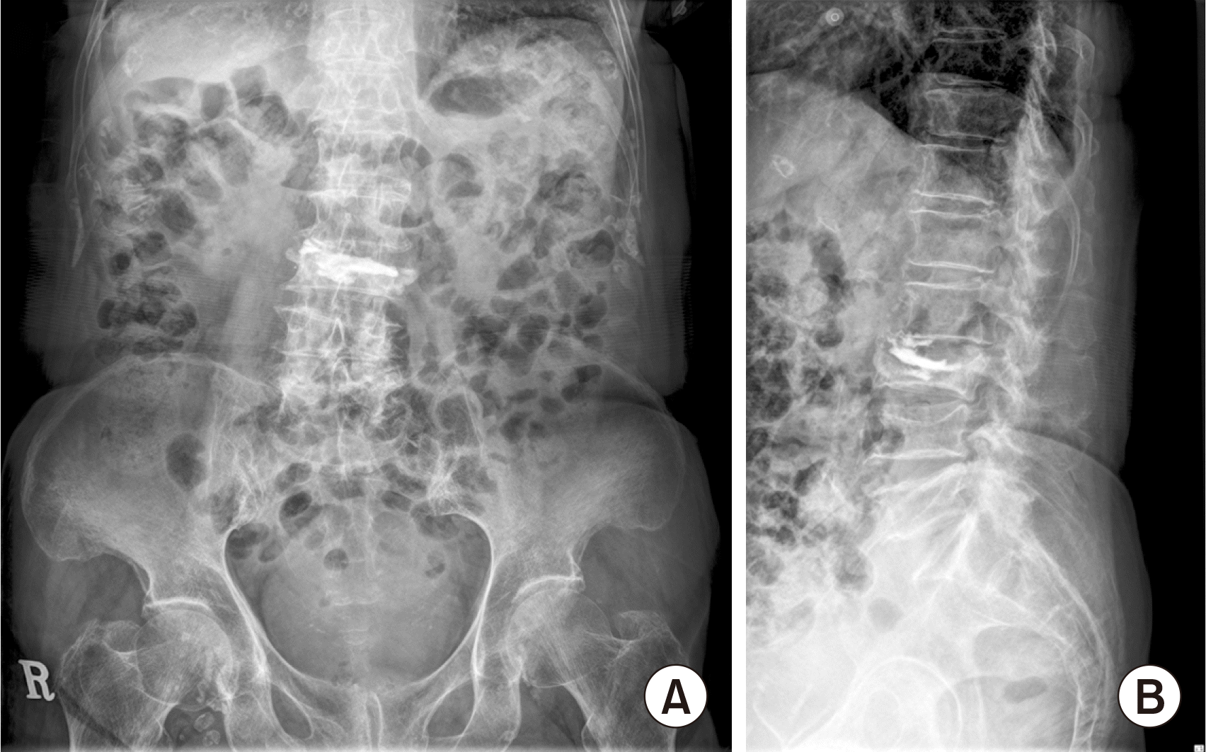
PVP was completed by fluoroscopically confirming that there was no epidural leakage or complication (Fig. 3); a dressing was applied at the injection site, and the patient was discharged on the day of the procedure without neurological complications. One week later, the patient visited our pain clinic and reported greater comfort while walking, and her radiculopathy was reduced from 7–8 to 2 according to NRS scores.
Go to :
Discussion
Kummell’s disease can also be referred to as ‘avascular necrosis disorder in intravertebral column’, ‘vertebral pseudarthrosis’, and ‘delayed disorder of collapsed vertebra’. It is typically indicated by low-density air shadow in lumbar spine plain radiographs, intravertebral vacuum in CT, and vertebral fractures with vacuum cleft filled with liquid or air on low-signal T1- and T2-weighted images in MRI [3,5,13]. Although its clinical manifestation is similar to that of osteoporotic vertebral fracture, Kummell’s disease is considered that the decrease of shear stress and causing tiny vertebral fracture by insufficient blood supply [1]. Apart from this, Kummell’s disease often causes low back pain and radiating pain as it is accompanied by intravertebral body instability and spinal canal or foraminal stenosis [1,4].
Conservative treatment is the primary treatment for patients with Kummell’s disease [14]; bracing, bed rest, and taking medications, including analgesics, NSAIDs, and anticonvulsants; however, despite these measures, our patient’s condition did not improve. Moreover, we performed epidural steroid injections and facet joint blocks; however, even this offered little pain relief. Therefore, we considered more invasive procedure. Our patient was extremely old and was not suitable for general anesthesia due to cerebrovascular or cardiovascular complications. Further, the patient refused to provide consent for open surgery. We planned to perform PVP to reduce pain and correct the vertebral body deformity for treatment.
PVP or PKP is a good treatment modality for patients with Kummell’s disease who are unsuitable for operation under general anesthesia like our patient was, and it may also have an analgesic effect and improve patients’ quality of life [3,4,6,15]. Although several studies have reported no statistically significant differences in pain relief between PVP and PKP, with the advantages of shorter operating time, less expensive than PKP [6,7], PVP is the preferred choice. In treating patients with Kummell’s disease, the goal is to restore the fractured vertebral height when performing the procedure [14]. Therefore, we considered several methods to increase the height of the vertebral body in PVP, and some reports were as follows.
First, based on reports stating spontaneous recovery of the vertebral body occurs with a popping sound as soon as the needle was inserted during PVP or PKP [5,8], we expected that effect of decompression by producing negative pressure as removing the inlet from needle into vertebral column where is filled with air or liquid the features of Kummell’s disease. Therefore, we did not inject bone cement immediately after inserting the needle and rather waited for 5 minutes for the vertebral body to spontaneously decompress via vacuum cleft occurring negative pressure while the air flow with the inlet is removed. We also observed an immediate effect and found that the height of the vertebral body spontaneously recovered on just inserting the needle without any bone cement injection. After checking the re-expansion of the height of the vertebral column, bone cement was very carefully injected and the injection was confirmed by fluoroscopic guided images. Hur et al. [5] described that spontaneous reduction requires a vacuum cleft with an intact vertebral wall, which maintains a negative pressure within the cleft. As mentioned in the previous case report, this was rare cases so there is no consensus on time to allow spontaneous recovery of vertebral body after removing stylet of cannula. We expect that this method can be apply to avascular necrosis such as Kummell’s disease, however, further studies need to be undertaken to verify the precise mechanism of this phenomenon and specific indication of this method.
Second, during PVP, Carlier et al. [12] reported that applying manual pressure directly on the top of the kyphosis may also help impose hyperlordosis. And, Wenger et al. [11] reported that manual pressure with cautious axial traction and posterior-to-anterior pressure on the spine may be a good option to effectively reduce fractures of the lumbar spine in prone and hyperlordotic positions. In addition, manual compression is usually applied to the puncture site for 5 to 10 minutes after needle removal to minimize bruising and hematoma formation [9,10]. In line with the recommendations of these studies, we gently performed manual compression with loading of operator’s weight to the patient in anticipation of bleeding control and vertebral body reexpansion.
In conclusion, we can treat Kummell’s disease effectively by performing PVP including combination therapy with negative pressure that re-expand intravertebral body and manual forceful compression at the needle insertion site. Although further research will be needed in the future, we recommend the mentioned combination therapy based on the successful execution of this case.
Go to :
 XML Download