

 PDF
PDF ePub
ePub Citation
 Print
Print


INTRODUCTION
Docosahexaenoic acid (DHA), an important n-3 long chain polyunsaturated fatty acid (LCPUFA), is involved in a range of physiological functions across tissues that affect the human lifespan. During the perinatal period, DHA indispensably contributes to brain, neuron and retinal development; irreversible defects occur in its absence [123]. Furthermore, DHA is essential for normal spermatogenesis, as a deficiency causes male infertility [4]. With the substantiated functions in hypo-triglyceridemia, anti-coagulation, vasodilation, and anti-inflammation, DHA protects against cardiovascular diseases [567]. Moreover, it has been consistently reported that decreased DHA concentrations in the human brain and peripheral tissues are associated with a range of brain disorders, including cognitive and learning impairment, depression, bipolar disease, and attention deficit hyperactivity [891011].
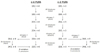
In addition to being derived from eating marine-based foods, DHA can be synthesized endogenously by conversion from the essential fatty acid α-linolenic acid (C18:3, n-3). This involves alternating desaturation and elongation reactions shared between n-6 and n-3 PUFA. Delta-6/delta-4 desaturase (D6D) and delta-5 desaturase (D5D), which are encoded by the Fads2 and Fads1 genes, respectively, dehydrogenate on the assigned carbon [12]. Elongase 5 (Elovl5), elongase 2 (Elovl2), and elongase 4 (Elovl4) target substrates with various chain lengths (C18/C20, C20-24 and C26-34, respectively) [13]. In 1991, a circuitous pathway (coupled microsomal-peroxisomal pathway) was suggested [14] and confirmed in fibroblast cultures from patients with peroxisome biogenesis defects [1516]. In that pathway, C20 was elongated consecutively to C24 and terminated by a single round of peroxisomal β-oxidation after delta-6 desaturation (Fig. 1).
Among the genes participating in LCPUFA biosynthesis, Fads1, Fads2, Elovl5, plus Scd1 which is responsible for introducing a double bond at the delta-9 position of C16:0 and C18:0, are positively regulated by peroxisome proliferator-activated receptor alpha (PPARα) and sterol regulatory element-binding transcription factor 1c (SREBP-1c) [171819]. In the last step in the circuitous pathway (peroxisomal β-oxidation), the rate limiting enzyme acyl-CoA oxidase (encoded by Acox), as well as enoyl-CoA hydratase/3-hydroxyacyl-CoA dehydrogenase and thiolase, are well-recognized PPARα target genes [20]. On the other hand, the importance of PPARα in DHA biosynthesis is unclear. Conflicting results for tissue DHA accretion have been achieved when PPARα was activated pharmacologically [212223]. Thus far, no DHA deficiency has been reported in PPARα KO mice. Despite the lack of consensus in the relative importance of these two apparently redundant pathways, the significance and non-compensation for the circuitous pathway are supported by the Zellweger syndrome decreasing the DHA concentrations dramatically [1415] and Elovl2 KO male mice being infertile due to a DHA deficiency [13]. Based on the notion that the PPARα activity is strongly correlated with peroxisomal β-oxidation, this study examined the role of PPARα on DHA biosynthesis, because DHA-containing food is not widely available for many persons.
To this aim, two experiments were conducted: mice with differential PPARα levels (+/+, +/− and −/− genotypes; Exp I) and activities (± PPARα agonist; Exp II). An n-3 PUFA depleted/replenished regimen was used to exclude the confounding effects of DHA passing from the mothers, via the placenta and milk. This n-3 PUFA depleted/replenished regimen has two advantages: 1) to ensure equal basal levels at the beginning (n-3 depletion); and 2) once the DHA precursor is provided, these depleted mice promptly start n3-LCPUFA synthesis. In addition to the hepatic mRNA levels of the enzymes involved in DHA biosynthesis, the tissue DHA and its associated functional proteins were measured as the outcome parameters.
MATERIALS AND METHODS
Study design
In Exp I, PPARα −/− (KO), +/− (HZ) and +/+ (WT) mice were used to test the effects of the PPARα protein levels on tissue DHA accretion. For groups KO, HZ, and WT, there were eight mice (males: females = 1:1) in each group. To deplete the tissue DHA concentrations in neonates, mice were born and nursed by dams eating a sunflower oil diet (deficient in the DHA precursor, α-linolenic acid). After weaning (3 weeks of age), the pups were fed a soybean oil diet (sufficient in α-linolenic acid) to promote DHA biosynthesis. Four weeks later (i.e. seven weeks of age), they were sacrificed by carbon dioxide asphyxiation. Aliquots of the liver and brain cortex were quick-frozen in liquid nitrogen and stored at −80℃ for RNA extraction. A portion of the liver and brain cortex were stored at −20℃ for fatty acid analysis.
In Exp II, to test the effects of PPARα activation on tissue DHA accretion, WT mice were used and an n-3 PUFA depleted/replenished regimen was applied. After weaning, the pups were fed a soybean oil diet, with or without 0.5% (wt./wt.) clofibrate (CF; TCI, Tokyo, Japan), a PPARα agonist. The control (C) and CF groups contained 16 mice (males: females = 1:1) in each group. The mice were sacrificed at seven weeks of age. The liver, brain cortex, and retina were collected and stored at −20℃ (for fatty acid analysis) or −80℃ (for RNA and protein extraction).
To verify n3-PUFA depletion by the sunflower oil diet (i.e., basal fatty acid profiles), no additional batches of animals were used considering the 3Rs of animal welfare. Instead, three neonates (each from WT, KO, and HZ group, respectively) selected randomly were sacrificed at weaning for fatty acid analysis in the liver. No α-linolenic acid, EPA and DHA were detectable with a detection limit ≥ 0.1%.
Mice breeding, genotyping and diet
Heterozygous PPARα mice were produced by mating homozygous PPARα-null (B6;129S4-Pparatm1Gonz/J) male mice with a pure C57BL/6J genetic background, and their wild-type female counterparts, which were both purchased from Jackson Lab (Bar Harbor, ME, USA). When these heterozygous mice were at least eight weeks of age, they were mated to produce KO, HZ and WT (expected ratio, 1:2:1) genotypes, which was confirmed by PCR of mouse tail DNA, according to the instructions from Jackson Lab (Fig. S1). The litter size was adjusted to 6–8 with equal numbers of both sexes.
The composition of diets followed the AIN93M [24] with dietary fat modified from 4 to 10% (wt./wt.) and a reciprocal reduction in corn starch. Dietary fat came from either sunflower oil (Taiwan Sugar Corporation, Tainan, Taiwan) with an n-6/n-3 ratio > 200, or soybean oil (President, Tainan, Taiwan), with a n-6/n-3 ratio ≅ 7 because a high n-6/n-3 ratio deteriorates the endogenous synthesis of DHA [25]. No dietary DHA was provided in the feed. Table S1 lists the feed composition. All mice were kept in a room maintained at 23±2℃ with a controlled 12-h light:dark cycle with access to food and drinking water ad libitum. The body weight was recorded weekly and the food intake was recorded every other day. The protocols for animal care and handling were approved by the Institutional Animal Care and Use Committee of China Medical University (IRB No. 104-130-N).
Lipid extraction, phospholipid isolation and fatty acid analysis
The total lipids were extracted from the tissues (liver, brain cortex, and retina) using a 2:1 mixture of methanol/chloroform. As phospholipids are dominant in the brain and neurons, the phospholipid fraction was isolated further from the total lipid of the brain cortex in Exp. II using an SPE column (CHROMABOND NH2) (Macherey-Nagel GmbH & Co. KG, Düren, Germany). This was not feasible for retina lipids because there were only trace amounts of samples, even when pooled from four mice within a group. Extracts were taken to dryness and weighed to estimate the total lipid content. The total lipids or phospholipid fraction was subjected to transesterification by methanol/dichloromethane = 3/1, as described elsewhere [26], and the resulting fatty acid methyl esters were dissolved in n-hexane for fatty acid analysis in a Hewlett-Packard 5890 gas chromatograph with flame ionization detection on a SP-2380 fused silica capillary column (30 m × 0.25 mm× 0.2 µm; Supelco, Bellefonte, PA, USA) using nitrogen as the carrier gas (1.5 mL/min). The oven temperature program was set to 110℃ for 5 min, which was then increased at 10℃/min to 170℃, then at 3℃/min to 230℃, and held at 230℃ for 5 min. The fatty acid peaks were identified by a comparison of the retention times with authentic standards.
RNA isolation and mRNA detection
The total RNA was extracted from homogenized tissues (liver, brain cortex and retina) using an RNeasy Mini Kit (Qiagen, Germantown, MD, USA) according to the manufacturer's instructions. The RNA concentrations were determined using a NanoDrop (Thermo Fisher Scientific, Carlsbad, CA, USA). The total RNA (1 µg) was reverse-transcribed into first-strand cDNA using an iScript™ cDNA Synthesis Kit (Bio-Rad, Hercules, CA, USA) according to the manufacturer's instructions. For real-time PCR, a SYBR system with self-designed primers was used (Table S2). The 15 µL reaction mixture contained 12.5 ng cDNA, 20 pmol primers, and 1× SYBRGreen. Amplification using 40 cycles of two steps, i.e., 95℃ for 15 s and the assigned annealing temperature (see Table S2) for 1 min, was performed on an ABI Prism 7900HT sequence detection system.
The measured genes associated with the functions of DHA in the brain and retina are explained below. Major facilitator superfamily domain-containing protein 2 (encoded by Mfsd2a) is a transporter for DHA across the blood-brain or blood-retinal barrier [2728]. Brain-derived neurotrophic factor (encoded by Bdnf) and nerve growth factor (encoded by Ngf) are neurotrophins with expression regulated differentially by n-3 PUFA [29], and tropomyosin receptor kinase B (encoded by Ntrk2) serves as a BDNF high-affinity receptor [30]. Transcription factors paired box 6 (encoded by Pax6) and cone-rod homeobox (encoded by Crx) are critical for retinal development [31]. Opn1sw, Opn1mw, and Opn1lw, encode short-, middle- and long-wavelength opsins in cone cells, respectively. Rho encodes rhodopsin in rod cells.
BDNF protein
The BDNF concentrations in the brain cortex were measured using ELISA kits (R&D, Minneapolis, MN, USA). Tissue homogenates were prepared in RIPA buffer [50 mM Tris buffer, pH7.4, containing 150 mM NaCl, 1 mM EDTA, 1% NP-40, 0.25% sodium deoxycholate, 0.1% sodium dodecyl sulfate] and 1% protease inhibitor cocktail (Sigma P8340, St. Louis, MO, USA).
Statistical analyses
All values are expressed as the mean ± standard deviation (SD). The differences among the groups in Exp I were determined using a one-way ANOVA and Duncan's multiple range test, whereas comparisons between the two groups in Exp II were analyzed using a Student's t-test (two-tailed). Data were checked for the normality of distribution. When the variances were not homogeneous, the data were log-transformed prior to statistical analysis. The General Linear Model package (SAS Institute, Cary, NC, USA) was used for all statistical analyses and P-values < 0.05 were considered significant.
RESULTS
PPARα deficiency on tissue DHA accretion (Exp I)
A PPARα deficiency had no influence on the growth and food intake; the body weight and food intake were similar in the three groups (WT, HZ and KO) across the experimental interval (data not shown).
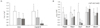
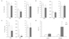
In accordance with a PPARα deficiency, the hepatic mRNA levels of the PPARα target genes, Acox and Cyp4a10 were reduced significantly in the HZ and KO mice compared to the WT mice, with a genotype-dependent effect evident for Cyp4a10 (Fig. 2A). For hepatic desaturases (Fads1, Fads2 and Scd1) and elongases (Elovl2 and Elovl5), the PPARα deficiency reduced the mRNA levels of Fads2, Fads1, and Scd1, but not Elovl2 and Elovl5 (Fig. 2B).
Regardless of whether it was in the liver or brain cortex, there was no difference in total lipid concentration among the WT, HZ and KO groups (22 ± 8 mg/g in liver and 55 ± 18 mg/g in brain cortex) (Table S3). The fatty acid composition of the total lipids extracted from the liver and brain cortex are shown (Table S3). Attention was paid to three LCPUFA, DHA, eicosapentaenoic acid (EPA), and arachidonic acid (AA)%. Hepatic DHA and AA were reduced significantly in KO but not in HZ mice; there were only 50% of those in the WT and HZ counterparts (Fig. 3A). On the other hand, in the brain cortex, KO reduced only AA, but there were no differences in DHA and EPA among the three genotypes (Fig. 3B).
PPARα activation on tissue DHA accretion and function (Exp II)
Clofibrate reduced the body weight and food intake during the treatment period (postweaning). Hepatomegaly, a hallmark of the peroxisome proliferator effect in rodents, was present in the CF group at the end point (6.7 ± 0.4 vs 4.1 ± 0.4%, P < 0.05; liver/body weight ×100% of CF and C group), confirming PPARα activation.
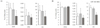
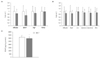
In accordance with PPARα activation, the hepatic mRNA levels of Acox and Cyp4a10 were increased significantly by CF with 1.8- and 3.4- fold induction, respectively (Fig. 4A). With the exception of Elovl2, CF increased the mRNA levels of all desaturases and elongases to varying degrees (Fig. 4B). In brain cortex, however, the mRNA levels of these enzymes were unaffected (data not shown).
The total lipid concentration in the liver of group CF was 40% lower than that of group C (Table S4). No difference in the total phospholipid concentration was found in the brain cortex of the two groups. The total lipid content in the retina was not measured because of the trace amounts of specimen. The fatty acid composition in the total lipids of the liver and retina, as well as in the phospholipid of the brain cortex, are shown (Table S4). Treatment with CF caused 18, 24, and 55% decreases in the DHA% in the liver (Fig. 5A), brain cortex (Fig. 5B), and retina (Fig. 5C), respectively. Hepatic EPA and AA% were also reduced by CF. To test the fatty acid catabolism, the expression of genes participating in mitochondrial or peroxisomal β-oxidation were measured (Fig. 5D). The hepatic mRNA levels of Cpt1a (encoding carnitine palmitoyltransferase 1A) and Ehhadh (encoding enoyl-CoA hydratase/3-hydroxyacyl-CoA dehydrogenase or L-bifunctional enzyme) were greater in the CF group than in the C group.
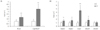
For the genes associated with DHA transportation and function, the mRNA levels of Mfsd2a, Ngf and Ntrk2 in the brain cortex (Fig. 6A), as well as Mfsd2a, Pax6, Crx, Opn1sw, Opn1mw, Opn1lw, and Rho in the retina (Fig. 6B) were similar in the control and CF groups. On the other hand, there was a significant decrease in the transcript (Fig. 6A) and protein levels (Fig. 6C) of BDNF in the brain.
DISCUSSION
Although the rate of α-linolenic acid conversion to DHA is quite low in humans (estimated to be < 0.1% in men and 9% in women) [32], it is important for people with limited access to marine foods or DHA supplements. This study focused on PPARα impacts on DHA (and other LCPUFA) accretion because the PPARα activity can be altered by factors in foods, medicines, and gene polymorphisms. As expected, expressions of the enzymes involved in LCPUFA synthesis were down- or up-regulated, respectively, with a PPARα deficiency or activation. On the other hand, both scenarios led to reduced DHA accretion. Among the proteins associated with DHA transport or function in the brain and retina, there were lower levels of neurotrophin BDNF mRNA and protein in an n-3 PUFA depleted/replenished model of CF-treated mice.
PPARα could be regarded as a cellular fatty acid sensor, which upon activation, increased not only transport and β-oxidation of fatty acids, but also lipogenesis (with collaboration with SREBP-1c) to protect the liver from cytotoxicity due to the influx of fatty acids under common stresses, e.g., fasting [21]. A battery of genes involved in LCPUFA synthesis, Fads1, Fads2 and Elovl5, plus Scd1, are upregulated by PPARα and SREBP-1c concomitantly [17181933]. Even the two transcription factors have reciprocal roles on the lipid metabolism (catabolism for the former and anabolism for the latter); this paradox was explained by a promise of LCPUFA production, regardless of the energy state [19]. Their mRNA change patterns were closely related to a PPARα deficiency or activation, even though the constitutive level of Elovl5 was not altered by PPARα ablation. In accordance with the suppressed Acox, Fads1, and Fads2 expression, liver n-6 and n-3 LCPUFA precursor (C18:2, n-6 and C18:3, n-3) accumulated, whereas the chain length ≥ C20 (C20:3, n-6; C20:4, n-6; C22:4, n-6; C22:5, n-6; C22:5, n-3 and C22:6, n-3) were depleted in HZ and KO mice (Table S3). In the case of PPARα activation, in response to up-regulated Acox, Fads1, Fads2, and Elovl5, C18:2, n-6 and C18:3, n-3 decreased, as expected, with variable changes in the down-stream LCPUFA (Table S4 and below).
Reduced tissue DHA accretion mediated by CF administration was in accordance with a previous report [23] showing that under the conditions of increased mitochondrial and peroxisomal proliferation, a relatively higher β-oxidation rate of n-3 LCPUFA in the mitochondria and peroxisome might decrease the hepatic concentrations of EPA and DHA. In accordance with this notion, 1.5-, 2-, 5.5-fold greater expression of Cpt1a (indicator of mitochondrial β-oxidation), Acox and Ehhadh (both indicators of peroxisomal β-oxidation), respectively, as well as 3.8-fold greater Cyp4a10 expression (indicator of microsomal ω-oxidation) were observed in the CF group than in the C group, suggesting that fatty acid clearance was accelerated by CF. DHA-CoA was a very poor substrate for mitochondrial carnitine acyltransferase-I and -II but the peroxisomal DHA-CoA oxidase increased dramatically after the peroxisome proliferator treatment [23]. Madsen et al. [23] reported the increased combustion of DHA upon PPARα activation beyond the increased biosynthesis, thereby contributing to lower accretion. On the other hand, more than simply improving the systemic lipid profile, Tian et al. [22] attributed the cardioprotective effects of CF partially to altered myocardial fatty acid composition, namely an increase in DHA and decrease in n-6 PUFA, resulting in a lower n-6/n-3 ratio. In contrast to CF-reduced concentrations of DHA in the liver, brain cortex, and retina detected in this study, whether CF has a distinct effect from other tissues on the cardiac DHA uptake or utilization remains unknown.
The conversion of α-linolenic acid to DHA is greater in women than in men, an effect generally attributed to estrogen [34]. The reduced partition of α-linolenic acid to β-oxidation (hence, more availability of LCPUFA substrates), greater translocation of C24 from the endoplasmic reticulum to peroxisome, or higher desaturase/elongase/peroxisomal β-oxidation enzyme activity in women compared to men was proposed [3235]. On the other hand, this sexual dimorphism on DHA accretion or enzyme activity was not apparent in the present study. A stratum of gender was conducted in this study but no significant gender effects were noted (data not shown). This might be due to the age at study termination (seven weeks, before full sexual maturity). Greater expression of Fads1 and Fads2 were reported in female rats than male rats at 14 weeks of age fed a standard chow diet [34].
DHA accounts for approximately 50–60% of the total fatty acid content within the rod outer segments of photoreceptors [36]. Although CF caused a 55% reduction in retinal DHA, the gene expression profile of the retina examined in this study was unaffected. PAX6 and CRE are indispensable for the proliferation and differentiation of photoreceptors [3738], and DHA is a molecular cue for achieving complete differentiation [31]. In this study, the lack of difference in the Pax6 and Cre transcripts between the C and CF groups may be due to the difficulty in detecting the differential expression of the two transcription factors beyond the early stages of retinal development (perinatal period). In studies conducted in vitro, DHA enhanced the level of opsin expression [31], even though in the present study, there was no reduction in mRNA levels of Opn1sw, Opn1mw, Opn1lw, and Rho in the CF-treated mice. DHA contributes to the brain, neuron, and retinal function by the structural and signaling roles. DHA not only influences retinal gene expression by acting as a ligand for nuclear hormone receptors, it also activates the membrane-bound retinal proteins via membrane fluidity, affects the rhodopsin regeneration/stability/kinetics partly by direct binding to rhodopsin (lipid-protein interaction), and influences cellular differentiation and survival in many ways [363940]. Mounting evidence suggests that reduced retinal DHA impaired the retinal function. Abnormal electroretinograms were recorded across species (mice, rats, guinea pigs, cats, and rhesus monkeys) when the retinal DHA decreased ≥ 30% [36].
The CF-mediated BDNF reduction in brain is believed to be secondary to a lower DHA concentration in the brain cortex, rather than a direct effect of CF on BDNF expression. DHA upregulates the expression of BDNF and other neurotrophins and their receptors [29]. The tight junction between capillary microvessels and the expression of ATP binding cassette efflux transporters in the blood-brain barrier make the brain impermeable to xenobiotics from circulation [41]. Clofibrate cannot cross this barrier. The reduced brain DHA accretion could be due to lower hepatic DHA levels. In line with the notion, the mRNA levels of the enzymes participating in LCPUFA synthesis (i.e. Fads1, Fads2, Elovl5, and Elovl2) in the brain cortex were unaffected by CF administration.
The CF-mediated reduction in BDNF in the brain cortex was consistent with previous clinical studies, suggesting a linkage between cholesterol-lowering agents and depression and suicidality [4243]. Epidemiological studies have described an association between lower serum cholesterol concentrations and increased suicide risk that could not be attributable entirely to depression-related malnutrition and weight loss [4445]. Meta-analysis suggested that lowering cholesterol could cause or worsen the depressive symptoms and increase the risk of suicide [46]. On the other hand, there were inconsistent findings in that low cholesterol concentrations in depressed patients and suicidal victims might not be a direct factor [47]. Indeed, the result appeared to be contradictory to the context that PPARα has neuroprotective and neurotrophic effects based on its requirement for the hippocampal expression of plasticity-related molecules [48]. Recently, it was suggested that PPARα agonists may benefit memory/learning or have antidepressantl-ike effects by promoting the BDNF signaling cascade [4950]. WY14643 and fenofibrate (PPARα agonists) individually were confirmed to restore, PPARα-dependently, the acetylcholine receptor antagonist- or chronic social defeat stress-induced reductions in hippocampus BDNF, phosphorylation of MAPK-ERK, PI3K-Akt and CREB, as well as adult hippocampal neurogenesis in mice. The discrepancy might be caused by differences in experimental models, treatment agents or duration. Of course, the impact of lower BDNF on BDNF signaling will require further study.
A particular strength of this study was the use of an n-3 PUFA deleted/replenished model to eliminate the influence of maternal DHA, thereby facilitating a determination of bioconversion and tissue accretion of DHA. On the other hand, care should be taken when interpreting the CF-reduced DHA accretions observed in this study. Because the CF effects among PPARα KO, HZ and WT mice were not compared, the reductions of DHA and BDNF mediated by CF could not be ascribed solely to PPARα activation. CF might have effects other than PPARα activation; at least in a rodent liver, CF has been demonstrated to induce Cyp2b family members that are known to be regulated by the constitutive-activated receptor [51].
For future perspectives, functional tests (e.g. electroretinograms, cognitive or behavior tests), particularly with suppressed DHA accretion across tissues in CF-treated mice, should be conducted to determine the physiological significance or relevance. Moreover, this study raised a concern that DHA concentrations should be monitored when a fibrate-class hypolipidemic medicine is prescribed chronically to people with a low DHA intake.
Overall, the current study confirmed that the transcription of enzymes participating in LCPUFA biosynthesis, including Fads1, Fads2, Scd1, Elovl5, and Acox were changed in accordance with PPARα. On the other hand, tissue DHA accretion decreased in two opposing PPARα scenarios, i.e., ablation and activation. The significance in functional impairments mediated with a PPARα deficiency or activation awaits further study.
 XML Download