

 PDF
PDF ePub
ePub Citation
 Print
Print


INTRODUCTION
Oxidative stress, inflammation and vascular dysfunction are major pathological features of the atherosclerotic process, distinctly observed in cerebro- and cardiovascular diseases [123]. Excess free radicals (i.e., reactive oxygen species, ROS) produced in the body cause lipid peroxidation, protein alterations and DNA damages leading to oxidative stress, thereby stimulating various cytokine productions from inflammatory and immune cells, and finally dysregulating the vascular function [4567].
Many studies have endeavored to reveal natural candidate molecules which are capable of regulating the oxidative stress and inflammatory processes [891011121314]. Among the natural candidates, the abundant phytochemicals present in plant sources have attracted great attention because of their pleiotropic functions including antioxidant capacity, enzyme modulation and gene transcription [11121314]. Recently, it is reported that leaves and branches of Carpinus turczaninowii (C. turczaninowii) have antioxidant capacity and anti-inflammatory effects [151617]. As reported by Ko et al. [1516] the extract from C. turczaninowii branches inhibits the production of interleukin (IL)-6 and nitric oxide (NO) in RAW264.7 cells. Kang et al. [17] also reported antioxidant, skin-whitening (reduced cellular melanogenesis in murine melanoma cells), anti-inflammation (reduced NO production in RAW264.7 cells) and antibacterial properties of the C. turczaninowii leaf extract. The roots and leaves of C. turczaninowii have been used for improvement of fatigue from excessive physical labor as well as the treatment of bruises or abscesses [from Illustration book of Korean medicinal herb]. The species C. turczaninowii is a deciduous tree belonging to the Betulaceae family, found endemic in Korea (Korean hornbeam) [1819]. Although few studies have identified chemical compounds from C. turczaninowii branches, these are limited to naringenin and quercetin glycosides which belong to flavonoids [18], and carpinontriols A and B which have a diarylheptanoid skeleton bearing on carbonyl and five hydroxyl functional groups. The compounds isolated from the extract are potential pharmaceuticals agents [1516].
Contrarily, there are no studies determining the regulatory effect of C. turczaninowii extract on the arterial inflammatory response, which is a major pathological process observed in atherosclerosis [123]. Therefore, this study was undertaken to investigate if C. turczaninowii extract regulates the arterial inflammatory response, using human aortic vascular smooth muscle cells (HAoSMCs).
MATERIALS AND METHODS
Preparation of Carpinus turczaninowii extract
Complete plants (branches, leaves, bodies and roots, etc.) of C. turczaninowii were collected in January 2015 from Suin Mountain in GangjinGun, Korea, and identified by Heung Su Lee, the plant taxonomist and curator at the Natural History Museum of Hannam University, Daejeon, Korea. A voucher specimen was deposited at a publicly available herbarium (specimen deposition number: NIBRVP0000519846). The plant material was air-dried, ground and extracted three times with 70% ethanol for 24 h at room temperature. The extract was filtered, evaporated under reduced pressure, and freeze-dried to obtain a powder. It was stored in a deep freezer (−80℃) until further testing. For experiments, the lyophilized extract powder was dissolved in 70% ethanol (final concentration: 30 mg/mL). The extract stock solution was filtered through 0.2 µm Minisart® syringe filter (Sartorius Stedim Biotech GmbH, Goettingen, Germany) for molecular experiments. The filtered extract stock solution was then aliquoted and stored at −80℃ till further analysis.
DPPH free radical scavenging assay
2,2-diphenyl-1-picrylhydrazyl (DPPH; Sigma-Aldrich Co., LLC., St. Louis, MO, USA), L-ascorbic acid (Tokyo Chemical Industry Co., Ltd., Tokyo, Japan) and ethanol (Duksan pure chemical Co., Ltd., Ansan, Korea) were used for the DPPH free radical scavenging assay. This assay was performed as per the method described by Brand-Williams et al., with slight modifications [20]. The antiradical activity of ascorbic acid (50 µM) was used as the positive control. Briefly, 1 mL control + 1 mL 70% ethanol was used as the control solution (Ac), whereas 1 mL DPPH solution (290 µM) + 1 mL 70% ethanol was used as the blank solution (A0). For the assay, 1 mL DPPH solution (290 µM) was mixed with 1 mL varying concentrations of the sample (As) (5–100 µg/mL). For the assay, all mixtures were shaken and stood at room temperature in the dark before measurement (up to 5 min). The absorbance was measured at 517 nm, every 5 min for 40 min, using the UV-Visible spectrophotometer (OPTIZEN POP, Mecasys Co., Ltd., Daejeon, Korea). All experiments were repeated four times. The scavenging activity of DPPH radicals was calculated by the following equation:
Quantification of total phenolic content
Folin-Ciocalteu reagent, gallic acid (Sigma-Aldrich Co., LLC., St. Louis, MO, USA) and sodium carbonate (Acros Organics Co., Ltd., NJ, USA) were applied for quantifying the total phenolic content (TPC) in the C. turczaninowii extract solution, using a modified method from that described by Deshpande & Cheryan [21]. Serial dilutions of gallic acid (0.5–1,000 µg/mL) in 70% ethanol were used as standard solutions. The absorbance was measured using a microplate absorbance reader (iMark™, Bio-Rad Laboratories, Hercules, CA, USA). The wavelength correction was set to 655 nm. Total phenolic content of the C. turczaninowii extract solution is expressed as gallic acid equivalents (mg of GAEs/g of extract). All experiments were repeated four times.
Cell culture
Primary human vascular aortic smooth muscle cells (HAoSMCs) (American Type Culture Collection, ATCC, Manassas, VA, USA) were maintained in a humidified atmosphere at 37℃ with 5% CO2, and cultured in VSMC basal medium supplemented with 5 ng/mL rhFGF-basic, 5 µg/mL rhInsulin, 5 ng/mL rhEGF, 10 mM L-glutamine, 50 µg/mL ascorbic acid, 5% fetal bovine serum, 10 µg/mL gentamicin, 10 Units/mL penicillin, 10 µg/mL streptomycin, 0.28 µg/mL amphotericin B, and 33 µM phenol red (ATCC, Manassas, VA, USA).
Cell viability assay
Cell viability was measured using the Cyto X™ Cell viability assay kit (LPS solution, Daejeon, Korea), with a slight modification of the manufacture's protocol. The absorbance (A) was measured at 450 nm using the microplate absorbance reader (iMark™, Bio-Rad Laboratories, Hercules, CA, USA). All experiments were repeated four times.
Quantitative real time-PCR
Quantitative real time-PCR (qPCR) was performed to determine if the extract modulates mRNA expression of IL-6 and tumor necrosis factor-α (TNF-α). Briefly, HAoSMCs were treated with the extract (1 and 10 µg/mL) and incubated for 6 h. Total cellular RNA was extracted from the treated HAoSMCs using Trizol reagent (Invitrogen, Carlsbad, CA), following the manufacturer's protocol. Poly (A) was subsequently added using poly (A) polymerase (Ambion, Austin, TX). One Step SYBR® Prime Script TM RT-PCR Kit II (Takara, Japan) was used to conduct qPCR. The PCR was performed using the following primers (5′ to 3′): TNF-α (F): CGT CAG CCG ATT TGC TAT CT, (R): CGG ACT CCG CAA AGT CTA AG; IL-6 (F): GTT GCC TTC TTG GGA CTG AT, (R): CTG GCT TTG TCT TTC TTG TTA T, and β-actin (F): TCT GGC ACC ACA CCT TCT A, (R): AGG CAT ACA GGG ACA GCA C. Denaturing was carried out at 95℃ for 3 min, 40 cycles of 95℃ for 20 sec, annealing at 60℃ for 20 sec, and extension at 72℃ for 20 sec. At each 72℃ extension step, the fluorescence was measured at 585 nm. Expression of each factor was measured using an ABI prism 7500 Real-Time PCR System (Life Technologies Corporation, CA), and analyzed with comparative Ct quantification. β-actin was amplified as an internal control. The values are presented as relative quantity (RQ). All experiments were repeated four times.
Assessment of inflammatory markers in cell supernatants
HAoSMCs were plated at a density of 1.3 × 10⁴ cells/well in 24-well plates. At approximately 80% cell confluency, the culture media was changed to serum-free medium. Cells were subsequently stimulated with or without LPS (10 ng/mL) for 15 min, after which varying concentrations of the extract (1, 5, and 10 µg/mL) were added to the wells, and incubated for either 2 h or 24 h. After the appropriate incubation time, cell-free supernatants were collected. Concentrations of IL-6, TNF-α and soluble intracellular adhesion molecule-1 (sICAM-1) were measured using enzyme linked immunosorbent assay kits (PEPROTECH, Inc., Seoul, Korea), as per the manufacturer's protocol. The absorbance was read at 450 nm using the microplate absorbance reader (iMark™, Bio-Rad Laboratories, Hercules, CA, USA). All experiments were repeated four times.
Statistical analysis
Statistical analyses were performed using the Win SPSS ver22.0 (Statistical Package for the Social Science, SPSS Inc., Chicago, IL, USA). Data are expressed as the means ± standard deviation (SD) of four independent experiments. For descriptive purposes, the mean values are presented as untransformed values. The differences between groups were determined by independent t-test. Pearson correlation analysis was also performed. A two-tailed value of P < 0.05 is considered to be statistically significant.
RESULTS
Antioxidant activity and total phenolic content of C. turczaninowii extract
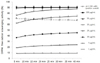
The antioxidant potential of C. turczaninowii extract was determined by the DPPH assay. As shown in Fig. 1, DPPH free radicals are scavenged by the C. turczaninowii extract in time-and dose-dependent manners. The average IC50 value for 40 min is 14.8 ± 1.97 µg/mL (i.e. from 17.5 IC50 value at 5 min to 12.4 µg/mL IC50 value at 40 min). Indeed, the radical scavenging activities of the extract at 100 and 50 µg/mL were found to be stably high at 40 min (average, 90.7 ± 0.03% and 89.7 ± 0.24%, respectively). The activities of the extract were similarly high and increasing in a time-dependent manner at 30 and 20 µg/mL (76.4–87.6% and 62.2–74.8%, respectively). Time-dependent increase in activity for 40 min was also observed for 10, 5 and 1 µg/mL (37.0–45.5%, 18.8–24.2%, and 8.8–9.4%, respectively). In addition, total phenolic content (TPC) was determined to be 225.6 ± 21.0 mg of GAE/g of the extract.
Cell viability
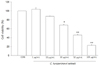
Cells viabilities were measured after 24 h incubation with varying concentrations of the C. turczaninowii extract (1–100 µg/mL). The cell viabilities of HAoSMCs incubated with 1 and 10 µg/mL of the extract were not significantly different from those of non-treated cells (control, CON) (about 104% and 88%, respectively) (Fig. 2), but cells incubated with 30, 50 and 100 µg/mL were dose-dependently reduced compared to CON (about 80%, 46%, and 23% viable, respectively). Based on the above result, further molecular experiments were performed using extract concentrations between 1 and 10 µg/mL.
Modulatory effect of C. turczaninowii extract on mRNA expression of pro-inflammatory cytokines in HAoSMCs
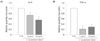
To examine if pro-inflammatory cytokine expressions are modulated by the C. turczaninowii extract, IL-6 and TNF-α mRNA levels expressed in HAoSMCs were measured at 6 h after the extract treatment (1 and 10 µg/mL). As shown in Fig. 3, exposure to the extract significantly suppresses the mRNA expressions of IL-6 and TNF-α in the cells.
Suppression of IL-6 and sICAM-1 production from LPS-stimulated HAoSMCs by C. turczaninowii extract
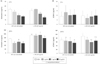
Fig. 4A and 4B present the effects of C. turczaninowii extract on the levels of IL-6 and sICAM-1 secreted from non-stimulated (CON) and LPS-stimulated HAoSMCs. Briefly, cells were treated with varying concentrations of the extract (1, 5, and 10 µg/mL) 15 min after stimulation with (or without) LPS (10 ng/mL), and further incubated for 2 h and 24 h. The IL-6 secretion was dose-dependently suppressed after 2 h incubation with the extract, at 1, 5 and 10 µg/mL in CON cells, and at 5 and 10 µg/mL in LPS-stimulated cells (upper panel, Fig. 4A). Similar patterns were observed after 24 h incubation with the extract (at 1, 5 and 10 µg/mL in CON cells, and at 10 µg/mL in LPS-stimulated cells) (lower panel, Fig. 4A). In addition, sICAM-1 production in both CON and LPS-stimulated cells was dose-dependently suppressed by the extracts after 24 h exposure, but not in 2 h incubation with the extract (Fig. 4B). Furthermore, TNF-α levels in CON or the LPS-stimulated cells were not detected at both 2 h and 24 h after exposure to the extract.
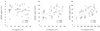
Relationship between IL-6 and sICAM-1 levels secreted from HAoSMCs treated with C. turczaninowii extract
Fig. 5 shows the relationship between sICAM-1 and IL-6 levels secreted from HAoSMCs after 2 h or 24 h incubation with the extract. After 2 h incubation, sICAM-1 levels did not significantly correlate with IL-6 levels (r = −0.094, P = 0.527). However, sICAM-1 levels after 24 h exposure to the extract were positively correlated to IL-6 levels at 2 h exposure (r = 0.418, P = 0.003) and 24 h (r = 0.524, P < 0.001).
DISCUSSION
Results from this study indicate that C. turczaninowii extract modulates the arterial inflammatory response in LPS-stimulated HAoSMCs, and can be a potential therapeutic agent for atherosclerosis. This is further supported by the fact that C. turczaninowii extract has a high amount of TPCs, and has a strong ability to scavenge the DPPH free radicals. Interestingly, the radical scavenging capacities show a time-dependent increase. Consequently, this high antioxidant capacity might be the basis of suppressing the inflammatory response in arterial cells, and thereby ameliorating the vascular function.
C. turczaninowii belongs to the Betulaceae family; it is a deciduous woody plant usually grown in the Jeju islands of Korea [19]. Very few recent studies have reported the antioxidant capacity and anti-inflammatory effects of leaves and branches of C. turczaninowii, thereby suggesting the potential use as a therapeutic agent for inflammatory diseases [15161722]. As reported in other studies [15161722], the current study also evaluates the antioxidant capacity of the C. turczaninowii extract. Oxidative stress caused by free radicals due to various intrinsic and extrinsic factors (cigarette smoking, metals, saturated fats, lipid peroxidation, glycated proteins, hyperglycemia, etc.) [34567] stimulate cytokine production from immune cells in circulation and body tissues [126], consequently leading to vascular dysfunction or directly harming the vascular endothelium, which thereby increases the risk of atherogenesis not only in the blood vessels, but also in the brain [14567].
Ko et al. [15] reported that IL-6 production from LPS-stimulated RAW264.7 cells (1 µg/mL LPS) was significantly reduced when treated with the extract of C. turczaninowii branches (50 to 500 µg/mL). They further suggested that from among the 8 compounds isolated from C. turczaninowii branches, carpinontriols A and B may be major contributors to higher antioxidant capacities [16]. Considering that NO is one of the inflammatory mediators, NO generation from the cells were slightly, but dose-dependently decreased, after 24 h C. turczaninowii extract treatment (25 to 200 µg/mL) [15]. Kang et al. [17] also showed dose-dependent reduction of NO production from LPS-stimulated RAW264.7 cells (1 µg/mL of LPS) after exposure to the extract (12.5 to 100 µg/mL). In addition, in their study of NO inhibition by 260 types of Jeju plant extracts, Yang et al. reported about 69% inhibition of NO and 80% viability of RAW264.7 cells after exposure to 100 µg/mL of C. turczaninowii extract [22].
The current study measured the mRNA levels and secreted concentrations of IL-6 and TNF-α from HAoSMCs. IL-6 and TNF-α are well-known pro-inflammatory cytokines that promptly respond to inflammatory stimuli [23]. In this study, increased productions of IL-6 from the non-stimulated cells were significantly and dose-dependently attenuated in 2 h after treatment of C. turczaninowii extracts, at concentrations of 1, 5 and 10 µg/mL. Interestingly, IL-6 production in the LPS-stimulated cells were also dose-dependently suppressed at 2 h after C. turczaninowii treatment (5 and 10 µg/mL). Furthermore, secretion of IL-6 from non-stimulated or LPS-stimulated cells were significantly suppressed at 24 h after treatment. Conversely, TNF-α levels from non-stimulated and LPS-stimulated HAoSMCs were not detected at both 2 h and 24 h after treatment, even though significant reduction of mRNA expressions of IL-6 and TNF-α were detected 6 h after exposure (1 and 10 µg/mL). These phenomena may be partly explained by the report of Schildberger et al. [24], which states that cytokine secretion patterns following LPS-stimulation differ according to the type of cytokine (i.e., TNF-α, IL-6, IL-8 and IL-1β) and cells (i.e., monocytes, peripheral blood mononuclear cells (PBMCs), and THP-1). For example, IL-6 secretion after LPS stimulation is not detected in THP-1 cells, but gradually increases in PBMCs (up to 500 pg/mL), and is gradually but promptly increased in monocytes (up to 600 pg/mL, peaks at 8 h and subsequently plateaus) for 24 h [24]. Secreted TNF-α levels were also greater in monocytes than in PBMCs and THP-1 cells. However, unlike IL-6, secreted TNF-α levels peaked at 4 h after stimulation, and subsequently decreased time-dependently, returning almost to baseline or non-detectable levels after 24 h. Furthermore, secreted TNF-α levels were not detected within 2 h after LPS stimulation [24]. Therefore, the report by Schildberger et al. [24] partly supports our results, where the secreted TNF-α levels were not detected at 2 h and 24 h after treatment, but mRNA levels of TNF-α were detected at 6 h post-exposure. This result indicates that the C. turczaninowii extract significantly suppresses the TNF-α production, together with IL-6 levels in human arterial cells. Secretion of pro-inflammatory cytokines may be mediated by nuclear factor kappaB (NF-κB) signaling in vascular cells [25]. Although we did not directly study the activation of NF-κB signaling in this study, our results may provide the possibility that C. turczaninowii extract modulates NF-κB activation, thereby attenuating pro-inflammatory cytokine expression in human arterial cells. Further studies are required for determining the modulation effect of C. turczaninowii extract on NF-κB signaling-mediated cytokine secretion in human arterial cells.
The circulating form of ICAM-1 (sICAM-1) is constitutively expressed or induced on the surface of various cells [26272829]. It is found in body fluids such as the blood stream, cerebrospinal fluids, bronchoalveolar lavage fluids and urine [303132]. In clinical settings, elevated levels of sICAM are associated with cardiovascular risk factors (hypertension, cigarette smoking and alcohol consumption) [333435]. Increased sICAM-1 levels are also observed in hyperlipidemic subjects (hypertriglyceridemia and hypercholesterolemia) [3637], and are positively correlated with triglycerides, fibrinogen and tissue-type plasminogen activator antigen, which lead to the development of atherosclerosis [3839]. Secretion of sICAM-1 is regulated by various factors including cytokines (increased by IL-6, TNF-α, IL-1, interferon-γ, angiotensin II, saturated fatty acids and alcohol; decreased by IL-10, insulin, n3-fatty acids and antioxidants) [404142434445]. Although two possible mechanisms for sICAM-1 production have been considered (reflection of ICAM-1 expressed on the cell surfaces, and direct-coding by mRNA transcripts), it has yet to be clearly elucidated. In our study, sICAM-1 production from non-stimulated cells and LPS-stimulated cells were dose-dependently suppressed by the C. turczaninowii extract at 24 h after treatment, but no significant alterations were observed at 2 h after exposure. Based on the previous reports and our results, we believe the sICAM-1 levels may be mediated by the inflammatory cytokines (i.e., increased by IL-6, TNF-α etc.) and modulated by C. turczaninowii extract.
This study has a few limitations. The secreted cytokines produced from the cells were measured only at 2 h and 24 h after treatments. In addition, the mRNA expressions of cytokines were measured only in non-stimulated cells at 6 h after treatment. Therefore, further studies are required to confirm if production of cytokines and vascular function markers in the non-stimulated and LPS-stimulated cells are modulated by C. turczaninowii extract between 2 h and 24 h after treatment. Furthermore, all previous studies have been performed only in RAW264.7 macrophages and not in vascular cells. Considering this, our study using human arterial cells demonstrates that C. turczaninowii extract has the potential to attenuate proinflammatory responses in human arterial conditions. However, to elucidate the anti-atherogenic effect of C. turczaninowii extract in the human artery, we need to undertake further studies using other parts of arterial tissue. Also, metabolite profiling of chemical compounds in the C. turczaninowii extract (antioxidants and anti-inflammation) is required, as well as comparison with other well-known bioactive compounds exerting anti-inflammatory effects.
Despite the study limitations, this present study indicates that C. turczaninowii extract has the ability to modulate the arterial inflammatory response, and is a potential therapeutic agent for atherosclerosis.
 XML Download