

 PDF
PDF ePub
ePub Citation
 Print
Print


Introduction
Melanin is a pigment that determines the color of human skin. The pigmentation of skin protects against irritation of sunlight and other physico-chemical exterior stimuli, hence functioning as a decisive defense line [1]. However, overproduced or abnormal distribution of melanin including freckles, black spots, and lentigo can cause aesthetic concerns [2]. Furthermore, melanogenesis is known to be associated with melanoma etiology [3]. For these reasons, regulation of melanogenesis, especially the development of skin bleaching or whitening agents, is a major stream in aesthetic industry as well as dermatological clinic [456].
Recent studies have actively searched for materials that possess whitening potential. Corticosteroids, hydroquinone, and tretinoin are widely used to treat hyperpigmentation in the clinic despite some side effects [7]. Arbutin, kojic, and ascorbic acid are also alternatively applied to treat hyperpigmentation issue within cosmetic products [8910]. However, along with the benefit of those compounds, they also have poor efficacy and undesirable results such as erythema, stinging sensations, contact eczema, and potential carcinogenicity [1112]. Therefore, there is a great need for developing novel molecules with high whitening potential and safety but minimal side-effects.
Barely is a commonly cultivated cereal grain globally. It is the second mostly consumed stable grain food in Korea. Barely and its young leaves, barely sprout, are familiar herbal remedy with great health benefits. In traditional medicine, barely sprout has been applied to treat inflammatory and cardiovascular diseases [1314]. Recent evidences have also proven that supplement with barley sprout extract (BSE) can improve blood lipid profile and glucose level [1516]. Barely and its sprout are high in vitamins as well as various antioxidants [1718]. Phenolic compounds including vanillic and ferulic acids, lutonarin, saponarin, and luteolin are phytochemicals commonly found in barely sprouts (BS) [1317]. Given the high contents of antioxidant, BSE could be a potential novel anti-melanogenic agent.
In this study, we investigated the anti-melanogenic effect of water extracted BS in murine melanocytes explant B16F10. The whitening potency of BSE was estimated with the altered melanin contents in cells stimulated by alpha-melanocyte stimulating hormone (α-MSH). The activity and expression of tyrosinase, a rate-limiting protein for melanin synthesis, as well as the expression of microphthalmia-associated transcription factor (MITF), a strong inducer of tyrosinase, were also determined. Additionally, to determine chemical compositions and the anti-oxidant capacity of BSE, HPLC technique was used.
Methods
Preparation of barley sprout extract
BS was obtained from Saetemwon (Yeonggwang-gun, Chonnam, Korea). Dried BS was grounded to prepare water extract. A total of 50 g of BS powder was soaked in 1,500 mL of deionized H2O at 40℃ for 4 h. The aqueous fraction was separated by filter paper (No. 1, Whatman, GE Healthcare, IL, USA) and then concentrated using a vacuum rotary evaporator (Benton Harbor, MI, USA). Samples were finally lyophilized to powder and stored at −20℃ until use.
Cell culture and in vitro differentiation
Murine B16F10 melanocyte explants were obtained from Korean Cell Line Bank (Seoul, Korea). They were grown in Dulbecco's Modified Eagle Medium (Gibco, Grand Island, NY, USA) containing 10% fetal bovine serum and 1% penicillin-streptomycin (Gibco) at 37℃ with 5% CO2 in an incubator. Cells were grown until reaching 100% confluency.
Cell viability assay
To determine cell viability after treatment with BSE, WST-1 [2-(4-iodophenyl)-3-(4-nitrophenyl)-5-(2,4-disulfophenyl)-2H-tetrazolium monosodium salt] (Takara Bio Inc., Shiga, Japan) was applied. B16F10 melanocytes were seeded into each well of 96-well plate at a density of 5 × 10⁴ cells/well. Six different concentrations (0, 50, 125, 250, 500, and 1,000 µg/mL) of BSE diluted with medium were added into each well and incubated at 37℃ with 5% CO2 for 72 h. After the incubation, 10 µl of WST-1 PreMix was added to each well at 1 to 10 dilution. After incubating at 37℃ for 0.5 h, the absorbance of the 96-well plate was measured at wavelength of 440 nm on a microplate reader (Sunrise, Tecan, Vienna, Austria).
Melanogenesis assay
Inhibitory effect of BSE on melanogenesis was determined using published method of Hosoi et al. [19] with modifications. Briefly, B16F10 cells were plated into a 6-well plate at density of 2 × 10⁴ cells/well and grown at 37℃ in a 5% CO2 incubator for 24 h. Cells were then treated with or without α-MSH (100 nM) (Sigma-Aldrich Co., St. Louis, MO, USA), BSE (50, 125 and 250 µg/mL), and arbutin (150 ppm) (Sigma). After 72 h of incubation, B16F10 cells were washed with 1x PBS (Sigma) and centrifuged at 14,000 rpm for 10 min. Obtained pellets were lysed with 1N NaOH containing 10% DMSO (Sigma) at 60℃ for 1h. The absorbance was read at 405 nm. Content of melanin in each sample was estimated as the percent to that of control sample using the following equation:
Tyrosinase activity assay
To determine tyrosinase activity, melanocytes were firstly treated with or without BSE, α-MSH, and arbutin at concentrations described above for 72 h. These cells were washed with 10 mM PBS (pH 6.6) containing 1% Triton x-100 (Sigma) for 5 min. Samples were centrifuged and 40 µL of supernatant was placed into each well of a 96-well plate. To each well, 200 µL of 10 mM L-DOPA (Sigma) dissolved in 0.1M sodium phosphate buffer was added and the plate was incubated at 37℃ for 30 min. DOPA chrome generated from tyrosinase was determined by measuring absorbance at 475 nm.
Western blot assay
B16F10 cells were treated with or without α-MSH, BS extract, and arbutin as described above and incubated at 37℃ in a 5% CO2 incubator for 72 h. These cells were then lysed with cell lysis buffer (Sigma). Protein concentration was determined using commercial kit with bicinchoninic acid procedure (Thermo Scientific, Sunnyvale, CA, USA). The identical amount of protein was subjected to SDS-PAGE (Sigma) and transferred onto a PVDF membrane (Millipore, Billerica, MA, USA). For primary antibody reaction, blots were blocked with 5% non-fat dried milk and 0.1% Tween 20 (Sigma) followed by incubation with primary antibody specific to tyrosinase and MITF at 4℃ overnight. For secondary antibody reaction, membrane was washed and then incubated with horseradish peroxidase conjugated secondary antibody. Antibody for tyrosinase was purchased from Santa Cruz Biotechnology (Texas, CA, USA). Antibodies for MITF and secondary reaction were obtained from Abcam (Cambridge, MA, USA). Antigen-antibody complexes were detected using an enhanced chemiluminescence (PerkinElmer Life Sciences, MA, USA). For normalization of tested sample, β-actin was utilized as internal control. Relative densities of western blots of tyrosinase and MITF were estimated with Supernova 1,800 (Centronic. Inc., Daejeon, Korea).
High-performance liquid chromatography analysis
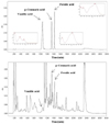
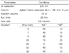
An analytical column (Agilent Zobax extended C18, 5 µm, 150 mm × 4.6 mm) with a mobile phase consisting of mixture of solvent A (water containing 0.2 % phosphoric acid) and B (acetonitrile), and a gradient elution (from 10:90 to 100:0, υ/υ) at a flow of 0.8 mL/min (Table 1) were applied for HPLC analysis (Alliance 2695 system, Waters, Milliford, MA, USA). The column temperature was maintained at 25℃. The detection wavelength was set at 270 nm for vanillic acid, p-coumaric acid, ferulic acid. The solvent was filtered through a 0.22 µm filter (Millipore) and degassed. The sample injection volume was 10 µL.
Statistical analysis
Data are presented as means ± standard deviation (SD). All tests were performed in triplicates. Differences between groups were estimated using one-way analysis of variance. Post-hoc comparison was made following Duncan's method. A two-sided p < 0.05 was considered statistically significant.
RESULTS
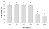
Cytotoxicity of water extract of barley sprout to B16F10 cells
Prior to treatment with BSE, cytotoxicity of BSE to B16F10 cells was determined employing WST-1 assay. B16F10 cells were treated with five different concentrations of BSE as well as a control (0 µg/mL) for 72 h. Results revealed that, at relatively lower concentrations (50, 125 and 250 µg/mL), the viability of murine melanocytes was not hindered by BSE (Fig. 1). However, higher levels of BSE (500 and 1,000 µg/mL) induced a decisive negative effect in the survival of B16F10 melanocytes. These results suggested that BSE was safe at concentration of 50, 125, or 250 µg/mL. Thus, these concentrations were selected for further analyses.
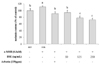
Effect of barley sprout extract on melanogenesis in B16F10 cells
To evaluate the inhibitory effect of BSE on melanogenesis, melanin contents in B16F10 cells treated with α-MSH were determined. As shown in Fig. 2, treatment with α-MSH (0.1 µM) increased melanin pigmentation by approximately 15% compared to normal control (cells not treated with α-MSH). However, treatment with BSE decreased melanin contents. At concentration of 250 µg/mL, BSE inhibited melanogenesis by 40% compared to the control (cells pre-treated with α-MSH) (p < 0.05). Arbutin exhibited inhibitory effect on melanin synthesis by 20% (150 ppm) compared to the control (p < 0.05) and the level of inhibition was statistically different compared to the effect mediated by BSE (250 µg/mL), as evidenced by Duncan's test. Therefore, BSE at the concentration of 250 µg/mL was observed to be more effective than arbutin, a common anti-melanogenesis compound.
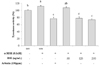
Effect of barley sprout extract on tyrosinase activity in B16F10 cells
The cellular activity of tyrosinase was also measured to determine the effect of BSE on melanogenesis in B16F10 cells (Fig. 3). In α-MSH treated melanocytes, tyrosinase activity was increased up to 15% compared to that in the normal control. However, similar to results observed in measurement of melanin, activity of tyrosinase was inhibited by BSE treatment at approximately 40%. Treatment with BSE at 125 and 250 µg/mL also exhibited similar levels of reduction in tyrosinase activity compared to arbutin.
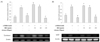
Effect of barley sprout extract on expression of tyrosinase and MITF proteins
Using Western blotting technique, the regulatory effect of BSE on melanogenic enzyme, tyrosinase, and MITF was estimated. Treatment with α-MSH increased the expression of tyrosinase in melanocytes. However, treatment with BSE decreased the expression of tyrosinase and MITF proteins with the concentration (Figs. 4A and 4B). Furthermore, the level of decrease in the expression of the two proteins after treatment with BSE at 250 µg/mL was lower or similar compared to the effect upon arbutin treatment (p < 0.05).
Identification of potential functional compounds in barley sprout extract
A mixture of three standards and aqueous fraction extract was measured at 270 nm to determine functional components of BSE. In the extract, vanillic acid, p-coumaric acid, and ferulic acid were detected, and contained 0.055, 0.089, and 0.094%, respectively (Fig. 5).
DISCUSSION
Barely and its sprout are commonly consumed food materials as good energy source with nutritious value. Previous studies have verified the mechanism of their health benefits and expanded their applications. In the current study, we examined the potency of BSE as a skin-whitening agent. Findings proved that compounds from BSE showed decisive inhibitory effects on melanogenesis by decreasing the activity and expression of MITF and tyrosinase.
Melanin production is a complicate biochemical pathway involving multiple biochemical and genetic regulation loci [3]. Various melanin-stimulating factors including UV, stress, and α-MSH can induce the expression of MITF, a melanocyte-specific transcription factor via melanocortin receptor 1 (MC1R) which transmits a signal to cAMP-PKA (cyclic AMP and protein kinase A) [311]. The activation of MITF subsequently leads to enhanced expression of master melanogenic enzyme tyrosinase. Activated protein finally catalyzes oxidation of L-tyrosine or L-DOPA to DOPA quinone. This is a common regulatory step in both synthesis of pheomelanin (red/yellow pigment) and eumelanin (brown/black pigment). Therefore, compounds with inhibitory effect on MITF and tyrosinase are important for the regulation of skin pigmentation [3].
Due to recent trends in cosmetic and pharmaceutical area, many natural materials have been screened and evaluated for their potential health applications [520]. Using simplified mushroom tyrosinase assay, Chaenomeles speciosa, Dryopteris crassirhizoma, and Gastrodia ellata have been found to able to inhibit tyrosinase activity [20]. Other plant extracts including Bupleurum falcatum, Caragana sinica, Morus alba, and Tussilago farfara can also decrease DOPA auto-oxidation activity [20]. Inhibitory effect of Gastrodia elata Blume on melanogenesis has also been reported using in vivo and in vitro models [45]. BSE has recently been highlighted because of its health benefits including its anti-pigmentation effect. Meng et al. have screened the molecules and their inhibition activity in methanol extract of BS for the biosynthesis of melanin with melanoma cells [17]. Here we report another evidence that BSE has potential application as an anti-pigmentation agent. The aqueous extract of BS presented a distinctive inhibition effect on melanogenesis with minimal cytotoxicity. Furthermore, the inhibitory effect of BSE on melanin biosynthesis was comparable to that of arbutin, a well-known whitening compound. Considering issues associated with known whitening agents, such depigmentation efficacy of natural food material sourced BSE could have potential application in the cosmetic industry.
Young BS are high in various polyphenols [1314]. Depigmentation potency of BSE could be due to its anti-oxidative compounds. Earlier studies have suggested that epicatechin, baicalein, hesperidin, luteolin, and kaempferol can inhibit melanin biosynthesis [3]. Hesperidin and catechins are known to inhibit the accumulation of MITF. Epigallocatechin gallate can also decrease the accumulation of tyrosinase [3]. Baicalein can decrease the accumulation of MITF protein. Furthermore, luteolin, genistein, and kaempferol can modify melanogenesis by targeting tyrosinase either directly or indirectly [3]. In the present study, to identify potential action compounds with anti-melanogenic effects, BSE was analyzed using HPLC method. Although the assay did not isolate all known health beneficial compounds in BSE, polyphenols, p-coumaric acid, ferulic acid, and vanillic acid were identified. Inhibitory effect on melanogenesis and potency as skin whitening agent for those molecules have been observed earlier. P-coumaric acid is a secondary metabolite in multiple herbs. In both in vivo and in in vitro models, p-coumaric acid can effectively inhibit tyrosinase and reduce melanin contents [21]. P-coumaric acid extracted from rice bran can reduce gene and protein expression of MITF and tyrosinase by suppressing CREB phosphorylation during cAMP signaling [2223]. Other studies with human and murine melanocytes explants have also reported that ferulic acid and vanillic acid possess potential as depigmenting agents. Ferulic acid shows significant skin whitening and anti-wrinkle effect [2425]. Therefore, it has potential as a favorable cosmetic agent, although its potential mechanism of action may differ between studies [2425]. Treatment with vanillic acid can also significantly decrease cellular melanin contents [26]. Vanillic acid can down-regulate the expression of MC1R and MITF, hence decreasing the activity of tyrosinase to generate such anti-melanogenic effect [26]. Our experimental results suggested that treatment with BSE could suppress cellular expression of MITF which subsequently affected the activity and expression of tyrosinase. Taken together, these results suggest that the polyphenols in BSE possibly suppress the activity and expression of MITF and tyrosinase, thus decreasing cellular melanin content in α-MSH stimulated B16F10 melanocytes.
Experimental findings of the current study suggest that aqueous extracted BS has considerable potential as a whitening agent. Treatment with BSE altered gene expression and activity of critical enzymes such as MITF and tyrosinase. However, more precise action molecules and the role of BSE in signaling pathways regulating those melanogenesis related enzymes remain unclear. Furthermore, findings were obtained from an in vitro model. Further animal and clinical studies are needed to confirm our findings to secure its application.
In summary, water extracted BS (250 µg/mL) is proposed as an effective depigmentation agent. As a natural food sourced material, BSE could appeal to the public in the consumer market with applications in cosmetic and pharmaceutical industries.
Summary
The present study investigated the anti-melanogenic effect of water extract of BS in a murine melanocytes model. Experimental results revealed that water extract of BS significant reduced melanin synthesis. It also decreased melanogenesis related markers, including the activity and expression of tyrosinase and MITF. These findings suggest that aqueous BS has potential to develop depigmentation agent as a natural food material.
 XML Download