Journal List > J Nutr Health > v.52(2) > 1122024

Bae, Kim, Bae, Kim, and Ly: Anti-inflammatory effects of fruit and leaf extracts of Lycium barbarum in lipopolysaccharide-stimulated RAW264.7 cells and animal model∗

Abstract

Purpose:

Medicinal herbs have recently attracted attention as health beneficial foods and source materials for drug development. Recent studies have demonstrated that extracts of Lycium's fruits and roots have a range of physiologically active substances. The extract of Lycium's leaves has been reported to have excellent anti-oxidant and anti-microbial activity, but its anti-inflammatory efficacy is not known. The chlorophyll present in the leaves can act as an anti-oxidant or pro-oxidant depending on the presence of light. Therefore, this study analyzed the anti-inflammatory effects of Lycium's fruit extract (LFE), leaf extract (LLE), and leaf extract with chlorophyll removal (LLE with CR).

Methods:

This study examined the inhibitory effects of LFE, LLE, and LLE with CR on pro-inflammatory mediator production as well as on the expression of iNOS and COX-2 in lipopolysaccharide (LPS)-stimulated RAW264.7 cells and BALB/c mice.

Results:

LFE, LLE, and LLE with CR inhibited the production of pro-inflammatory mediators (NO, TNF-α, IL-6, and IL-1β) and the expression of iNOS and COX-2 in LPS-stimulated RAW 264.7 cells in a dose-dependent manner. Furthermore, the administration of LLE and LLE with CR inhibited the serum pro-inflammatory cytokine levels and suppressed DNA damage in BALB/c mice. In particular, LLE with CR exhibited the highest anti-inflammatory activity.

Conclusion:

These results suggest that the fruit and leaves of Lycium are potential therapeutic agents against inflammation.

REFERENCES

1.Zedler S., Faist E. The impact of endogenous triggers on trauma-associated inflammation. Curr Opin Crit Care. 2006. 12(6):595–601.
crossref
2.Isomäki P., Punnonen J. Pro- and anti-inflammatory cytokines in rheumatoid arthritis. Ann Med. 1997. 29(6):499–507.
3.Vane JR., Mitchell JA., Appleton I., Tomlinson A., Bishop-Bailey D., Croxtall J, et al. Inducible isoforms of cyclooxygenase and nitric-oxide synthase in inflammation. Proc Natl Acad Sci U S A. 1994. 91(6):2046–2050.
crossref
4.Higuchi M., Higashi N., Taki H., Osawa T. Cytolytic mechanisms of activated macrophages. Tumor necrosis factor and L-arginine-dependent mechanisms act synergistically as the major cytolytic mechanisms of activated macrophages. J Immunol. 1990. 144(4):1425–1431.
5.Nathan C. Nitric oxide as a secretory product of mammalian cells. FASEB J. 1992. 6(12):3051–3064.
crossref
6.Guzik TJ., Korbut R., Adamek-Guzik T. Nitric oxide and superoxide in inflammation and immune regulation. J Physiol Pharmacol. 2003. 54(4):469–487.
7.Zhang R., Kang KA., Piao MJ., Kim KC., Kim AD., Chae S, et al. Cytoprotective effect of the fruits of Lycium chinense Miller against oxidative stress-induced hepatotoxicity. J Ethnopharmacol. 2010. 130(2):299–306.
crossref
8.Kim TS., Park WJ., Ko SB., Kang MH. Development of extracts of Lycii folium having high antioxidant activity. J Korean Soc Food Sci Nutr. 2008. 37(10):1318–1322.
crossref
9.Xiao J., Liong EC., Ching YP., Chang RC., So KF., Fung ML, et al. Lycium barbarum polysaccharides protect mice liver from carbon tetrachloride-induced oxidative stress and necroinfla-mmation. J Ethnopharmacol. 2012. 139(2):462–470.
crossref
10.Chen H., Olatunji OJ., Zhou Y. Anti-oxidative, anti-secretory and anti-inflammatory activities of the extract from the root bark of Lycium chinense (Cortex Lycii) against gastric ulcer in mice. J Nat Med. 2016. 70(3):610–619.
crossref
11.Luo Q., Cai Y., Yan J., Sun M., Corke H. Hypoglycemic and hypolipidemic effects and antioxidant activity of fruit extracts from Lycium barbarum. Life Sci. 2004. 76(2):137–149.
crossref
12.Mocan A., Vlase L., Vodnar DC., Bischin C., Hanganu D., Gheldiu AM, et al. Polyphenolic content, antioxidant and antimicrobial activities of Lycium barbarum L. and Lycium chinense Mill. leaves. Molecules. 2014. 19(7):10056–10073.
13.Liu SC., Lin JT., Hu CC., Shen BY., Chen TY., Chang YL, et al. Phenolic compositions and antioxidant attributes of leaves and stems from three inbred varieties of Lycium chinense Miller harvested at various times. Food Chem. 2017. 215:284–291.
crossref
14.Albishi T., John JA., Al-Khalifa AS., Shahidi F. Antioxidative phenolic constituents of skins of onion varieties and their activities. J Funct Foods. 2013. 5(3):1191–1203.
crossref
15.Olatunde OO., Benjakul S., Vongkamjan K. Antioxidant and antibacterial properties of guava leaf extracts as affected by solvents used for prior dechlorophyllisation. J Food Biochem. 2018. 42(5):e12600.
16.Park SH., Kim JM., Kim JH., Oh YS., Joo DH., Lee EY, et al. Antioxidative effects and component analysis of graviola (Annona muricata) leaf extract/fractions. J Soc Cosmet Sci Korea. 2017. 43(4):309–320.
17.Kim JE., Bae SM., Nam YR., Bae EY., Ly SY. Antioxidant activity of ethanol extract of Lycium barbarum's leaf with removal of chlorophyll. J Nutr Health. 2019. 52(1):26–35.
crossref
18.Ko YE., Oh SR., Song HH., Ryu HW., Ly SY., Kim JW. The effect of 4α,5α-epoxy-10α,14-dihydro-inuviscolide, a novel immunosuppressant isolated from Carpesium abrotanoides, on the cytokine profile in vitro and in vivo. Int Immunopharmacol. 2015. 25(1):121–129.
crossref
19.Cho HY., Noh KH., Cho MK., Jang JH., Lee MO., Kim SH, et al. Anti-oxidative and anti-inflammatory effects of genistein in BALB/c mice injected with LPS. J Korean Soc Food Sci Nutr. 2008. 37(9):1126–1135.
crossref
20.Tice RR., Agurell E., Anderson D., Burlinson B., Hartmann A., Kobayashi H, et al. Single cell gel/comet assay: guidelines for in vitro and in vivo genetic toxicology testing. Environ Mol Mutagen. 2000. 35(3):206–221.
crossref
21.Oh YC., Cho WK., Im GY., Jeong YH., Hwang YH., Liang C, et al. Anti-inflammatory effect of Lycium fruit water extract in lipopolysaccharide-stimulated RAW 264.7 macrophage cells. Int Immunopharmacol. 2012. 13(2):181–189.
crossref
22.Cho EJ., Kim YE., Lee DE., Sung NY., Byun EH., Park WJ. Comparison of the antioxidative and anti-inflammatory activities of Lycium chinense leaves and fruits extracts according to the harvest time. J Korean Soc Food Sci Nutr. 2018. 47(7):717–724.
crossref
23.Vilcek J., Lee TH. Tumor necrosis factor. New insights into the molecular mechanisms of its multiple actions. J Biol Chem. 1991. 266(12):7313–7316.
24.Olmos G., Lladó J. Tumor necrosis factor alpha: a link between neuroinflammation and excitotoxicity. Mediators Inflamm. 2014. 2014:861231.
crossref
25.Gabay C., Lamacchia C., Palmer G. IL-1 pathways in inflammation and human diseases. Nat Rev Rheumatol. 2010. 6(4):232–241.
crossref
26.Scheller J., Garbers C., Rose-John S. Interleukin-6: from basic biology to selective blockade of pro-inflammatory activities. Semin Immunol. 2014. 26(1):2–12.
crossref
27.Xie JH., Tang W., Jin ML., Li JE., Xie MY. Recent advances in bioactive polysaccharides from Lycium barbarum L., Zizyphus jujuba Mill, Plantago spp., and Morus spp.: structures and functionalities. Food Hydrocoll. 2016. 60:148–160.
crossref
28.Zhu J., Zhang Y., Shen Y., Zhou H., Yu X. Lycium barbarum polysaccharides induce Toll-like receptor 2- and 4-mediated phenotypic and functional maturation of murine dendritic cells via activation of NF-κB. Mol Med Rep. 2013. 8(4):1216–1220.
crossref
29.González-Gallego J., García-Mediavilla MV., Sánchez-Campos S., Tuñón MJ. Fruit polyphenols, immunity and inflammation. Br J Nutr. 2010. 104(Suppl 3):S15-S27.
crossref
30.Chapple IL. Reactive oxygen species and antioxidants in inflammatory diseases. J Clin Periodontol. 1997. 24(5):287–296.
crossref
31.Shibata H., Sakamoto Y., Oka M., Kono Y. Natural antioxidant, chlorogenic acid, protects against DNA breakage caused by monochloramine. Biosci Biotechnol Biochem. 1999. 63(7):1295–1297.
crossref
32.Benjakul S., Kittiphattanabawon P., Sumpavapol P., Maqsood S. Antioxidant activities of lead (Leucaena leucocephala) seed as affected by extraction solvent, prior dechlorophyllisation and drying methods. J Food Sci Technol. 2014. 51(11):3026–3037.
crossref

Fig. 1
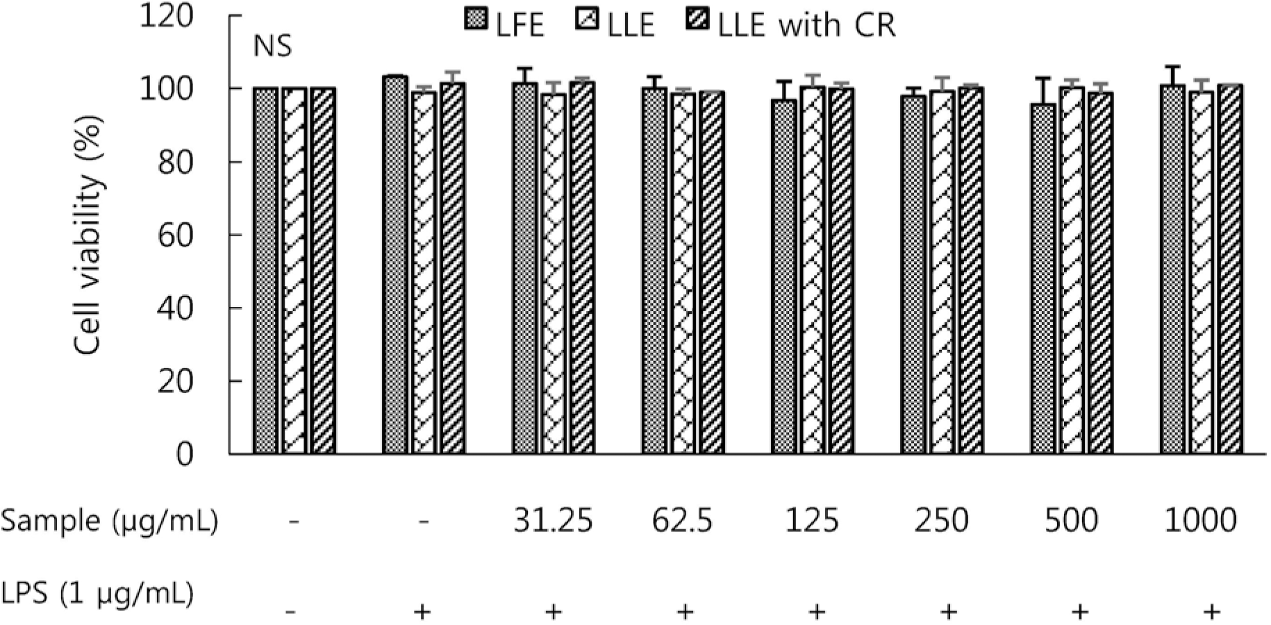
Effect of Lycium's fruit extracts (LFE) and leaf extracts (LLE), leaf extracts chlorophyll removal (LLE with CR) on cell viability of lipopolysaccharide (LPS)-induced RAW264.7 cells. Cells were treated with LFE, LLE, LLE with CR (31.25, 62.5, 125, 250, 500, 1,000 μg/mL), then with or without LPS (1 μg/mL) for 24 h. The cell proliferation was estimated by the MTT assay with WST system. Each bar represents the mean ± SD. NS: not significantly different by Duncan's multiple range test (p < 0.05).
jnh-52-129f1.tif
Fig. 2
Effect of Lycium's fruit extracts (LFE) and leaf extracts (LLE), leaf extracts chlorophyll removal (LLE with CR) on the production of nitric oxide (NO) in lipopolysaccharide (LPS)-induced RAW264.7 cells. Cells were treated with LFE, LLE, LLE with CR (31.25, 62.5, 125, 250, 500, 1,000 μg/mL), then with or without LPS (1 μg/mL) for 24 h. The culture supernatant of the treated cells were used to measure NO level. Levels of nitric oxide were determined by Griess reagent. Each bar represents the mean ± SD. Significant values are represented by an asterisk (∗) (p < 0.05 compared to the group treated with LPS alone).
jnh-52-129f2.tif
Fig. 3
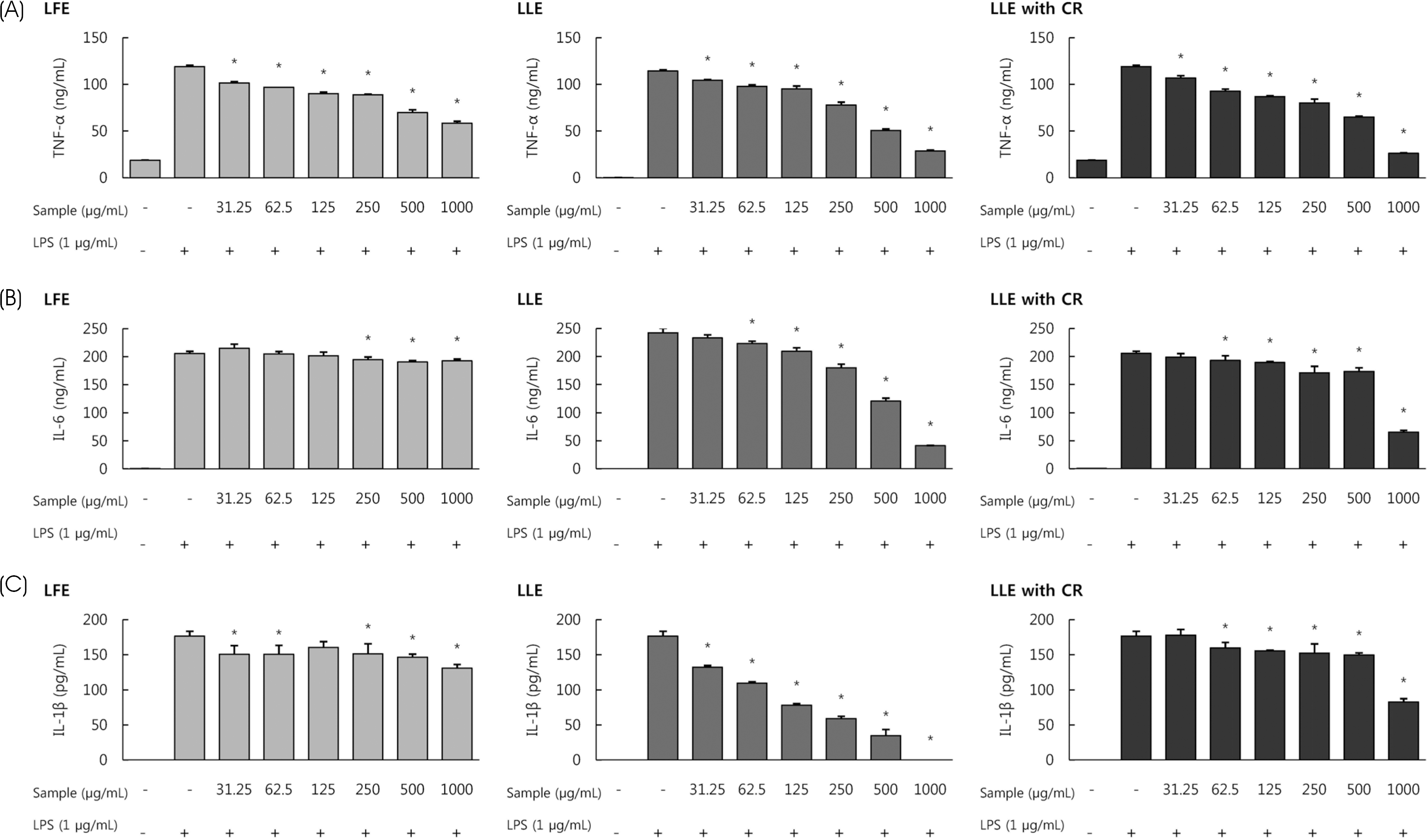
Effect of Lycium's fruit extracts (LFE) and leaf extracts (LLE), leaf extracts chlorophyll removal (LLE with CR) on the production of TNF-α (A), IL-6 (B), IL-1β (C) in lipopolysaccharide (LPS)-induced RAW264.7 cells. Cells were treated with LFE, LLE, LLE with CR (31.25, 62.5, 125, 250, 500, 1,000 μg/mL), then with or without LPS (1 μg/mL) for 24 h. The levels of pro-inflammatory cytokines in the cell culture supernatant were determined by ELISA. Each bar represents the mean ± SD. Significant values are represented by an asterisk (∗) (p < 0.05 compared to the group treated with LPS alone).
jnh-52-129f3.tif
Fig. 4
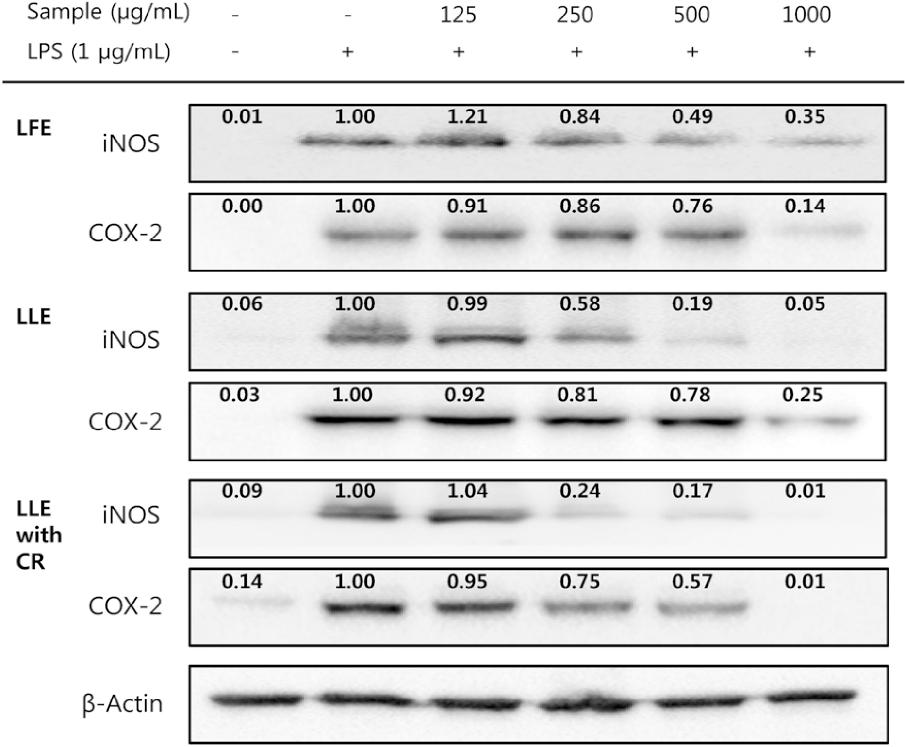
Effect of Lycium's fruit extracts (LFE) and leaf extracts (LLE), leaf extracts chlorophyll removal (LLE with CR) on iNOS and COX-2 protein expression in lipopolysaccharide (LPS)-induced RAW264.7 cells. Cells were treated with LFE, LLE, LLE with CR (125, 250, 500 and 1,000 μg/mL), then with or without LPS (1 μg/mL) for 24 h. Cell lysates were used for western blot analysis. The levels of protein expression in iNOS and COX-2 were normalized to the β-actin signals. The relative band intensities are indicated above each band.
jnh-52-129f4.tif
Fig. 5
Effect of Lycium's leaf extracts (LLE), leaf extracts chlorophyll removal (LLE with CR) on the production of serum TNF-α (A), IL-6 (B), IL-1β (C) in LPS-induced BALB/c mice. LLE and LLE with CR (200 mg/kg body weight) was administered orally in mice for seven days, and then challenged intraperitoneally with LPS (5 mg/kg body weight). The levels of pro-inflammatory cytokines in the serum were determined by ELISA. Serum TNF-α, IL-6, IL-1β levels were analyzed 8 h after the last LPS challenge. Each bar represents the mean ± SD. Significant values are represented by an asterisk (∗) (p < 0.05 compared to the group treated with LPS alone).
jnh-52-129f5.tif
Table 1.
Body weight of mice before and after oral administration
Group1) Body weight (g)  
Before oral administration After oral administration t-value4)
N 24.17 ± 1.532) 23.70 ± 1.01 1.468NS3)
C 24.73 ± 1.25 23.63 ± 1.34 19.053∗4)
LLE 24.57 ± 2.46 23.37 ± 2.39 20.785
LLE with CR 25.37 ± 0.64 23.83 ± 0.38 5.890

1) N (normal), treated with PBS for 8 h; C (control), only treated with LPS 5 mg/kg body weight for 8 h; LLE, treated with Lycium's leaf extracts (200 mg/kg body weight) for seven days and LPS 5 mg/kg body weight for 8 h; LLE with CR, treated with Lycium's leaf extracts chlorophyll removal (200 mg/kg body weight) for seven days and LPS 5 mg/kg body weight for 8 h

2) Values represent mean ± SD (n = 3).

3) Not significantly different

4) p < 0.05 for body weight after oral administration vs baseline, paired t-test

Table 2.
Levels of DNA damage expressed as tail DNA, tail length and tail moment in BALB/c mice lymphocyte
Group1) Tail DNA (%) Tail length (μm) Tail moment
N 5.40 ± 0.652)3)a 10.21 ± 1.75a 0.82 ± 0.17a
C 15.64 ± 5.14b 46.61 ± 14.17b 9.48 ± 6.06b
LLE 8.42 ± 1.40a 20.23 ± 3.91a 2.29 ± 0.74a
LLE with CR 8.57 ± 1.63a 14.77 ± 1.29a 1.93 ± 0.42a
F-value 7.176 14.438 4.996

1) N (normal), treated with PBS for 8 h; C (control), only treated with LPS 5 mg/kg body weight for 8 h; LLE, treated with Lycium's leaf extracts (200 mg/kg body weight) for seven days and LPS 5 mg/kg body weight for 8 h; LLE with CR, treated with Lycium's leaf extracts chlorophyll removal (200 mg/kg body weight) for seven days and LPS 5 mg/kg body weight for 8 h

2) Values represent mean ± SD (n = 3).

3) Means with different letters (a-b) are significantly different by Duncan's multiple range test (p < 0.05). 4)

4)∗ p < 0.05

TOOLS
Similar articles