

 PDF
PDF ePub
ePub Citation
 Print
Print


Abstract
Anterior interosseous nerve is purely a motor nerve and supplies flexor pollicis longus, flexor digitorum profundus to the index finger, and pronator quadratus. The etiology and treatment option of anterior interosseous nerve syndrome remain controversial. Bilateral involvement of the anterior interosseous nerve have been described rarely; however, we found no reported case of non-simultaneous bilateral anterior interosseous nerve palsy associated with the entrapment neuropathy. We present the unique case of delayed anterior interosseous nerve syndrome, 3 years 5 months following an identical event in the opposite extremity and literature review.
Go to : 
References
1. Sunderland S. The intraneural topography of the radial, median and ulnar nerves. Brain. 1945; 68:243–99.

2. Nagano A. Spontaneous anterior interosseous nerve palsy. J Bone Joint Surg Br. 2003; 85:313–8.
3. Tinel J. Nerve wounds: symptomatology of peripheral nerve lesions caused by war wounds. London: Balliere, Tindall and Cox;. 1918.
4. Kiloh LG, Nevin S. Isolated neuritis of the anterior interosseous nerve. Br Med J. 1952; 1:850–1.
5. Fearn CB, Goodfellow JW. Anterior interosseous nerve palsy. J Bone Joint Surg Br. 1965; 47:91–3.
6. Barbour JR, Gontre G, Mackinnon SE. Delayed bilateral anterior interosseous neuritis: an atypical presentation. J Hand Surg Eur Vol. 2013; 38:206–7.
7. Spinner M. The anterior interosseous-nerve syndrome, with special attention to its variations. J Bone Joint Surg Am. 1970; 52:84–94.
8. Collins DN, Weber ER. Anterior interosseous nerve syndrome. South Med J. 1983; 76:1533–7.
9. Sood MK, Burke FD. Anterior interosseous nerve palsy: a review of 16 cases. J Hand Surg Br. 1997; 22:64–8.
10. Park IJ, Roh YT, Jeong C, Kim HM. Spontaneous anterior interosseous nerve syndrome: clinical analysis of eleven surgical cases. J Plast Surg Hand Surg. 2013; 47:519–23.
Go to :
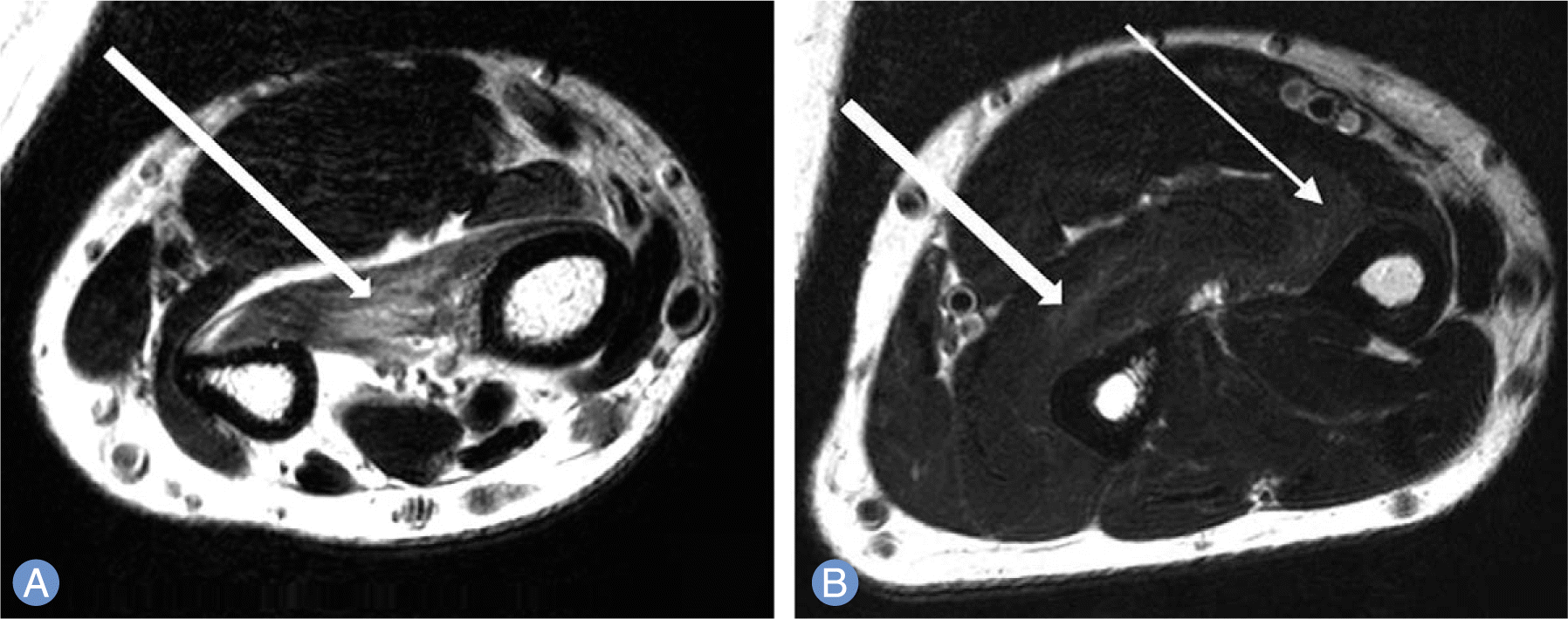 | Fig. 1.Preoperative magnetic resonance imaging of the left forearm. T2-weighted image coronal view shows (A) increased signal intensity, atrophy and edematous change of the pronator quadratus (arrow), (B) increased signal intensity, mild atrophy and edematous change of the flexor digitorum profundus (thick arrow) and flexor pollicis longus (thin arrow). |
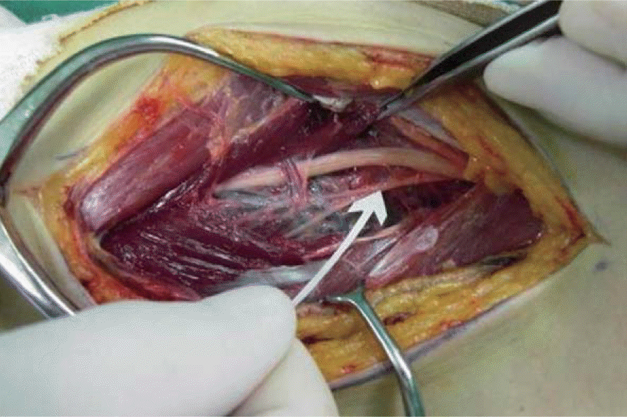 | Fig. 2.The fibrous arch of flexor digitorum superficialis was removed and adjacent tissue was exfoliated. Compression of the anterior interosseous nerve (arrow) and adhesion with the adjacent tissue were observed in the operation theatre. |
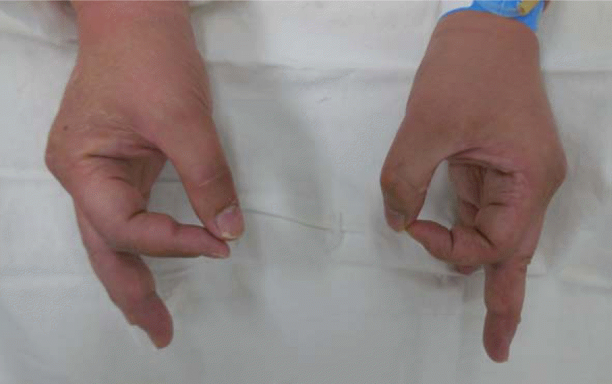 | Fig. 3.The patient was unable to perform ‘O’ due to the paralysis of right flexor pollicis longus and flexor digitorum profundus to the index finger. On the other hand, the left side recovered paralysis of the flexor pollicis longus due to anterior interosseous nerve compression for 3 years 5 months after surgical decompression. |
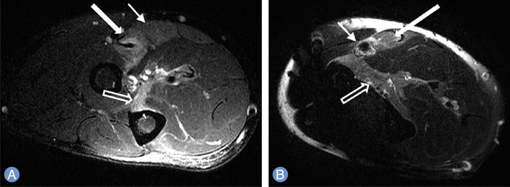 | Fig. 4.Preoperative right forearm magnetic resonance imaging T2-weighted image coronal view shows (A) increased signal change, atrophy and edematous change of the pronator teres (thick arrow), flexor carpi radialis (thin arrow), flexor digitorum profundus (empty arrow), and (B) increased signal change, atrophy and edematous change of the flexor carpi radialis (thick arrow), flexor pollicis longus (thin arrow), pronator quadratus (empty arrow). |
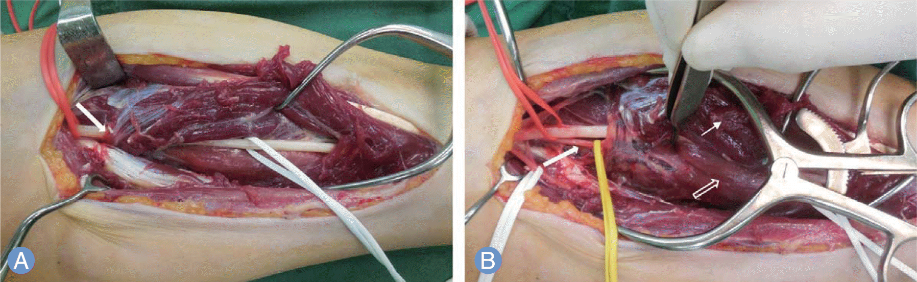 | Fig. 5.Intraoperative clinical photo of right forearm. (A) The entrapment of anterior interosseous nerve which lies radial, inferior to the median nerve was observed before removing the fibrous arch of flexor digitorum superficialis (arrow). (B) The atrophy of anterior interosseous nerve (thin arrow) after removing the fibrous arch of flexor digitorum superficialis, atrophy and pale color of flexor digitorum profundus (empty arrow) compared to flexor digitorum superficialis (thin arrow) was observed. |
 XML Download