

 PDF
PDF ePub
ePub Citation
 Print
Print


Abstract
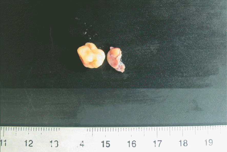
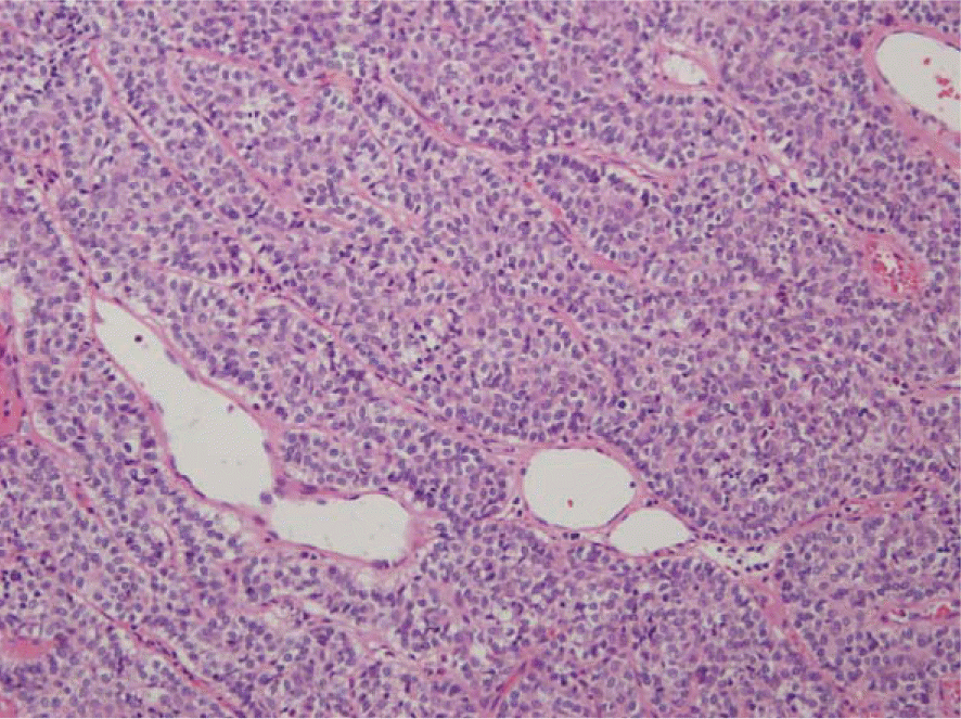
A 36-year-old man presented with a hard mass on the right forearm. He had no specific symptom, but wanted surgical excision for cosmetic purpose. On the physical examination, the mass was located on the ulnar side of forearm, presented mild tenderness. Ultrasound examination showed a hypoechoic mass, 1.3×1 cm mass in the subcutaneous tissues. Under the local anesthesia, the patient underwent an excisional biopsy of the lesion. The histopathology and the immunohistochemical analysis confirmed the tumor to be a glomus tumor. The extradigital glomus tumors are sparsely reported apart from pain. The patients can present with subcutaneous nodule, or with discoloration of the skin. These atypical symptoms make difficult to diagnose extradigital glomus tumors. In the current study, we report the case of a patient with asymptomatic glomus tumor in a extradigital lesion of forearm.
References
1. Lee SK, Song DG, Choy WS. Intravascular glomus tumor of the forearm causing chronic pain and focal tenderness. Case Rep Orthop. 2014; 2014:619490.

2. Schiefer TK, Parker WL, Anakwenze OA, Amadio PC, Inwards CY, Spinner RJ. Extradigital glomus tumors: a 20-year experience. Mayo Clin Proc. 2006; 81:1337–44.
3. Kim SH, Suh HS, Choi JH, Sung KJ, Moon KC, Koh JK. Glomus tumor: a clinical and histopathologic analysis of 17 cases. Ann Dermatol. 2000:95–101.
4. Lee CH, Byeon JH, Rhie JW, Kang YJ, Cho MJ, Lim P. Clinical analysis of twenty cases of glomus tumor in the digits. J Korean Soc Plast Reconstr Surg. 1995; 22:169–78.
5. Takei TR, Nalebuff EA. Extradigital glomus tumour. J Hand Surg Br. 1995; 20:409–12.
6. Nigam JS, Misra V, Singh A, Karuna V, Chauhan S. A glomus tumour arising from the flexor aspect of the forearm: a case report with review of the literature. J Clin Diagn Res. 2012; 6:1559–61.
7. Schoenleber SJ, Rosenberg AE, Temple HT. Painful forearm mass in a 75-year-old man. Clin Orthop Relat Res. 2014; 472:776–80.
Fig. 1.
Preoperative clinicial photo shows protruding mass localized to the proximal dorsal portion of forearm.
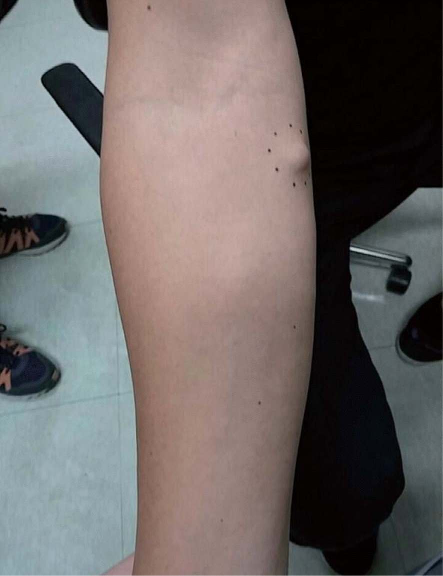
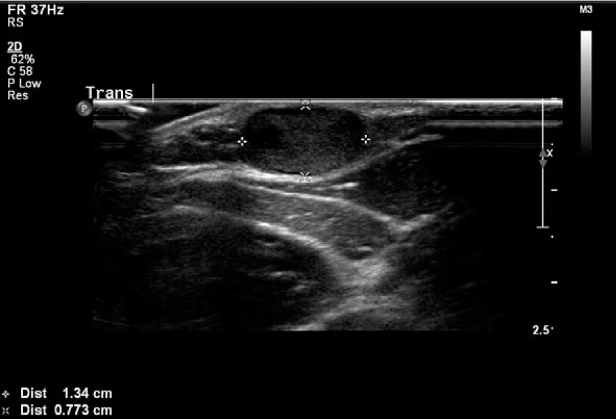
Fig. 2.
Ultrasonography of the right forearm. About 1.34×0.7 cm sized hypoechoic lesion is located on subcutaneous fat layer.
 XML Download