Abstract
Purpose:
Groin or abdominal flap, anterolateral thigh free flap, and radial forearm flap can typically be performed in large defects, however satisfactory results in functional recovery and aesthetic aspect have not been achieved using these methods. Medial sural artery perforator free flap is recommended as a complement to these disadvantages, therefore we report the functional and aesthetic results of this flap for reconstruction of large finger defects.
Methods:
From January 2008 to December 2013, 10 patients with large soft tissue defect of the fingers were treated with medial sural artery perforator free flap. Six months after the final surgery, metacarpophalangeal joint and proximal interphalangeal joint range of motion was measured, and the circumference of the reconstructed finger was compared with that of the contralateral side. In addition, for assessment of the aesthetic satisfaction, the patients and three physicians compared the color of the reconstructed finger with that of adjacent skin on a five-point scale.
Results:
The flaps survived without complications in all ten cases. Average flexion was 77 degrees in the metacarpophalangeal joint and 84 degrees in the proximal interphalangeal joints. The average circumference of the reconstructed finger was measured as 12 percent larger than contralateral. The patient’s subjective satisfaction (4.1) and physicians’ objective satisfaction (4.2) regarding aesthetic aspect were very good.
Conclusion:
Medial sural artery perforator free flap is a very thin, stable, fascio-cutaneous flap which has a tendon gliding effect and produces aesthetically good results. Therefore we consider medial sural artery perforator free flap as the flap which can solve the drawbacks of other techniques associated with large finger defect reconstruction.
Go to : 
References
1. de Korte N, Dwars BJ. van der Werff JF. Degloving injury of an extremity. Is primary closure obsolete? J Trauma. 2009; 67:E60–1.
2. Cavadas PC, Sanz-Gimenez-Rico JR, Gutierrez-de la Camara A, Navarro-Monzonis A, Soler-Nomdedeu S, Martinez-Soriano F. The medial sural artery perforator free flap. Plast Reconstr Surg. 2001; 108:1609–15.

3. Slattery P, Leung M, Slattery D. Microsurgical arterialization of degloving injuries of the upper limb. J Hand Surg Am. 2012; 37:825–31.

4. Hahn SB, Kim SH, Jung JH, Kang HJ. Free flap transfer for the reconstruction of injured hands. J Korean Soc Surg Hand. 2005; 10:234–41.
5. Daniel RK, May JW Jr. Free flaps: an overview. Clin Orthop Relat Res. 1978; (133):122–31.
6. Song YG, Chen GZ, Song YL. The free thigh flap: a new free flap concept based on the septocutaneous artery. Br J Plast Surg. 1984; 37:149–59.

7. Ali RS, Bluebond-Langner R, Rodriguez ED, Cheng MH. The versatility of the anterolateral thigh flap. Plast Reconstr Surg. 2009; 124(6 Suppl):e395–407.

8. Koshima I, Nanba Y, Tsutsui T, Takahashi Y. New anterolateral thigh perforator flap with a short pedicle for reconstruction of defects in the upper extremities. Ann Plast Surg. 2003; 51:30–6.

9. Ahn HC, Choi MS, Hwang WJ, Sung KY. The transverse radial artery forearm flap. Plast Reconstr Surg. 2007; 119:2153–60.

Go to : 
 | Fig. 1.
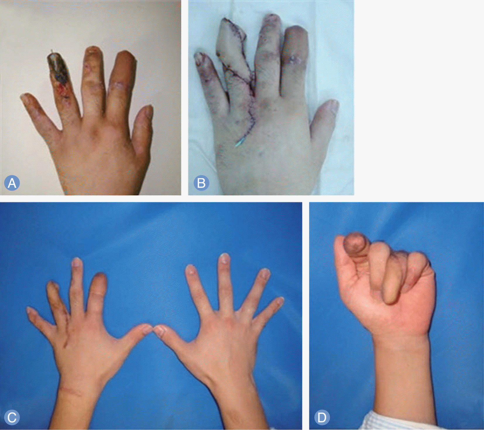
(A) Preoperative view. (B) Circumferential coverage using medial sural artery perforator free flap. (C) Compare the circumference of reconstructed finger to contralateral normal ring finger on postoper-ative 6 months. (D) Compare the joint flexion of reconstructed ring finger to index finger of groin flap on postoperative 6 months. |
 | Fig. 2.
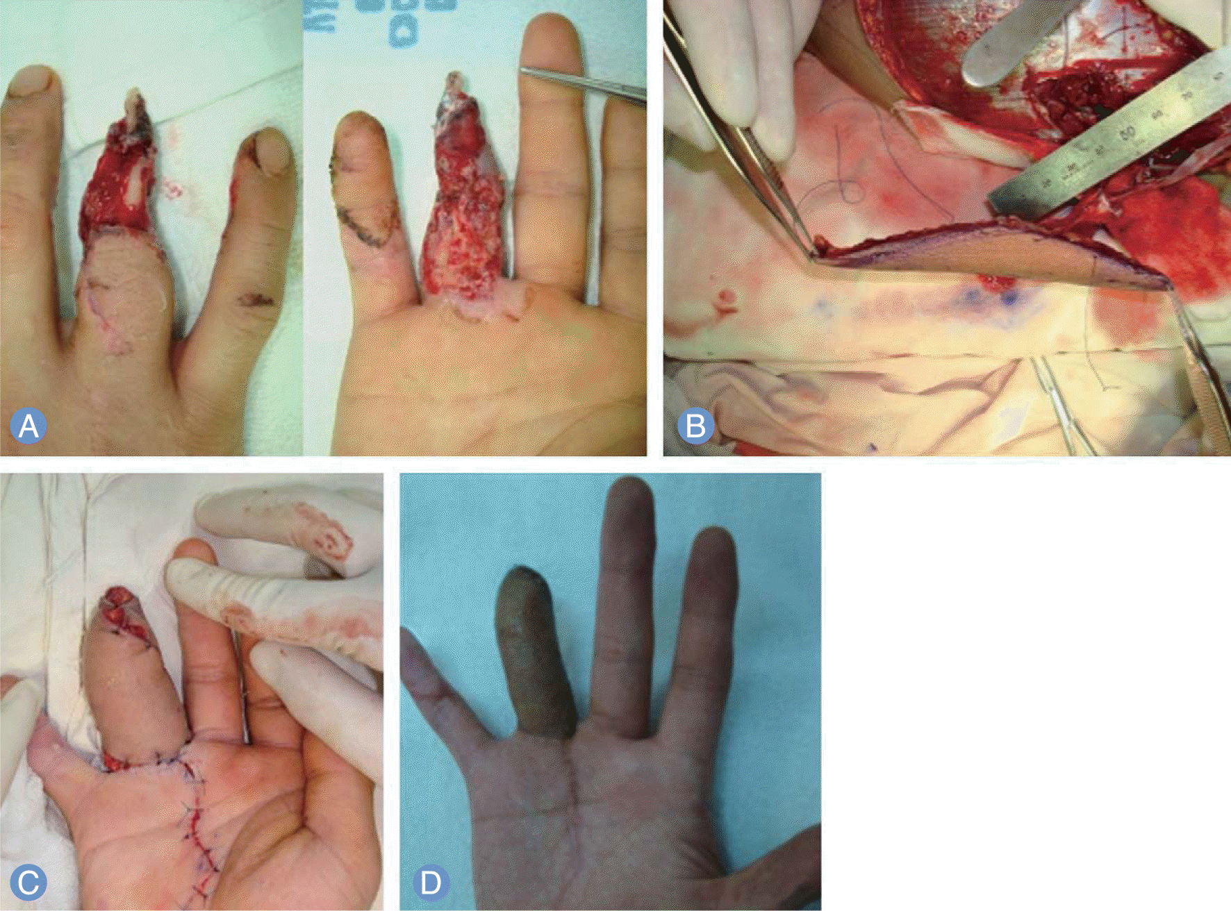
(A) Preoperative view. (B) Intraoperative finding of medial sural artery perforator free flap harvesting. (C) Circumferential coverage using medial sural artery perforator free flap. (D) Postoperative 12 months view. |
Table 1.
Patient summary and results
Table 2.
Aesthetic satisfaction
| Assessment | Patient | Physicians* |
|---|---|---|
| Very well matching (5 point) | 2 | 3 |
| Well matching (4 point) | 7 | 6 |
| Moderate matching (3 point) | 1 | 1 |
| Poor matching (2 point) | 0 | 0 |
| Very poor matching (1 point) | 0 | 0 |
| Average | 4.1 | 4.2 |


 PDF
PDF ePub
ePub Citation
 Print
Print


 XML Download