

 PDF
PDF ePub
ePub Citation
 Print
Print


INTRODUCTION
Folate plays an essential role as a major coenzyme in onecarbon metabolism. It has been suggested that inappropriate folate nutritional status can result in carcinogenesis via aberrant DNA methylation. This process arises because of decreased S-adenosylmethionine (SAM) synthesis and hence an alteration in gene expression. Folate shortage can also impair DNA stability via misincorporation of uridylate into DNA.1 As part of the molecular process of carcinogenesis, folate deficiency in normal tissue may predispose cells to neoplastic transformation23 or suppress the progression of pre-existing neoplasms in model systems.2456 By contrast, recent research has found that high folate levels may actually increase cancer incidence.78 These intriguing, yet contradictory findings relating to the development and progression of cancer imply that a defined and moderate level of folate intake and blood status is critical to maintaining health.9
Studies now suggest that folate intake and blood folate level are related to cancer risk particularly of colon and breast cancer.11011 Large studies indicate folate plays a role as a major aetiological factor in colorectal cancer risk with high folate intake reducing colorectal risk by about 40% compared to low folate intake, although results are inconsistent. For instance, low folate level was reported to have a protective effect against colorectal cancer,7 while high dose folic acid supplements might increase the risk of colorectal neoplasia.1012 Folate has also been considered as one of the factors involved in breast cancer aetiology. Recent studies suggest that folate has a protective effect against breast cancer,1314 with high concentrations of homocysteine and cysteine affording greater risk for breast cancer when folate levels were low.15 However, a number of studies reported conflicting results such as no relationship between folate intake or blood folate levels and breast cancer risk,16 or that high folate intake may increase the risk of breast cancer.817 To date, there is no clear explanation for this conflicting role of folate status on cancer risk.
Since the commencement of mandatory folic acid fortification with pteroylmonoglutamic acid (PteGlu) in several countries from the late 1990s, the blood folate status of many populations has been enhanced.18 As a consequence of this, there has been a striking reduction in congenital malformation rates including neural tube defects.19 Despite this major public health achievement, emerging evidence shows that mandatory folic acid fortification and elevated folate level might be responsible for several adverse effects, including the accumulation of unmetabolized PteGlu in blood, a changed distribution of cellular folyl vitamers,20 reduced natural killer cell cytotoxicity,21 a rise in twin births22 and the mean dose for methotrexate therapy.23 It has also been speculated that the increase of colorectal cancer incidence since mandatory folic acid fortification may be due to the higher folate intake, or exposure to altered folyl vitamers.10242526 For many of these phenomena we do not have any clear explanation. However, folic acid fortification using PteGlu as a main vehicle does seem to elicit an effect on health.
Therefore, we examined the effects of various experimental concentrations of PteGlu on the growth of colorectal (Caco-2) and breast cancer (MCF7) cell lines to explore the potential effects that PteGlu might have on health outcomes including carcinogenesis. The genotype of a common variant in a key protein involved in PteGlu metabolism; dihydrofolate reductase 19bp deletion/insertion (DHFR 19-del), was also determined in the two cell lines. We were particularly interested to elucidate findings in the context of population level mandatory folic acid fortification.
MATERIALS AND METHODS
Chemicals and Reagents
The following chemicals for cell culture and cell growth measurement were purchased from Sigma (CA, USA): Rosewell Park Memorial Institute medium 1640 (RPMI 1640), folate free RPMI 1640, L-glutamine, foetal bovine serum (FBS), phosphate buffered saline, EDTA-trypsin solution, thiazolyl blue tetrazolium bromide (MTT) and dimethyl sulfoxide. PteGlu was purchased from Schircks laboratories (Jona, Switzerland). For genotype scoring of DHFR 19-del variant, DNA was extracted using QIAamp DNA blood mini kit purchased from Qiagen (Hilden, Germany), for DNA amplification. Oligonucleotides were synthesized by Invitrogen (CA, USA). Other chemicals for genotyping determination including restriction enzymes were purchased from Promega (WI, USA). Reagents for electrophoresis (agarose and tris-borate EDTA buffer) were purchased from Sigma (CA, USA).
Preparation of Cell Lines
The human colon cancer cell line Caco-2, and breast cancer cell line MCF7 were purchased from the European Collection of Cell Cultures (Salisbury, UK). Cells were initially cultured in normal RPMI 1640 medium containing 10% FBS. For adjusting cells to folate free medium, cells were cultured in media, slowly changing the ratio of folate free and standard RPMI 1640 for three weeks. Folate free medium contains only a small amount of folate derived from FBS (~1 ng/ml), this level of folate in the growth medium is able to maintain growth of cell lines at a physiologically sustainable level.27 The medium was changed every other day, and cell lines were passaged every four days. Cell lines were incubated at 37℃ in 95% humidity and 5% CO2.
Cell culture
Caco-2 and MCF7 cells pre-cultured in folate free medium were seeded at a density of 3 × 10³cell/well in 24-well plate. These cells were cultured in the media with 5 concentrations of PteGlu, as well as a control (0 µg/ml), for 6 days: relatively low concentration (at and below the level of normal RPMI 1640) - 0.1 µg/ml and 1 µg/ml, and high concentration - 50 µg/ml, 250 µg/ml and 500 µg/ml. The pH value of the PteGlu standard was 6.5.
Cell Growth determination
Cell growth was determined using modified MTT assay according to Alley et al.28 The cell growth of each plate was read on day 2, 4 and 6 using a microplate reader (Benchmark plus, Bio-Rad, CA, USA). All experiments were done in triplicate. Cell growth change was calculated using the average absorbance of three wells for each concentration, and presented as percentage growth compared to the water control. To examine the variation between each test, the coefficient of variation was calculated, which was below 10% in all cases.
Genotyping for the DHFR polymorphism
Genomic DNA from Caco-2 and MCF7 cells was extracted, and DHFR 19-del genotype ascertained according to Johnson et al..29
RESULTS
DHFR 19-del genotype
The Caco-2 and MCF7 cell lines have a different DHFR 19-del genotype. The Caco-2 cell line was homozygous recessive with two deletion alleles, but the MCF7 cell line was heterozygous, reflecting carriage of both the deletion and non-deletion allele.
Caco-2 cell growth
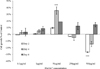
Figure 1 shows the effect of PteGlu on Caco-2 cell growth. PteGlu resulted in a differential response in Caco-2 cells at each concentration. At 0.1 µg/ml and 1 µg/ml PteGlu, there were only minor and non-significant growth differences compared to the control. The cells were also cultured in lower PteGlu concentrations – 1 ng/ml and 10 ng/ml but again cells did not show any significant difference (data not shown). This may be due to the high baseline levels of PteGlu in the normal RPMI 1640 initially used to culture the cells (1 µg/ml). However, Caco-2 cells exhibited significant differences in growth at higher PteGlu concentrations. 50 µg/ml PteGlu enhanced Caco-2 cell growth, peaking on day 4 being enhanced by 36% (p = 0.008). By contrast, Caco-2 growth was suppressed by 250 µg/ml and 500 µg/ml on day 2 and 4. With concentration of 500 µg/ml, cell growth was inhibited by approximately 30% on day 2 (p = 0.005) but this inhibition was reversed by day 4. All concentrations promoted Caco-2 cell growth by day 6, but in all cases this was not statistically significant.
MCF7 growth
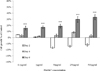
PteGlu conferred a different growth pattern in MCF7 cells in response to PteGlu concentrations compared to Caco-2 cells (Fig. 2). Although it was not statistically significant, on day 2, cell growth was increased at concentrations of 0.1 µg/ml and 1 µg/ml. The remaining three higher concentrations of PteGlu (50 µg/ml, 250 µg/ml and 500 µg/ml) inhibited MCF7 cell growth by an inverse proportion on day 2. Interestingly, 50 µg/ml of PteGlu increased cell growth of Caco-2 cells on day 2, however it caused contrasting cell growth inhibition with MCF7 on the same day. This MCF7 growth inhibition with above 50 µg/ml PteGlu was no longer apparent on day 4. All PteGlu concentrations significantly increased growth of MCF7 cells compared to the control on day 6 (p < 0.0001), yet this noticeable cell growth was not observed in the Caco-2 cell line. This MCF7 cell growth with each PteGlu concentration did not differ within groups, however it did occur in a dose dependant manner with 500 µg/ml enhancing cell growth up to 33% (p < 0.0001).
DISCUSSION
Folate is one of the best studied nutrients in relation to human health and has been extensively researched since the discovery that it can prevent NTD3031 and reduce homocysteine.1832 However, emerging evidence suggests that folate is also a significant nutrient in oncogenesis. Inappropriate folate intake and status are key risk factors for cancer due to the vitamin's critical role in DNA methylation and synthesis. Low folate nutrition is known to be associated with increased risk for cancer via aberrant DNA methylation and impaired stability. On the other hand, excess folate could also be a concern, which may prompt the progression of pre-existing neoplasms via alteration in purine and/or pyrimidine synthesis.9 The effect of folate on health, as a consequence, has been described as a double-edged sword.9 Timing and doses are therefore critical in using folic acid while treating cancer.33
The current study investigated the effects of PteGlu concentrations on the growth of both colon and breast cancer cell lines. Each cancer cell line was grown in media with different PteGlu concentrations for 6 days. At relatively low concentrations (0.1 µg/mL and 1 µg/mL), there was no significant difference of Caco-2 cell growth compared to the control. Even though Caco-2 cells had an adaption period to folate free medium for 3 weeks prior to the main experiment, high levels of baseline PteGlu within the culture medium may have resulted in a 'threshold effect' for the Caco-2 cells in relation to PteGlu metabolism. Therefore, this may explain why no significant change of cell growth was observed with a comparatively low PteGlu level.
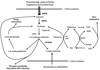
Interestingly, Caco-2 cells showed significantly different growth patterns at high concentrations – 50 µg/ml and 500 µg/ml of PteGlu. Caco-2 cell growth was enhanced, peaking on day 4 at 50 µg/ml. At a PteGlu concentration of 500 µg/ml, however, Caco-2 growth was suppressed significantly on day 2 (p < 0.005), this suppression effect was no longer apparent on day 6. It may be hypothesized that the effect of high PteGlu concentration is related to PteGlu metabolism which differs from the metabolism of natural folate coenzymes (Fig. 3).
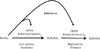
PteGlu is a synthetic form of folate which does not exist naturally and needs an additional step to enter into human folate metabolism. This additional step is via DHFR. DHFR mainly catalyzes the reduction of dihydrofolate (H2PteGlu) to tetrahydrofolate (H4PteGlu). It is also responsible for conversion of PteGlu to H2PteGlu but with a very high Km (Fig. 4). In addition, H2PteGlu allosterically modulates activity of methylentetrahydrofolate reductase (MTHFR), the central enzyme of folate metabolism.34 PteGlu from fortified food and supplements is turned into H2PteGlu and is metabolized into H4PteGlu by DHFR. Considering these characteristics of folate metabolism, the increased growth of Caco-2 cells at 50 µg/ml PteGlu could possibly be explained as follows: H2PteGlu inhibits MTHFR activity sparing 5,10-methylenetetrahydrofolate, which is shunted towards purine and pyrimidine biosynthesis instead of the methylation process. In this way PteGlu promotes cell proliferation, and therefore Caco-2 cells in 50ug/ml may show dominant growth compared to control cells (Fig. 3).34
The high Km of PteGlu for DHFR has also led to the idea that PteGlu may be an antimetabolite for DHFR, conferring competitive substrate inhibition on DHFR's activity towards H2PteGlu (Fig. 4).35 In the context of this substrate inhibition, 500 µg/ml may be a critical concentration that could result in a decrease of DHFR activity, thereby reducing H2PteGlu product formation and leading to the presence of less H4PteGlu to act as a thymidilate precursor for DNA synthesis. This mechanism might theoretically lead to a decrease in cell growth.36
MCF7 breast cancer cells showed a different response pattern from Caco-2 cells. MCF7 cells exhibited only a minor growth difference on day 2 and 4, yet all concentrations showed significantly increased growth when compared to the control on day 6 (p < 0.0001). It is possible to speculate that the DHFR enzyme activity of MCF7 is different from that of Caco-2 and, as a result MCF7 responded to PteGlu with a different pattern of growth. Such a discrepancy for DHFR activity has been reported previously.3537 DHFR activity varies between species, with the activity of DHFR in the rat being higher than in humans. Furthermore, approximately 5-fold differences in DHFR activity between individuals have been reported.35 DHFR also has various inhibition levels depending on the type of antifolate present.37 Additionally, the ratio of H2PteGlu/PteGlu and presence of genetic variants in DHFR could possibly affect enzyme activity.293839 The Caco-2 and MCF7 cell lines used for this study were confirmed as having different DHFR 19-del genotypes. It has already been reported that the DHFR 19-del homozygous recessive genotype exhibits altered DHFR mRNA expression compared to the heterozygous genotype.2940 In terms of DHFR activity, these findings could explain the differential cell growth response found between the two cell lines, raising questions related to the catalytic effect of PteGlu on DHFR in the context of subject, tissue and genotype.
In terms of mandatory folic acid fortification, the differential response of each cancer cell line may possibly imply other adverse effects of the fortification programme: Especially when considering the above results and difference in enzyme activities between individuals. Although it was mainly intended to target women of child-bearing age, the implementation of mandatory folic acid fortification has increased the total dietary folate intake of the entire population as a blanket intervention. The level of mandatory fortification has been adjusted for each country, taking into account nutritional intake status of its population and disease risk. However, the diverse characteristics of the population which could alter disease risk such as the presence of undiagnosed diseases, prescribed medications, supplement intake and genetic characteristics were not considered.41 In addition, the chronic exposure to excess PteGlu may lead to a range of unexpected effects. Junaid et al. have reported that in lymphoblastoid cells, high PteGlu in culture medium caused dysregulation of expression in more than 1,000 genes.42 We speculate that the DHFR-PteGlu interaction could be relevant: Excess PteGlu may effect DHFR activity, and its response may be differential, depending on genotype or individual, as observed in two different cell lines in the present study. In particular, excess PteGlu may have an influence on the progression of undiagnosed malignancies and potentially result in interactions with anti-cancer medicines,43 as DHFR is a major target for multiple antifolate drugs.44 Therefore, further research with respect to the safety and efficacy of mandatory folate fortification, taking these factors into account, is required. The findings of current study however are required to be interpreted with caution. The effect of PteGlu on cell proliferation was only determined by alteration of growth rate compared to control. Additionally, such differential growth trends may mainly present the tissue or disease specific characteristics in in vitro culture system.
CONCLUSION
Present data suggests that long-term exposure to high concentration PteGlu may potentially have a baseline effect in colon cancer cells, and that elevated PteGlu can lead to a contrast in the growth pattern of two different cancer cell lines, with the effect depending on exposure time and level of PteGlu, type of cancer and tissue. These findings are likely to be relevant when considering folic acid fortification in disease prevention.
 XML Download