Abstract
This study was conducted in order to compare the biological activities of leaf and root water extracts of Smilax china L. (SC) by measuring the total polyphenol and flavonoid contents, anti-oxidant activity, inhibitory effect on α-glucosidase, and anti-inflammatory gene expression. The total polyphenol and flavonoid contents of SC leaf (SCLE) and root (SCRE) water extracts were 127.93 mg GAE/g and 39.50 mg GAE/g and 41.99 mg QE/g and 1.25 mg QE/g, respectively. The anti-oxidative activities of SCLE and SCRE were measured using the 1,1-diphenyl-2-picrylhydrazyl (DPPH) and 2,2'-azino-bis(3-ethylbenzothiazoline-6-sulfonic acid) diammonium salt (ABTS) radical scavenging activity assay and reducing power assay. Both SCLE and SCRE scavenged radicals in a concentration-dependent manner, and SCLE showed stronger radical scavenging activity and reducing power than SCRE; however, both SCLE and SCRE exhibited lower activities than ascorbic acid. Compared to the anti-diabetic drug acarbose, which was used as a positive control, SCLE and SCRE exhibited low α-glucosidase inhibition activities; nevertheless, the activity of SCLE was 3.7 fold higher than that of SCRE. Finally, SCLE caused significantly decreased expression of the LPS-induced cytokines, iNOS, and COX-2 mRNA in RAW264.7 cells, indicating anti-inflammatory activity. These results indicate that SCLE might be a potential candidate as an anti-oxidant, anti-diabetic, and anti-inflammatory agent.
Figures and Tables
Fig. 1
Effects of water extract from Smilax china L. leaf and root on cell viability. Raw264.7 cells were cultured for 24 hr with various concentration of leaf and root extract. Cytotoxicity was determined by MTT assay. Results are presented as Mean ± SD of three independent experiments. SCLE: Smilax china L. leaf extract, SCRE: Smilax china L. root extract.

Fig. 2
Content of total polyphenol (A) and total flavonoid (B) of Smilax china L. leaf and root extract. As standard compounds garlic acid and quercetin, respectively, were used for measurement of polyphenol and flavonoid. Results are presented as Mean ± SD of three independent experiments.

Fig. 3
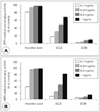
Anti-oxidative activity of Smilax china L. leaf and root extract. DPPH (A) and ABTS (B) radical scavenging activity assay was carried out according to concentration dependent manner. Ascorbic acid was used as positive control. Results are presented as Mean ± SD of three independent experiments.

Fig. 4
Anti-diabetes activity of Smilax china L. leaf and root extract. α-Glucosidase inhibitory activity assay was carried out according to concentration (A) and time (B) dependent manner. Acarbose was used as positive control. Results are presented as Mean ± SD of three independent experiments.

Fig. 5
Inhibition by water extract from Smilax china L. leaf on IL-1β, IL-6, iNOS and COX-2 mRNA expression in LPS-induced Raw 264.7 macrophage. Raw264.7 cells (1×10⁶ cells/mL) were pre-incubate for 24 hr, and the cells were stimulated with lipopolysacchride (LPS, 1 µg/mL) in the presence of Smilax china L. leaf extract (0.5 mg/mL) for 24 hr. Each value is expressed as Mean ± SD in triplicate experiments. *: p < 0.05, **: p < 0.01 compared with Con. #: p < 0.05, ##: p < 0.01 compared with LPS group. Con: non-treated (control) group, LPS: LPS alone treatment group, LPS + SCE: LPS induction in Smilax china L. leaf extract.

Table 2
DPPH and ABTS radical scavenging activity of water extracts obtained from Smilax china L. Leaf and root

Table 3
Reducing power of water extracts obtained from Smilax china L. Leaf and root Abbreviations: See Table 2

References
1. Song HS, Park YH, Jung SH, Kim DP, Jung YH, Lee MK, Moon KY. Antioxidant activity of extracts from Smilax china root. J Korean Soc Food Sci Nutr. 2006; 35(9):1133–1138.
2. Cha BC, Lee EH. Antioxidant activities of flavonoids from the leaves of Smilax china Linne. Korean J Pharmacogn. 2007; 38(1):31–36.
3. Jeong HJ, Lee SG, Lee EJ, Park WD, Kim JB, Kim HJ. Antioxidant activity and anti-hyperglycemic activity of medicinal herbal extracts according to extraction methods. Korean J Food Sci Technol. 2010; 42(5):571–577.
4. Kim EJ, Choi JY, Yu M, Kim MY, Lee S, Lee BH. Total polyphenols, total flavonoid contents, and antioxidant activity of Korean natural and medicinal plants. Korean J Food Sci Technol. 2012; 44(3):337–342.

5. Park MH, Choi C, Bae MJ. Effect of polyphenol compounds from persimmon leaves (Diospyros kaki folium) on allergic contact dermatitis. J Korean Soc Food Sci Nutr. 2000; 29(1):111–115.
6. Zhu M, Gong Y, Yang Z, Ge G, Han C, Chen J. Green tea and its major components ameliorate immune dysfunction in mice bearing Lewis lung carcinoma and treated with the carcinogen NNK. Nutr Cancer. 1999; 35(1):64–72.
7. Lee JM, Park JH, Chu WM, Yoon YM, Park E, Park HR. Antioxidant activity and alpha-glucosidase inhibitory activity of stings of Gleditsia sinensis extracts. J Life Sci. 2011; 21(1):62–67.

8. Park YM, Jeong JB, Seo JH, Lim JH, Jeong HJ, Seo EW. Inhibitory effect of red bean (Phaseolus angularis) hot water extracts on oxidative DNA and cell damage. Korean J Plant Resour. 2011; 24(2):130–138.

9. Shin JG, Park JW, Pyo JK, Kim MS, Chung MH. Protective effects of a ginseng component, maltol (2-Methyl-3-Hydroxy-4-Pyrone) against tissue damages induced by oxygen radicals. Korean J Ginseng Sci. 1990; 14(2):187–190.
10. Kwon JW, Lee EJ, Kim YC, Lee HS, Kwon TO. Screening of antioxidant activity from medicinal plant extracts. Korean J Pharmacogn. 2008; 39(2):155–163.
12. Gregg EW, Cadwell BL, Cheng YJ, Cowie CC, Williams DE, Geiss L, Engelgau MM, Vinicor F. Trends in the prevalence and ratio of diagnosed to undiagnosed diabetes according to obesity levels in the U.S. Diabetes Care. 2004; 27(12):2806–2812.

13. Lee SH, Lee JK, Kim IH. Trends and perspectives in the development of antidiabetic drugs for type 2 diabetes mellitus. Korean J Microbiol Biotechnol. 2012; 40(3):180–185.

14. Lee EB, Na GH, Ryu CR, Cho MR. The review on the study of diabetes mellitus in oriental medicine journals. J Korean Orient Med. 2004; 25(3):169–179.
15. Taylor-Fishwick DA. NOX, NOX who is there? The contribution of NADPH oxidase one to beta cell dysfunction. Front Endocrinol (Lausanne). 2013; 4:40.

16. Sasaki S, Inoguchi T. The role of oxidative stress in the pathogenesis of diabetic vascular complications. Diabetes Metab J. 2012; 36(4):255–261.

17. Tsujimoto T, Shioyama E, Moriya K, Kawaratani H, Shirai Y, Toyohara M, Mitoro A, Yamao J, Fujii H, Fukui H. Pneumatosis cystoides intestinalis following alpha-glucosidase inhibitor treatment: a case report and review of the literature. World J Gastroenterol. 2008; 14(39):6087–6092.

18. Kihara Y, Ogami Y, Tabaru A, Unoki H, Otsuki M. Safe and effective treatment of diabetes mellitus associated with chronic liver diseases with an alpha-glucosidase inhibitor, acarbose. J Gastroenterol. 1997; 32(6):777–782.

19. Sarkar D, Fisher PB. Molecular mechanisms of aging-associated inflammation. Cancer Lett. 2006; 236(1):13–23.

20. Fierro IM, Serhan CN. Mechanisms in anti-inflammation and resolution: the role of lipoxins and aspirin-triggered lipoxins. Braz J Med Biol Res. 2001; 34(5):555–566.

21. Higuchi M, Higashi N, Taki H, Osawa T. Cytolytic mechanisms of activated macrophages. Tumor necrosis factor and L-arginine-dependent mechanisms act synergistically as the major cytolytic mechanisms of activated macrophages. J Immunol. 1990; 144(4):1425–1431.
23. Sunyer T, Rothe L, Kirsch D, Jiang X, Anderson F, Osdoby P, Collin-Osdoby P. Ca2+ or phorbol ester but not inflammatory stimuli elevate inducible nitric oxide synthase messenger ribonucleic acid and nitric oxide (NO) release in avian osteoclasts: autocrine NO mediates Ca2+-inhibited bone resorption. Endocrinology. 1997; 138(5):2148–2162.

24. Kim JY, Jung KS, Jeong HG. Suppressive effects of the kahweol and cafestol on cyclooxygenase-2 expression in macrophages. FEBS Lett. 2004; 569(1-3):321–326.

25. Zarghi A, Arfaei S. Selective COX-2 inhibitors: a review of their structure-activity relationships. Iran J Pharm Res. 2011; 10(4):655–683.
26. McDaniel ML, Kwon G, Hill JR, Marshall CA, Corbett JA. Cytokines and nitric oxide in islet inflammation and diabetes. Proc Soc Exp Biol Med. 1996; 211(1):24–32.

27. Chung MJ, Walker PA, Brown RW, Hogstrand C. ZINC-mediated gene expression offers protection against H2O2-induced cytotoxicity. Toxicol Appl Pharmacol. 2005; 205(3):225–236.
28. Kim JM, Baek JM, Kim HS, Choe M. Antioxidative and anti-asthma effect of Morus bark water extracts. J Korean Soc Food Sci Nutr. 2010; 39(9):1263–1269.

29. Moreno MI, Isla MI, Sampietro AR, Vattuone MA. Comparison of the free radical-scavenging activity of propolis from several regions of Argentina. J Ethnopharmacol. 2000; 71(1-2):109–114.

30. Blois MS. Antioxidant determination by the use of a stable free radical. Nature. 1958; 181:1199–1200.
31. Re R, Pellegrini N, Proteggente A, Pannala A, Yang M, Rice-Evans C. Antioxidant activity applying an improved ABTS radical cation decolorization assay. Free Radic Biol Med. 1999; 26(9-10):1231–1237.

32. Arabshahi-Delouee S, Urooj A. Antioxidant properties of various solvent extracts of mulberry (Morus indica L.) leaves. Food Chem. 2007; 102(4):1233–1240.

33. Ryu HW, Lee BW, Curtis-Long MJ, Jung S, Ryu YB, Lee WS, Park KH. Polyphenols from Broussonetia papyrifera displaying potent α-glucosidase inhibition. J Agric Food Chem. 2010; 58(1):202–208.

34. Ko MS, Yang JB. Antioxidant and antimicrobial activities of Smilax china leaf extracts. Korean J Food Preserv. 2011; 18(5):764–772.

35. Kim YS, Lee SJ, Hwang JW, Kim EH, Park PJ, Jeong JH. Anti-inflammatory effects of extracts from Ligustrum ovalifolium H. leaves on RAW 264.7 macrophages. J Korean Soc Food Sci Nutr. 2012; 41(9):1205–1210.
36. Kang HW. Antioxidant and anti-inflammatory effect of extracts from Flammulina velutipes (Curtis) Singer. J Korean Soc Food Sci Nutr. 2012; 41(8):1072–1078.

37. Yoo KH, Jeong JM. Antioxidative and antiallergic effect of persimmon leaf extracts. J Korean Soc Food Sci Nutr. 2009; 38(12):1691–1698.

38. Oh YL. Protective effect of Smilax china L. extract on the cytotoxicity induced by chromium of environmental pollutant. J Korean Soc Plants People Environ. 2011; 14(1):29–34.
39. Lee SI, Lee YK, Kim SD, Kang YH, Suh JW. Antioxidative activity of Smilax china L. leaf teas fermented by different strains. Korean J Food Nutr. 2012; 25(4):807–819.

40. Pryor WA, Houk KN, Foote CS, Fukuto JM, Ignarro LJ, Squadrito GL, Davies KJ. Free radical biology and medicine: it's a gas, man! Am J Physiol Regul Integr Comp Physiol. 2006; 291(3):R491–R511.

41. Matough FA, Budin SB, Hamid ZA, Alwahaibi N, Mohamed J. The role of oxidative stress and antioxidants in diabetic complications. Sultan Qaboos Univ Med J. 2012; 12(1):5–18.
42. Pratt DE, Miller EE. A flavonoid antioxidant in Spanish peanuts (Arachia hypogoea). J Am Oil Chem Soc. 1984; 61(6):1064–1067.
43. Joo SY. Antioxidant activities of medicinal plant extracts. J Korean Soc Food Sci Nutr. 2013; 42(4):512–519.

44. Bischoff H. The mechanism of alpha-glucosidase inhibition in the management of diabetes. Clin Invest Med. 1995; 18(4):303–311.
45. Hwang JY, Han JS. Inhibitory effects of Sasa borealis leaves extracts on carbohydrate digestive enzymes and postprandial hyperglycemia. J Korean Soc Food Sci Nutr. 2007; 36(8):989–994.

46. Xu ML, Wang L, Xu GF, Wang MH. Antidiabetes and angiotensin converting enzyme inhibitory activity of Sonchus asper (L) hill extract. Korean J Pharmacogn. 2011; 42(1):61–67.


 PDF
PDF ePub
ePub Citation
 Print
Print


 XML Download