

 PDF
PDF ePub
ePub Citation
 Print
Print


Abstract
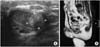
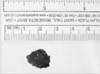
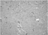
Fibrotic lesions occurring in the corpus cavernosum are usually cases of Peyronie's disease that originate from the tunica albuginea, or they are the fibrotic result of inflammatory processes. The lesion involving the corpus cavernosum, but not tunica albuginea is rare. We present here a case of fibrotic nodule arising in the corpus cavernosum with the sonographic and magnetic resonance imaging features. A 38-year-old man complained a small nodular mass in the left corpus cavernosum at the level of penoscrotal junction without abnormal curvature of the organ. We performed ultrasonography and magnetic resonance imaging to determine exactly what the lesion was. The lesion was removed and it was pathologically found to be a localized fibrotic nodule of the corpus cavernosum with some narrow-channeled vascular structures.
Figures and Tables
References
1. Lopes M, Lanzafame S, Magro G. Localized fibrosis of the corpus cavernosum: an example of fibrosis arising from the vascular smooth muscle cells. Report of a case with histogenetic considerations. Urol Int. 2000. 64:173–177.
2. Smith BH. Peyronie's disease. Am J Clin Pathol. 1966. 45:670–678.
3. Enzinger FM, Weiss S. Soft tissue tumors. 2001. 4th ed. St Louis: Mosby;45–102.
4. Bernardino ME, Jing BS, Thomas JL, Lindell MM Jr, Zornoza J. The extremity soft tissue lesion: a comparative study of ultrasound, computed tomography, and xeroradiography. Radiology. 1981. 139:53–59.
5. Hughes TM, Spillane AJ. Imaging of soft tissue tumours. Br J Surg. 2000. 87:259–260.
6. Crim JR, Seegar LL, Yao L, Chandnani V, Eckardt JJ. Diagnosis of soft tissue masses with MR imaging: can benign masses be differentiated from malignant ones? Radiology. 1992. 185:581–586.
7. Egund N, Ekelund L, Sako M, Persson B. CT of soft-tissue tumors. Am J Roentgenol. 1981. 137:725–729.
8. Schurch W, Seemayer T, Gabbiani G, Sternberg SS. Histology for pathologists. 1992. New York: Raven Press;118–125.
9. Gabbiani G. The cellular derivation and the life span of the myofibroblast. Pathol Res Pract. 1996. 192:708–711.
10. Sappino AP, Schurch W, Gabbiani G. Differentiation repertoire of fibroblastic cells: expression of cytoskeletal proteins as marker of phenotypic modulations. Lab Invest. 1990. 63:144–161.
 XML Download