

 PDF
PDF ePub
ePub Citation
 Print
Print


References
1. Baastrup CI. On the spinous processes of the lumbar vertebrae and the soft tissues between them, and on pathological changes in that region. Acta Radiol. 1933; 14:52–5.

2. Meleger AL, Krivickas LS. Neck and back pain: musculoskeletal disorders. Neurol Clin. 2007; 25:419–38.
3. Resnick DL. Diagnosis of bone and joint disorder. 4th ed.p. 1411–4. Philadelphia: W.B. Saunders;2002.
4. Bywaters EG, Evans S. The lumbar interspinous bursae and Baastrup's syndrome. An autopsy study. Rheumatol Int. 1982; 2:87–96.
5. Lin E. Baastrup's disease (kissing spine) demonstrated by FDG PET/CT. Skeletal Radiol. 2008; 37:173–5.
Go to : 
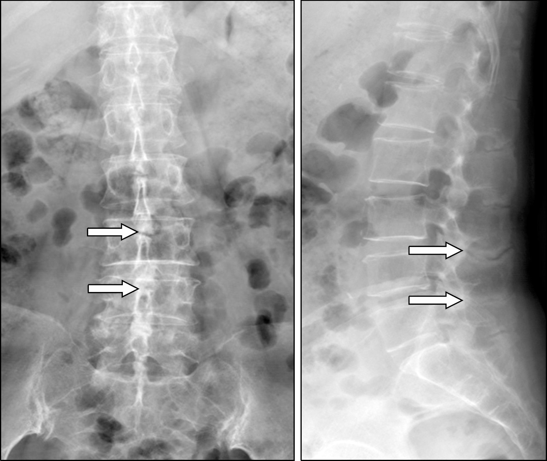 | Figure 1.Radiographies of the lumbar spine. The anteroposterior and lateral radiographs of the lumbar spine show abnormal contact of the apposing spinous processes and enlarged spinous processes that are flattened and sclerotic in their superior and inferior portions (arrows) of the whole lumbar spine. |
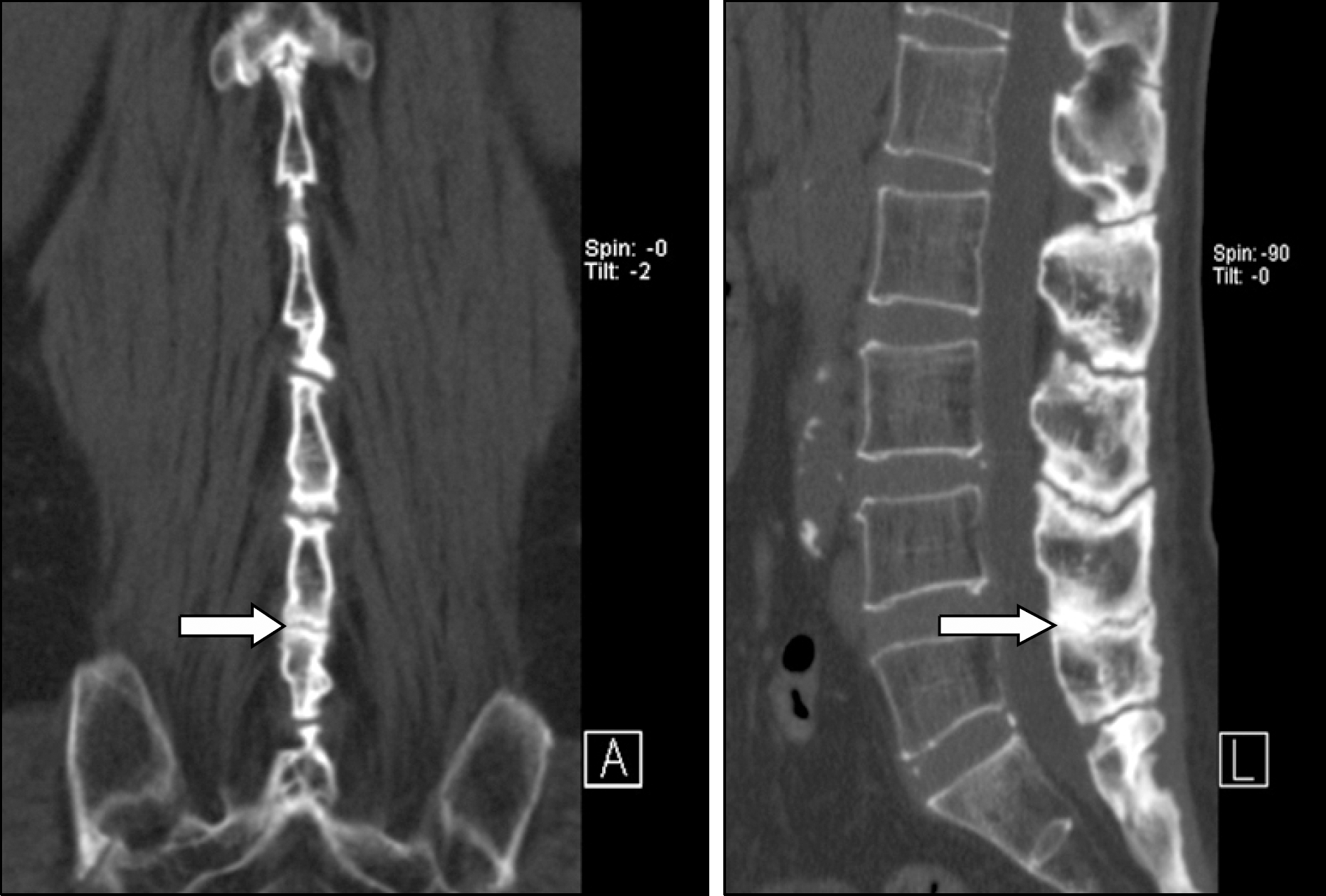 | Figure 2.CT of the lumbar spine. The coronal and sagittal reformatted CT scans also show enlarged spinous processes of the whole lumbar spine with abnormal contact of apposing spinous processes (arrow). |
 | Figure 3.Focal increased uptake in the spinous process areas (arrows) of the lower L-spine (L4 and L5 level) on the posterior whole body bone scan image (A). The right posterior oblique (RPO) and left posterior oblique (LPO) regional images show more clearly defined focal uptake lesions in the spinous process areas (arrows) and not only in the L4 and L5 levels, but also in the whole L-spine level (B). The focal uptake lesions on the bone scan images seem to match with the sclerotic lesions between the spinous processes, as detected on CT images, and the focal uptake lesions were possibly associated with active disease processes, including bony erosion and inflammatory changes, and these were most severe in the L4-5 level in this patient. |
 XML Download