Abstract
Rheumatoid arthritis is associated with an increased risk of hematological malignancy as a result of the RA itself or its treatment. We report here on an unusual case of a 55-year-old female with longstanding rheumatoid arthritis and who was treated with low dose methotrexate and hydrochloroquine. She was diagnosed with chronic myelogenous leukemia that manifested with severe throm-bocytosis and basophilia, and this was treated with imatinib mesylate. After 6 months, she achieved a complete cytogenetic response of the CML and a complete reso-lution of all the RA symptoms without DMARDs.
References
1. Williams WV, VonFeldt JM, Ramanujam T, Weiner DB. Tyrosine kinase signal transduction in rheumatoid synovitis. Semin Arthritis Rheum. 1992; 21:317–29.

2. Askling J, Fored CM, Baecklund E, Brandt L, Backlin C, Ekbom A, et al. Haematopoietic malignancies in rheumatoid arthritis: lymphoma risk and characteristics after exposure to tumour necrosis factor antagonists. Ann Rheum Dis. 2005; 64:1414–20.

3. Bernatsky S, Clarke AE, Suissa S. Hematologic malig-nant neoplasms after drug exposure in rheumatoid arthritis. Arch Intern Med. 2008; 168:378–81.

4. Asten P, Barrett J, Symmons D. Risk of developing cer- tain malignancies is related to duration of immunosuppressive drug exposure in patients with rheumatic diseases. J Rheumatol. 1999; 26:1705–14.
5. Baker GL, Kahl LE, Zee BC, Stolzer BL, Agarwal AK, Medsger TA Jr. Malignancy following treatment of rheumatoid arthritis with cyclophosphamide. Longterm case-control followup study. Am J Med. 1987; 83:1–9.
6. Wolfe F, Michaud K. Lymphoma in rheumatoid arthritis: the effect of methotrexate and antitumor necrosis factor therapy in 18,572 patients. Arthritis Rheum. 2004; 50:1740–51.

7. Pointud P, Prudat M, Peron JM. Acute leukemia after low dose methotrexate therapy in a patient with rheumatoid arthritis. J Rheumatol. 1993; 20:1215–6.
8. Georgescu L, Quinn GC, Schwartzman S, Paget SA. Lymphoma in patients with rheumatoid arthritis: association with the disease state or methotrexate treatment. Semin Arthritis Rheum. 1997; 26:794–804.
9. Cibere J, Sibley J, Haga M. Rheumatoid arthritis and the risk of malignancy. Arthritis Rheum. 1997; 40:1580–6.

10. Piper H, Mulherin D, Hardwick N. Multiple haemato-logical malignancies in a patient with rheumatoid arthritis without exposure to disease modifying therapy. Ann Rheum Dis. 2006; 65:268–9.

11. D'Aura Swanson C, Paniagua RT, Lindstrom TM, Robinson WH. Tyrosine kinases as targets for the treatment of rheumatoid arthritis. Nat Rev Rheumatol. 2009; 5:317–24.
12. Kameda H, Ishigami H, Suzuki M, Abe T, Takeuchi T. Imatinib mesylate inhibits proliferation of rheumatoid synovial fibroblast-like cells and phosphorylation of Gab adapter proteins activated by platelet-derived growth factor. Clin Exp Immunol. 2006; 144:335–41.

13. Ando W, Hashimoto J, Nampei A, Tsuboi H, Tateishi K, Ono T, et al. Imatinib mesylate inhibits osteoclastogenesis and joint destruction in rats with collagen-induced arthritis (CIA). J Bone Miner Metab. 2006; 24:274–82.

Figure 2.
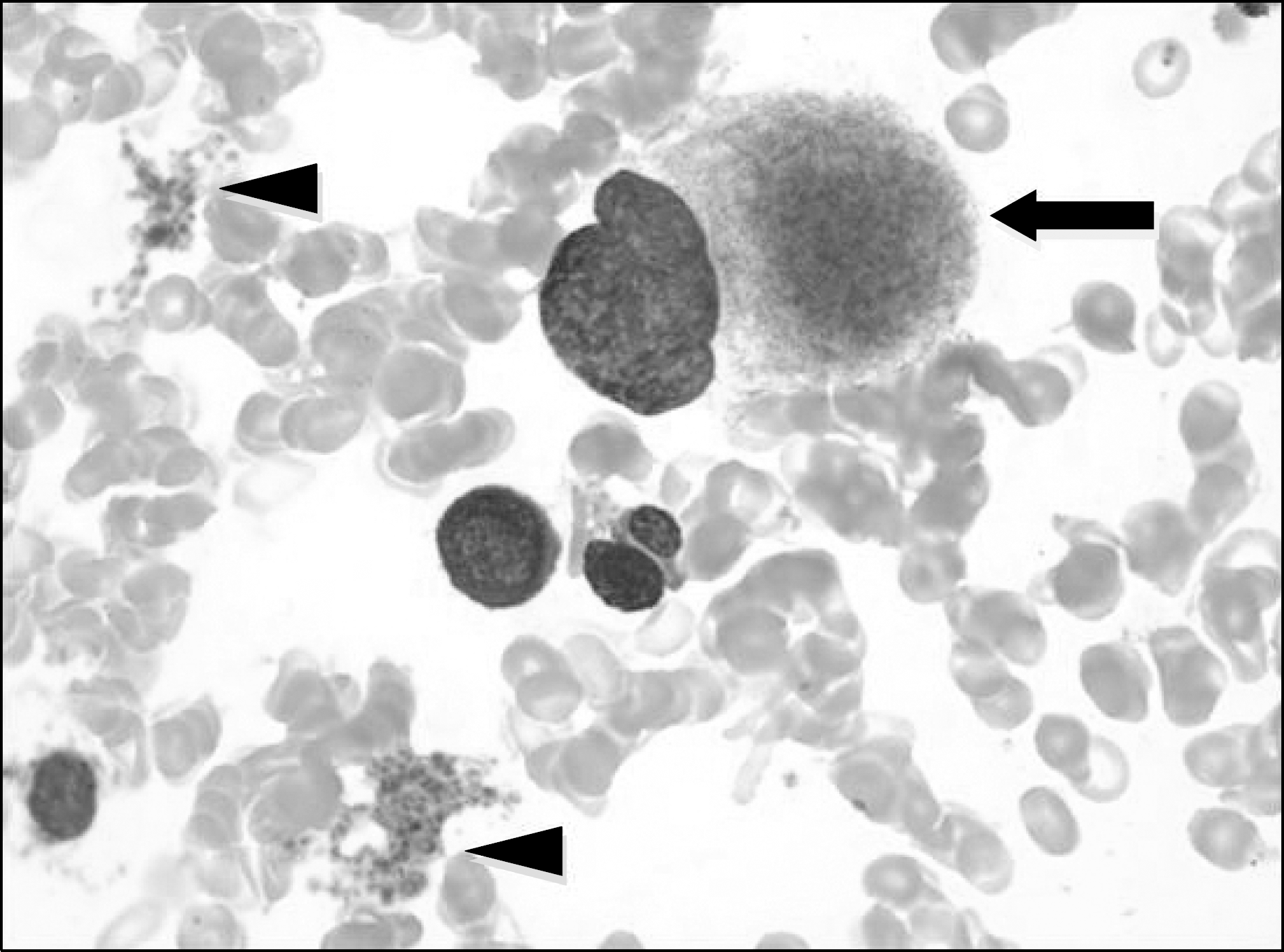
BM aspiration: The aspirate smear of the marrow and the touch preparation of the biopsy show mildly hypercellular marrow with a marked proliferation of dysplastic megakaryocytes (arrow) and many platelet clumpings (arrowhead). The blasts and pronormoblasts are mildly increased in number.


 PDF
PDF ePub
ePub Citation
 Print
Print


 XML Download