

 PDF
PDF ePub
ePub Citation
 Print
Print


Introduction
Many human diseases that appear in adulthood are related to growth patterns during early life. It is now recognized that early-life nutrition and other environmental factors play key roles in the pathogenesis and in the predisposition of specific human diseases. In this regard, in 1995 Dr. Barker wrote: "The fetal origins hypothesis states that fetal undernutrition in middle to late gestation, which leads to disproportionate fetal growth, programs later coronary heart disease."[1]. The word "program" illustrates the idea that the environmental stimuli received during critical periods of early fetal development can generate permanent changes in body structure and function that affect the homeostasis of specific organs in the adult life [2]. The responsiveness of a growing body to external cues is defined as developmental plasticity. Developmental plasticity derives from the ability of our genes to organize different ranges of physiological or morphological states in response to environmental conditions during fetal development. In this review we focus on particular situations in human and experimental animal models to underline the link between nutritional and environmental stimuli during critical periods of the early development and the susceptibility to disease development in the adult life.
Dutch Famine Birth Cohort
Insights in the importance of nutritional supply during a critical period on long-term disease outcome were gained from the Dutch Famine cohort. The Dutch Famine cohort is one of the most well-known cohorts that have been used to investigate the effects of prenatal undernutrition in humans. During the Nazi occupation from November 1944 to May 1945, food supply was extremely limited in some parts of the Netherlands. Daily rations during this famine period started at less than 1,000 kcal in November 1944, and decreased to about 500 kcal by April 1945. This six month food shortage had a major impact on particular situations, such as ongoing pregnancies. From 1998, the Dutch Famine Birth Cohort Study began reporting the outcome of pregnancies that occurred during this famine period and the consequences of massive maternal undernutrition on the offspring on a long-term scale [3-5]. Results showed an increased risk of cardiovascular diseases 40-50 years later in those children born to mothers who experienced extremely severe undernutrition during the first trimester of pregnancy [6]. The incidence of cardiovascular diseases was 2 times higher in this group compared to the control cohort [6]. Also, the serious nutritional deprivation increased the risk of metabolic disorders and breast cancer decades later in this cohort [7-9]. Notably, depending on the period of starvation (early versus late pregnancy, and preconceptional versus postnatal undernutrition) marked differences in disease outcomes were observed, indicating that the first trimester of pregnancy is particularly vulnerable to disease outcome in adulthood.
Why Your DNA Isn't Your Destiny
Another interesting epidemiological study pointed out the importance of nutritional status during puberty and its impact on the offspring's health, by investigating if the availability of food to one generation affected longevity and health of the descendants. This study conducted by Dr. Bygren, investigated the long-term effects of feast and famine on children growing up in the isolated Swedish county of Norrbotten, with the goal to unravel whether "Parents' experiences early in their lives change the traits they passed to their offspring" [10]. In the 19th century, Norrbotten was so isolated that if the harvest failed, people starved. Harvest failures occurred erratically, causing famines in 1800, 1812, 1821, 1836 and 1856. Bygren and colleagues found that if an individual went through a famine as a teenager, his or her grandchildren would have shown higher mortality risk ratio, but only if the grandchildren were the same sex as the grandparent who starved [10,11]. Starvation of a male only changed his grandson's mortality risk and starvation of a female only changed her granddaughter's mortality risk [10,11].
Early nutrition and onset of metabolic phenotype in adult
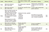
The three main insights of a number of studies from the Dutch and Norrbotten's cohorts are: 1) there may be critical developmental time windows where overnutrition or undernutrition may promote or impede disease development; 2) epigenetic factors can be modified by hunger exposure during these critical time windows; and 3) acquired epigenetic alterations are passed on to offspring. To better address these points, various animal models have been developed to explore whether nutritional or environmental modifications over critical developmental windows affect lifelong disease risk. It has been shown, for example, that overfeeding dams during gestation and lactation with high-caloric diet or reducing the number of pups in a litter could indirectly overnourish pups. Table 1 and Table 2 summarize some of the results observed in the offspring's phenotype induced by specific maternal dietary effects in different developmental time windows and in different experimental models. From these animal studies we can conclude that maternal overnutrition or undernutrition may act as an early mechanism of programming of the developing embryo/fetus, and may instruct a status of 'metabolic stress', restricting early embryonic cell proliferation and the generation of appropriately sized stem-cell pools. These observed phenotypic changes are dictated by the epigenetic-based molecular mechanisms that govern stability, transfer and expression of the eukaryotic genome.
Epigenetics
Since the Human Genome Project completed sequencing the 3 billion chemical base pairs that make up human DNA in April 2003, it became clear not only that we are able to read nature's complete genetic blueprint, but also that the information stored in the sequence of the DNA is not enough per se to completely explain human development, physiology and disease. The field dedicated to decipher the heritable features that complements the genetic information stored in the DNA sequence is termed "epigenetics". The prefix epi- is derived from the Greek preposition έπί, meaning above, on or over. Formally, epigenetics refers to the study of heritable changes in gene expression that are not caused by changes in the DNA sequence [12]. In a more practical sense, epigenetics includes the study of DNA modifications, the post-translational changes of the protein constituents of the chromatin, namely the histones, the chromatin modifications that appear to define biologic states in local regions of chromosomes, and the interaction of microRNAs with the genome [13]. Recent epigenetic studies have, for example, highlighted the mechanistic nature of the interactions between DNA and the enzymes that perform DNA replication, transcription, recombination, and repair [14].
DNA methylation
DNA methylation is a common modification in mammalian genomes and is considered a stable epigenetic mark transmitted through DNA replication and cell division [15]. Methylation is a covalent binding of a chemical methyl group to the cytosine (C) base in DNA sequence, specifically at CpG (cytosine-phosphate-guanine) dinucleotides. Methylation of C is typically associated with the 5' end of gene sequences, where CpG dinucleotides islands are particularly dense. Hypermethylation of the CpG islands is associated with transcriptional repression, while their hypomethylation is associated with the transcriptional activation of surrounding genes [16]. DNA methylation established during development and early postnatal life plays critical roles to regulate cell- and tissue-specific gene expression and genomic imprinting. Genomic imprinting is an epigenetic phenomenon in which the expression of a gene copy depends on its parent of origin [15,17], the significance of DNA methylation in genomic imprinting was highlighted in CpG islands of imprinted genes such as IGF2 and H19 [18]. In normal cells, the paternal IGF2 and maternal H19 gene are expressed while maternal IGF2 and paternal H19 are silenced by DNA methylation [19]. De novo DNA methylation is catalyzed by the DNA methyltransferases (DNMT) 3A and 3B, whereas DNA methylation is maintained after the replication of the DNA by DNMT1, which methylates hemi-methylated DNA [17]. Once established, DNA methylation is essentially maintained throughout life; however, a gradual hypomethylation occurs during aging, and it has been linked with some types of cancer [20].
Histone modification
The DNA in the cells is packaged as chromatin. The basic unit of chromatin is a nucleosome, which comprises 147 bp of DNA wrapped around a core of 8 histone proteins (two copies of histone H2A, H2B, H3 and H4) [21]. The N-terminal tails of the histones are subject to post-translational modifications including acetylation, methylation, ubiquitination, sumoylation, and phosphorylation [22]. Histone modifications lead to structural changes of the chromatin, and to the recruitment of effector proteins, for example transcription factors, which in turn bring about specific cellular processes by modulating target gene expression. Histone acetylation is exclusively associated with active and open chromatin states, while the methylation of lysine residues in the N-terminal of histones can either be an active or repressive mark depending on the specific lysine involved [23]. Many families of histone-modifying enzymes have been identified, including histone acetyl transferases, deacetylases, methyltransferases, and demethylases [24].
Non-coding RNAs
Non-coding RNAs (ncRNAs) have been implicated in the epigenetic regulation of gene expression by gene silencing or target mRNA degradation. Recent studies have shown that human miRNAs can induce chromatin remodeling [25,26], suggesting that DNA methylation, histone modification and miRNAs may work in concert to regulate gene expression.
Developmental plasticity
During the development of multicellular organisms, different cells and tissues acquire different programs of gene expression. It is well recognized that a series of precisely timed and regulated epigenetic changes are required to ensure the proper development of complex organisms like humans [15,27]. At the morula stage, an early step of embryonic development, DNA demethylation occurs to erase all of the parent-of-origin methylation marks, except those of the imprinted genes. This allows for the inheritance of parental-specific monoallelic expression in somatic tissues throughout adulthood. This demethylation phase is followed by the de novo DNA methylation of the genome to establish the proper methylation patterns of the growing organism [28]. In such a way, as the embryo grows the offspring acquires their appropriate epigenetic features. In parallel to these changes occurring in somatic cells, another set of genomic reprogramming takes place in the cells of the germ line during gametogenesis [29]. In fact, during gonadal sex determination, primordial germ cells undergo genome-wide demethylation, which erases previous parental-specific methylation marks. Afterward, for example in the male germ line, paternal methylation marks occur in specific genes in the gonocytes that subsequently will develop into spermatogonia. Conversely, the female germ line establishes maternal methylation marks of imprinted genes at a later stage. In addition to DNA methylation, histone modifications are thought to play a role in the establishment of both sex-specific and non sex-specific marks because an extensive loss of histone methylation and acetylation occurs along with the loss of DNA methylation at the morula stage [30]. Epigenetic marks assure the proper expression of the imprinted genes throughout the embryonic development. For example, genomic imprinting results in the monoallelic, parent-of-origin dependent expression of genes specifically required for key developmental steps. It is predictable from this scenario that the aberrant methylation of imprinted genes, mostly loss of imprinting (LOI) in the early stages of development, can alter the expression of critical genes, and then may bring out birth defects and adulthood diseases such as cancer [31]. In this regard, given the nature of the monoallelic expression, imprinted genes are particularly susceptible to the effect of epigenetic aberrations. In addition, for the strict dependence of these early steps of development to nutritional support, inherited and acquired epigenetic marks are particularly vulnerable to the interference coming from environmental stimuli [32].
Early life nutrition and altered epigenetic regulation
Due to the dynamic changes of the epigenetic regulation in development, particularly during gametogenesis and early embryogenesis, the epigenome displays labile nature, which allows it to respond and adapt to environmental stressors, including nutritional modification. For instance, periconceptional supplementation or restriction of the maternal diet with betaine, choline, folic acid, methionine, or vitamin B-12 in experimental models have been shown to affect the establishment of DNA methylation patterns, altering the gene expression and phenotype of the offspring [33]. Three main experimental approaches are in use to better understand the underlying mechanism by which nutritional modifications affect the epigenetic profile during critical developmental windows: 1) the study of epigenetically labile study of epigenetically labile genes, such as the insulin-like growth factor 2 (IGF2) gene; 2) the use of specific, natural animal models, such as the agouti mice; and 3) the targeting of a specific organ that is central for energy metabolism using dietary modification.
Insulin-like growth factor II (IGF2)
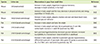
IGF2 is a key protein in human growth and development [34]. The IGF2 gene is maternally imprinted and is one of the best-characterized epigenetically regulated loci [32]. The imprinting of this locus is maintained through the methylation of a DNA sequence named the differentially methylated region (DMR), the hypomethylation of which leads to bi-allelic expression of the IGF2 gene. As illustrated by retrospective studies from the Dutch famine cohort [35] as well as in experimental animal studies [36-38], the IGF2 gene has been shown to be characterized by a labile methylation pattern depending on the nutritional or environmental stimuli received by the growing organism during the early life. Table 3 summarizes some data that support the observed developmental plasticity of the IGF2 locus. Notably, post-weaning mice fed a methyl-deficient diet exhibit permanent LOI and dysregulated expression of the IGF2 gene [37], suggesting that both childhood and maternal diet contribute to the LOI in the IGF2 locus in humans.
Agouti model
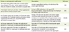
The agouti mouse has been extensively used to investigate the phenotypic impact of the nutritional modification during critical developmental periods, and the environmental influence on the fetal epigenome [39]. The agouti viable yellow (Avy) locus regulates mouse coat color; more importantly, the product of the agouti gene interferes with the regulation of body weight at the level of the hypothalamus. DNA hypomethylation of the agouti gene promoter results in the accumulation of the agouti protein; as consequence, the mouse develops a yellow coat color as well as obesity. Conversely, hypermethylation of the promoter reduces the level of the agouti protein, and this, consequently, results in mice with lean phenotype and brown coat color. Interestingly, feeding agouti female pregnant mice with a diet enriched with methyl donor supplementation such as folic acid, vitamin B12, choline and betaine has been shown to modify the phenotype of their heterozygote offspring [40]. This effect was confirmed to be mediated by the hypermethylation of the Avy promoter in the offspring of supplemented dams [41]. Table 4 provides additional evidences that specific maternal dietary treatments or environmental factors affect the phenotype of the agouti offspring through epigenetic mechanism.
Hypothalamic DNA methylation
The central brain region integrates various peripheral signals to regulate energy balance [42]. The hypothalamus plays a key role in the regulation of food intake and energy expenditure. For these reasons, the hypothalamus is an excellent organ candidate to study the epigenetic changes that mediate the developmental reprogramming of energy metabolism. Not surprisingly, maternal overnutrition [43-46] and undernutrition [47,48] have been shown to alter hypothalamic DNA methylation. These changes in DNA methylation have been reported to persist from early life to adulthood [43,46], affecting the overall metabolism in the adult.
Conclusion
Developing organisms seem to have a wide range of susceptibility to epigenetic changes [49]. Appropriate dynamics in epigenetic modifications are essential for embryogenesis, early fetal development and early postnatal growth. Consequently, the inadequate establishment of epigenetic modifications during critical developmental periods due to changes in the maternal diet or other environmental factors may induce pediatric developmental diseases and even affect health in adulthood. Since much of the reprogramming that occurs during early life may go unrecognized until adulthood, a better understanding of the interplay between genetic and epigenetic interaction in critical time windows of development would improve our ability to determine individual susceptibility to a wide range of diseases.

 XML Download