

 PDF
PDF ePub
ePub Citation
 Print
Print


Abstract
The aim of this study was to compare the effects of anticurvature filing with stainless steel k-file versus nickel-titanium ProFile in the shaping of mesial root canals of extracted mandibular molars.
A total of 60 canals from 30 mesial roots of mandibular molar teeth were randomly assigned to three groups with n=20 each. They were prepared with different instruments and methods: The first group with stainless steel k-file and circumferential filing, the second with precurved stainless steel k-file and anticurvature filing and the third with ProFile (.06 taper) and anticurvature filing. Using a micro-computed tomography system (skyscan-1076, SKYSCAN, Antwerpen, Belgium), pre-and post-operative specimens were scanned. Subsequently, canal images were superimposed and changes in root dentin thickness were measured at distal side (danger zone) of the canal. The data was analyzed using a one-way ANOVA and the comparison of means was conducted using a post hoc multiple comparison Tukey test.
There were significant differences in the change of root dentin thickness at the 7.5~8.5mm level between group 1 and 2, 3.5~6mm level between group 1 and 3 and 3.5~6mm level between group 2 and 3(n=20, P<0.05).
Figures and Tables
Figure 1
Pre-and Post-instrumentation canal images were superimposed and changes in root thickness were measured at distal side (danger zone) of the canal using the CTAn. Blue line is distance to external root surface pre-op and red line is post-op.
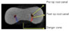
Figure 2
Change of root dentin thickness(mm) by canal preparation at danger zone before and after. At 7.5-8.5mm, significant differences were shown between group 1 and group 2(n=20, P<0.05). At 3.5-6mm, significant differences were shown between group 1 and group 3(n=20, P<0.05). At 3.5-6mm, significant differences were shown between group 2 and group 3(n=20, P<0.05).
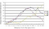
Figure 3
Comparison of group 1 and 2. The reconstructed 3-D root canal system before and after preparation is shown and superimposed cross-section images, which shows significant difference at 7.5-8.5mm level, are also shown. In group 2, more dentin was removed in the safe zone than in the danger zone at coronal 1/3 level.
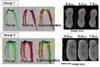
Figure 4
Comparison of group 1 and 3. The reconstructed 3-D root canal system before and after preparation is shown and superimposed cross-section images, which shows significant difference at 3.5-6mm level, are also shown.
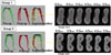
Table 1
Change of root dentin thickness(mm) by canal preparation at danger zone (mean ± S.D.). At 7.5~8.5mm, significant differences were shown between group1 and group2 (n=20, P<0.05). At 3.5~6mm, significant differences were shown between group1 and group3, 3.5~6mm level between group 2 and 3 (n=20, P<0.05).
(B): thickness of root canal dentin at danger zone before instrumentation.
(A): thickness of root canal dentin at safe zone after instrumentation.
(V): Change values of root dentin thickness by canal preparation at danger zone.
References
1. Schilder H. Clean and shaping the root canal. Dent Clin North Am. 1974. 18:269–296.
2. Bishop K, Dummer PM. A comparison of stainless steel Flexofiles and nickel-titanium NiTiFlex files during the shaping of simulated canals. Int Endod J. 1997. 30:25–34.

3. Schneider SW. A comparison of the canal preparations in straight and curved root canals. Oral surg. 1971. 32:271–275.
4. Weine F, Kelly RF, Lio PJ. The effect of proparation procedures on original canal shape and on apical foramen shape. J Endod. 1975. 1:255–262.
5. Meister F Jr, Lommel TJ, Gerstein H. Endodontic perforations which resulted in alveolar bone loss. Report of five cases. Oral Surg Oral Med Oral Pathol. 1979. 47(5):463–470.
6. Abou-Rass M, Frank AL, Glick DH. The anticurvature filing method to prepare the curved root canal. J Am Dent Assoc. 1980. 101(5):792–794.
7. Goerig AC, Michelich RJ, Schultz H. Instrumentation of root canals in molar using step-down technique. J Endod. 1982. 8:550–554.
8. Walia HM, Brantley WA, Gerstein H. An initial investigation of the bending and torsional properties of Nitinol root canal files. J Endod. 1988. 14:346–351.
9. Glossen CR, Haller RH, Dove SB, del Rio CE. A comparison of root canal preparation using Ni-Ti hand, Ni-Ti engine-driven, and K-Flex endodontic instruments. J Endod. 1995. 21(3):146–151.
10. Thompson SA, Dummer PM. Shaping ability of Hero 642 rotary nickel-titanium instruments in simulated root canals: Part 1. Int Endod J. 2000. 33:248–254.
11. Tachibana H, Matsumoto K. Application of x-ray computed tomography in endodontics. Endod Dent Traumatol. 1990. 6(1):16–20.
12. Nielsen RB, Alyassin AM, et al. Microcomputed tomography: An advanced system for detailed endodontic researc. J Endod. 1995. 21:561–568.
13. Gambill JM, Alder M, del Rio CE. Comparison of nikeltitanium and stainless steel hand file instrumentation using computed tomography. J Endod. 1996. 22:369–375.
14. Rhodes JS, Ford TR, et al. Micro-computed tomography: a new tool for experimental endodontology. Int Endod J. 1999. 32:165–170.
15. Gluskin AH, Brown DC. A reconstructed computerized tomographic comparison of Ni-Ti rotary GT files versus traditional instruments in canals shaped by novice operators. Int Endod J. 2001. 34(6):476–484.
16. Schäfer E. Shaping ability of Hero 642 rotary nickel-titanium instruments and stainless steel hand K-Flexofiles in simulated curved root canals. Oral Surg Oral Med Oral Pathol Oral Radiol Endod. 2001. 92:215–220.
17. Esposito PT, Cunningham CJ. A comparison of canal preparation with nickel-titanium and stainless steel instrumentation. J Endod. 1995. 21:173–176.
18. Peters OA, Laib A, Ruegsegger P, Barbakow F. Threedimensional analysis of root canal geometry by high resolution computed tomography. J Dent Res. 2000. 79(6):1405–1409.
19. Bergmans L, Van Cleynenbreugel J, Wevers M. A methodology for quantitative evaluation using microcomputed tomography. Int Endod J. 2001. 34(5):390–398.
20. Lim SS, Stock CJ. The risk of perforation in the curved canal : anticurvature filing compared with the step-back technique. Int Endod J. 1987. 20(1):33–39.
21. Kessler JR, Peters DD, Lorton L. Comparison of the relative risk of molar root perforation using various endodontic instrumentation techniques. J Endod. 1983. 9:439–447.
22. Kosa DA, Marshall G, Baumgartner JC. An analysis of canal centering using mechanical instrumentation techniques. J Endod. 1999. 25:441–445.
 XML Download